. 2016 Jul 21;535(7612):444-7.
doi: 10.1038/nature18622. Epub 2016 Jul 6.

The mechanism of RNA 5′ capping with NAD+, NADH and desphospho-CoA

The mechanism of RNA 5′ capping with NAD+, NADH and desphospho-CoA

Jeremy G Bird et al. Nature. .

Abstract

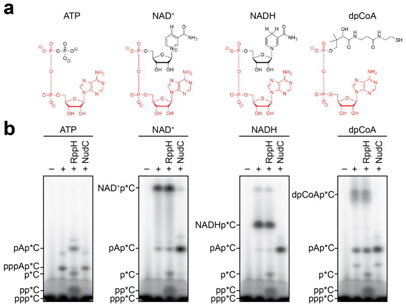
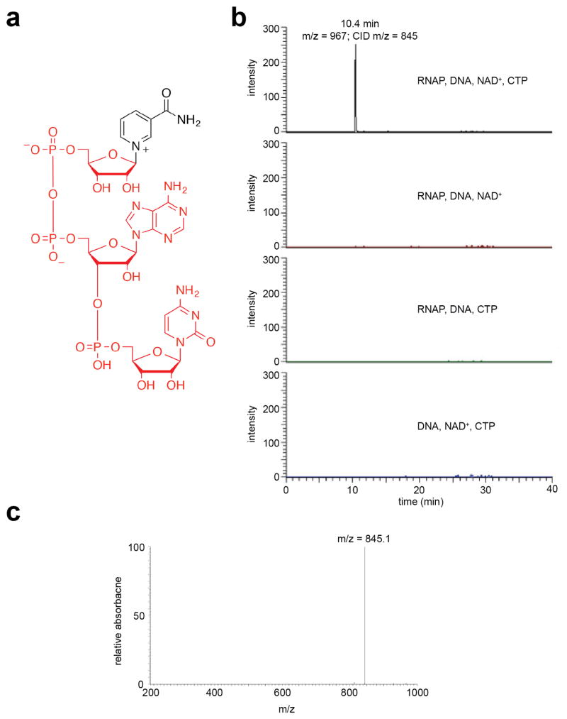
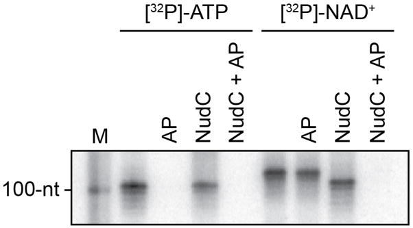
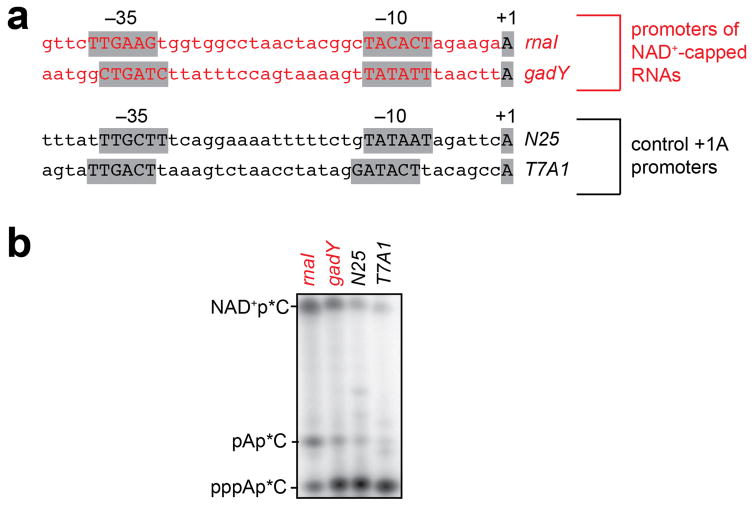
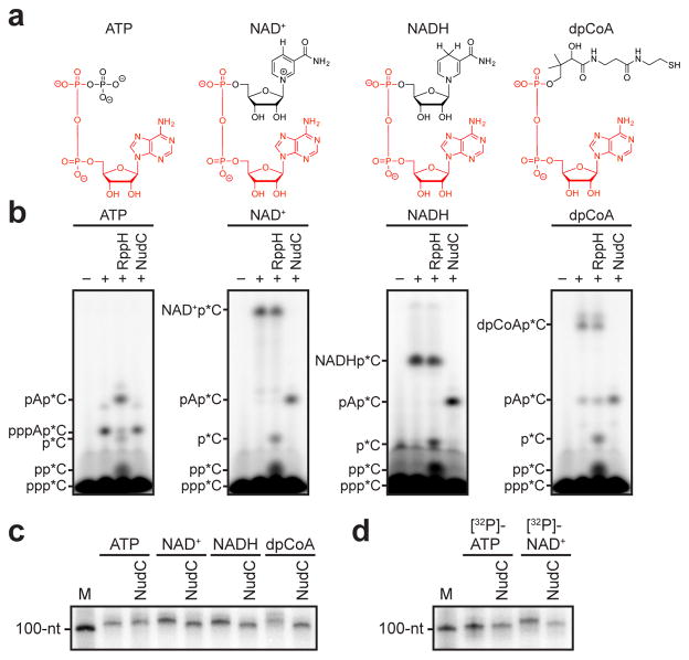
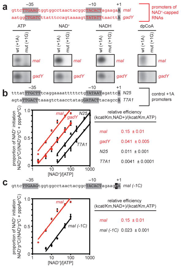
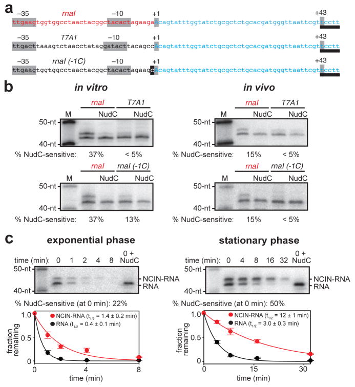
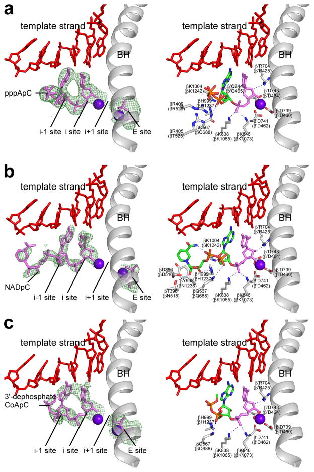
The chemical nature of the 5′ end of RNA is a key determinant of RNA stability, processing, localization and translation efficiency, and has been proposed to provide a layer of ‘epitranscriptomic’ gene regulation. Recently it has been shown that some bacterial RNA species carry a 5′-end structure reminiscent of the 5′ 7-methylguanylate ‘cap’ in eukaryotic RNA. In particular, RNA species containing a 5′-end nicotinamide adenine dinucleotide (NAD+) or 3′-desphospho-coenzyme A (dpCoA) have been identified in both Gram-negative and Gram-positive bacteria. It has been proposed that NAD+, reduced NAD+ (NADH) and dpCoA caps are added to RNA after transcription initiation, in a manner analogous to the addition of 7-methylguanylate caps. Here we show instead that NAD+, NADH and dpCoA are incorporated into RNA during transcription initiation, by serving as non-canonical initiating nucleotides (NCINs) for de novo transcription initiation by cellular RNA polymerase (RNAP). We further show that both bacterial RNAP and eukaryotic RNAP II incorporate NCIN caps, that promoter DNA sequences at and upstream of the transcription start site determine the efficiency of NCIN capping, that NCIN capping occurs in vivo, and that NCIN capping has functional consequences. We report crystal structures of transcription initiation complexes containing NCIN-capped RNA products. Our results define the mechanism and structural basis of NCIN capping, and suggest that NCIN-mediated ‘ab initio capping’ may occur in all organisms.


Figures

Extended Data Figure 1
Extended Data Figure 1. De novo transcription initiation by ATP and NCINs
a. Structures of ATP, NAD+, NADH, and dpCoA. Red, identical atoms. b. Initial RNA products of in vitro transcription reactions with ATP, NAD+, NADH, or dpCoA as initiating nucleotide and [α32P]-CTP as extending nucleotide (E. coli RNAP; PrnaI; see analogous data for PgadY in Figure 1b ). Products were treated with RppH (processes 5′-triphosphate RNA to 5′-monophosphate RNA and 5′-NTP to 5′-NDP/5′-NMP,) or NudC (processes 5′-NAD+/NADH-capped RNA to 5′-monophosphate RNA) as indicated. For gel source data, see Supplementary Figure 1.
Extended Data Figure 2
Extended Data Figure 2. LC/MS/MS analysis of initial RNA products of in vitro transcription reactions with NAD+ as initiating nucleotide and CTP as an extending nucleotide
a. Structure of NAD+pC (red, atoms corresponding to CID-generated fragment ion). b. Extracted ion chromatogram (signal derived from detection of parent ion of m/z = 967 and CID fragment of m/z = 845 corresponding to NAD+pC minus nicotinamide). Reactions contained the indicated components. c. Mass spectrum of CID fragment.
Extended Data Figure 3
Extended Data Figure 3. Sensitivity of full-length RNA products to alkaline phosphatase treatment
Full-length RNA products of in vitro transcription reactions with [γ32P]-ATP or [α32P]-NAD+ as initiating nucleotide and CTP, GTP, and UTP as extending nucleotides (E. coli RNAP; PrnaI fused to an A-less cassette ). Products were treated with alkaline phosphatase (AP; processes 5′ phosphates) or NudC (processes 5′-NAD+/NADH-capped RNA to 5′-monophosphate RNA) as indicated. Results indicate that full-length RNA products generated in reactions with [α32P]-NAD+ as initiating nucleotide are not sensitive to AP until they are processed by NudC. M, 100-nt marker. For gel source data, see Supplementary Figure 1.
Extended Data Figure 4
Extended Data Figure 4. Promoter-sequence effects on efficiency of NCIN-mediated transcription initiation: NAD+
a. Templates having rnaI, gadY, N25, and T7A1 promoters used in the assays. b. Representative raw data from experiments of Figure 2b. Initial RNA products of in vitro transcription reactions performed in the presence of 50 μM ATP and 1 mM NAD+ as initiating nucleotides and [α32P]-CTP as extending nucleotide (E. coli RNAP; PrnaI, PgadY, PN25, or PT7A1). (We note that contaminating AMP in the NAD+ stock results in production of pAp*C.) For gel source data, see Supplementary Figure 1.
Extended Data Figure 5
Extended Data Figure 5. Promoter-sequence effects on efficiency of NCIN-mediated transcription initiation: NADH and dpCoA
a. Left, dependence of NADH capping on [NADH]/[ATP] ratio (mean±SEM of 3 determinations). Right, relative efficiencies of NADH capping. (E. coli RNAP; PrnaI, PgadY, PN25, or PT7A1). b. Left, dependence of dpCoA-capping on [dpCoA]/[ATP] ratio (mean±SEM of 3 determinations). Right, relative efficiencies of dpCoA capping. (E. coli RNAP; PrnaI, PgadY, PN25, or PT7A1).
Extended Data Figure 6
Extended Data Figure 6. NCIN-mediated de novo transcrption initiation by eukaryotic RNAP II
Initial RNA products of in vitro transcription reactions with ATP, NAD+, or NADH as initiating nucleotide and [α32P]-UTP as extending nucleotide. Reactions were performed with yeast RNAP II and an artificial-bubble transcription initiation template. Products were treated with RppH or NudC as indicated. For gel source data, see Supplementary Figure 1.
Extended Data Figure 7
Extended Data Figure 7. Structural basis of NCIN-mediated transcription initiation: stereoviews
a–c. Crystal structures of RPo-pppApC, RPo-NAD+pC, and RPo-dpCoApC. Stereoviews of density and fit for initial RNA product. Green mesh, Fo-Fc omit map (contoured at 2.5σ in A–B and 2.2σ in c); red, DNA; pink, RNA product and diphosphate in “E site” (see); violet spheres, Mg2+(I) and Mg2+(II); gray, RNAP bridge helix.
Extended Data Figure 8
Extended Data Figure 8. AMP content of dpCoA stock
HPLC chromatogram of dpCoA stock (Sigma-Aldrich, lot SLBJ2886V; 50 nmol). Green: HPLC chromatogram of AMP (20 nmol). Comparison of chromatograms indicates that the dpCoA stock contains ~2% AMP. The observation that the dpCoA stock contains ~2% AMP in the dpCoA stock accounts for the formation of pApC in reactions performed with dpCoA (Figure 1b).
Figure 1
Figure 1. De novo transcription initiation by ATP and NCINs
a. Structures of ATP, NAD+, NADH, and dpCoA. Red, identical atoms. b. Initial RNA products of in vitro transcription reactions with ATP, NAD+, NADH, or dpCoA as initiating nucleotide and [α32P]-CTP as extending nucleotide (E. coli RNAP; PgadY). Products were treated with RppH (processes 5′-triphosphate RNA to 5′-monophosphate RNA and 5′-NTP to 5′-NDP/5′-NMP,) or NudC (processes 5′-NAD+/NADH-capped RNA to 5′-monophosphate RNA) as indicated. c, d. Full-length RNA products of in vitro transcription reactions with ATP, NAD+, NADH, or dpCoA as initiating nucleotide and [α32P]-CTP, GTP, and UTP as extending nucleotides (c), or with [γ32P]-ATP or [α32P]-NAD+ as initiating nucleotide and CTP, GTP, and UTP as extending nucleotides (d) (E. coli RNAP; PrnaI fused to an A-less cassette ). M, 100-nt marker. For gel source data, see Supplementary Figure 1.
Figure 2
Figure 2. Promoter-sequence effects on efficiency of NCIN-mediated transcription initiation
a. NCIN capping requires A+1. Top, promoters of NAD+-capped RNAs (promoter elements and start sites in gray). Bottom, initial RNA products of in vitro transcription reactions with ATP, NAD+, NADH, or dpCoA as initiating nucleotide and [α32P]-CTP as extending nucleotide [E. coli RNAP; wt (+1A), PrnaI (upper) or PgadY (lower); mut (+1G), +1G derivative of PrnaI (upper) or PgadY (lower)]. b. Promoter sequence determinants in addition to A+1 affect NCIN capping. Top, control +1A promoters. Bottom left, dependence of NAD+ capping on [NAD+]/[ATP] ratio (mean±SEM of 4 determinations). Bottom right, relative efficiencies of NAD+ capping. c. Promoter position −1 affects NCIN capping. Top, PrnaI (-1C) (-1 in black). Other features as in b. (mean±SEM of 3 determinations). For gel source data, see Supplementary Figure 1.
Figure 3
Figure 3. NCIN-mediated transcription initiation in vivo
a. Templates having rnaI, T7A1, and rnaI (-1C) promoters fused to identical transcribed regions (promoter elements, start sites, and position of RNA 3′-end in gray; DNA that directs synthesis of reference RNA in blue; site for MazF-mt3 endoribonuclease used to generate RNA products having uniform RNA 3′-ends, underlined). b. NCIN capping in vitro (left; 1 mM NAD+, 200 μM ATP, CTP, UTP, and GTP) and in vivo (right; RNA isolated from cells, treated with MazF-mt3 or MazF-mt3 plus NudC, and detected by hybridization). M, markers (40-nt, 50-nt). c. Effects of NCIN capping on RNA stability in vivo (rnaI template; left, exponential-phase cells; right, stationary-phase cells; times, minutes after addition of RNA-synthesis inhibitor rifampin; half-life values are the mean±SEM of 3 determinations for exponential phase and 5 determinations for stationary phase). For gel source data, see Supplementary Figure 1.
Figure 4
Figure 4. Structural basis of NCIN-mediated transcription initiation
a–c. Crystal structures of RPo-pppApC, RPo-NAD+pC, and RPo-dpCoApC. Left, electron density and atomic model for initial RNA product. Green mesh, Fo-Fc omit map (contoured at 2.5σ in a–b and 2.2σ in c); red, DNA; pink, RNA product and diphosphate in “E site” (see); violet spheres, Mg2+(I) and Mg2+(II); gray, RNAP bridge helix. Right, contacts between RPo and initial RNA product. Green and orange, carbon and phosphorus atoms derived from initiating nucleotide; pink, atoms derived from extending nucleotide; red, DNA atoms and non-DNA oxygen atoms; blue, nitrogen atoms; gray sticks, RNAP carbon atoms; violet sphere, Mg2+(I); gray ribbon, RNAP bridge helix.

Comment in

References

    1. Topisirovic I, Svitkin Y, Sonenberg N, Shatkin A. Cap and cap-binding proteins in the control of gene expression. RNA. 2011;2:277–298. - PubMed
    1. Hui M, Foley P, Belasco J. Messenger RNA degradation in bacterial cells. Annu Rev Genet. 2014;48:537–559. - PMC - PubMed
    1. Jaschke A, Hofer K, Nubel G, Frindert J. Cap-like structures in bacterial RNA and epitranscriptomic modification. Curr Opin Microbiol. 2016;30:44–49. - PubMed
    1. Kowtoniuk W, Shen Y, Heemstra J, Agarwal I, Liu D. A chemical screen for biological small molecule-RNA conjugates reveals CoA-linked RNA. Proc Natl Acad Sci USA. 2009;106:7768–7773. - PMC - PubMed
    1. Chen Y, Kowtoniuk W, Agarwal I, Shen Y, Liu D. LC/MS analysis of cellular RNA reveals NAD-linked RNA. Nature Chem Biol. 2009;5:879–881. - PMC - PubMed

Publication types

MeSH terms