Review
. 2016 Jul 7;535(7610):85-93.
doi: 10.1038/nature18849.

Interactions between the microbiota and pathogenic bacteria in the gut

Affiliations
Review

Interactions between the microbiota and pathogenic bacteria in the gut

Andreas J Bäumler et al. Nature. .

Abstract

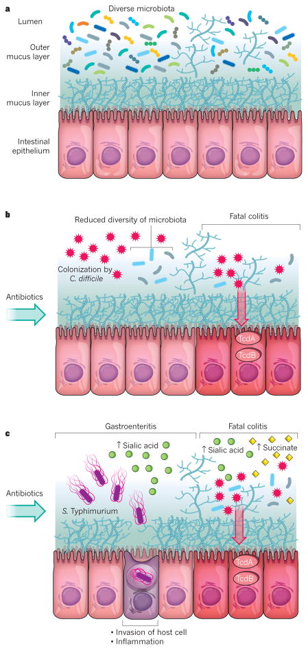
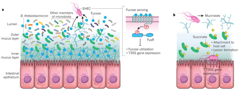
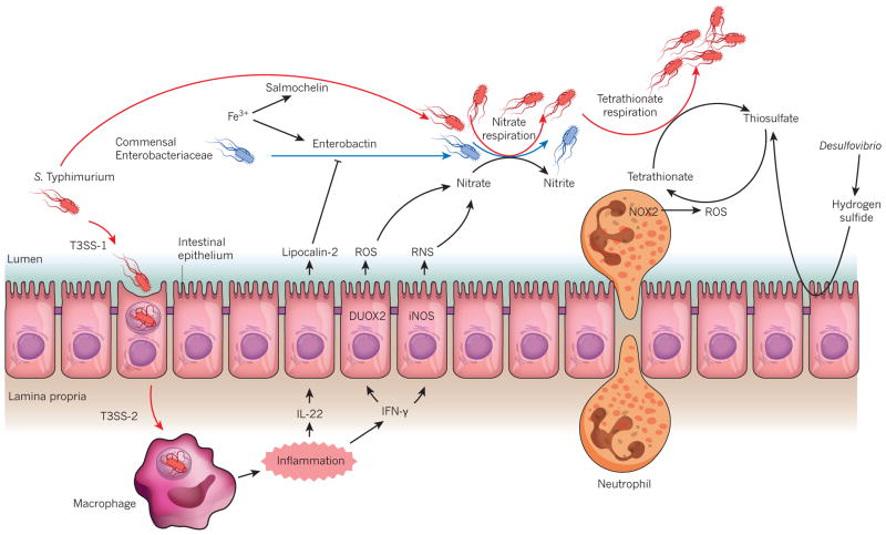
The microbiome has an important role in human health. Changes in the microbiota can confer resistance to or promote infection by pathogenic bacteria. Antibiotics have a profound impact on the microbiota that alters the nutritional landscape of the gut and can lead to the expansion of pathogenic populations. Pathogenic bacteria exploit microbiota-derived sources of carbon and nitrogen as nutrients and regulatory signals to promote their own growth and virulence. By eliciting inflammation, these bacteria alter the intestinal environment and use unique systems for respiration and metal acquisition to drive their expansion. Unravelling the interactions between the microbiota, the host and pathogenic bacteria will produce strategies for manipulating the microbiota against infectious diseases.


Conflict of interest statement

The authors declare no competing financial interests.

Figures

Figure 1
Figure 1. The impact of antibiotics on the microbiota and the expansion of enteric pathogens
a, A diverse and non-disturbed microbiota confers resistance to colonization by enteric pathogens in the intestinal epithelium. b, Treatment with antibiotics decreases the diversity of the microbiota and leads to expansion of the C. difficile population. Toxins that are released from C. difficile (TcdA and TcdB) enter and damage the cells of the epithelium, which leads to inflammation (colitis) and cell death. c, Treatment with antibiotics also leads to an increase in the levels of free sialic acid (from the host) and succinate (from the microbiota) in the lumen of the intestine. Elevated sialic acid promotes the expansion of the S. Typhimurium population, which can lead to inflammation (gastroenteritis) if the bacterium invades the cells of the intestinal epithelium. Elevated levels of sialic acid and succinate further promote the expansion of the C. difficile population and the development of colitis and cell death.
Figure 2
Figure 2. Modulation of enterohaemorrhagic E. coli virulence through nutrients provided by the microbiota
a, The microbiota resides in the lumen and outer mucus layer of the intestine. The saccharolytic bacterium Bacteroides thetaiotaomicron is a prominent member of the microbiota. It can release fucose from the mucus and makes the sugar available to other bacteria. When EHEC senses fucose through the FusKR signalling system, it represses both its use of the sugar and the expression of genes that encode the T3SS, a protein-translocation apparatus that enables the bacterium to secrete effector proteins into host cells. This repression prevents EHEC from competing for fucose with commensal E. coli and from expending energy unnecessarily on T3SS expression. b, Metabolites that are provided by B. thetaiotaomicron, such as succinate, lead to an increase in the expression by EHEC of the enzyme mucinase, which obliterates the mucus layers of the intestine. EHEC is then able to reach the intestinal epithelium. B. thetaiotaomicron then begins to secrete succinate and other metabolites that are required for gluconeogenesis into the now nutrient-poor environment. The compounds are sensed by EHEC, which upregulates its expression of the T3SS to enable the bacterium to attach to the epithelial cells of the host intestine and form lesions that cause diarrhoea.
Figure 3
Figure 3. The effect of intestinal inflammation on nutrient availability
S. Typhimurium uses its virulence factors (T3SS-1 and T3SS-2) to trigger intestinal inflammation. Cytokines that are released during inflammation, such as IL-22 and IFN-γ, trigger the release of antimicrobial molecules lipocalin-2, reactive oxygen species (ROS) and reactive nitrogen species (RNS) from the intestinal epithelium. Lipocalin-2 can block the growth of commensal Enterobacteriaceae that rely on the siderophore enterobactin for the acquisition of iron (Fe3+). It does not bind to the S. Typhimurium siderophone salmochelin, however, which confers the bacterium with resistance to its effects on growth. RNS and ROS react to form nitrate, which drives the growth of Enterobacteriaceae through nitrate respiration. Microbiota-derived hydrogen sulfide is converted to thiosulfate by colonic epithelial cells. Neutrophils that migrate into the lumen of the intestine during inflammation generate ROS that convert endogenous sulfur compounds (thiosulfate) into an electron acceptor (tetrathionate) that further boosts the growth of S. Typhimurium through tetrathionate respiration.

References

    1. Eckburg PB, et al. Diversity of the human intestinal microbial flora. Science. 2005;308:1635–1638. - PMC - PubMed
    1. Frank DN, et al. Molecular-phylogenetic characterization of microbial community imbalances in human inflammatory bowel diseases. Proc Natl Acad Sci USA. 2007;104:13780–13785. - PMC - PubMed
    1. The Human Microbiome Project Consortium. Structure function and diversity of the healthy human microbiome. Nature. 2012;486:207–214. - PMC - PubMed
    1. Ley RE, Peterson DA, Gordon JI. Ecological and evolutionary forces shaping microbial diversity in the human intestine. Cell. 2006;124:837–848. - PubMed
    1. Yurist-Doutsch S, Arrieta MC, Vogt SL, Finlay BB. Gastrointestinal microbiota-mediated control of enteric pathogens. Annu Rev Genet. 2014;48:361–382. - PubMed

Publication types

MeSH terms