. 2016 Jul 7;535(7610):153-8.
doi: 10.1038/nature18629.

Inflammasome-activated gasdermin D causes pyroptosis by forming membrane pores

Affiliations

Inflammasome-activated gasdermin D causes pyroptosis by forming membrane pores

Xing Liu et al. Nature. .

Abstract

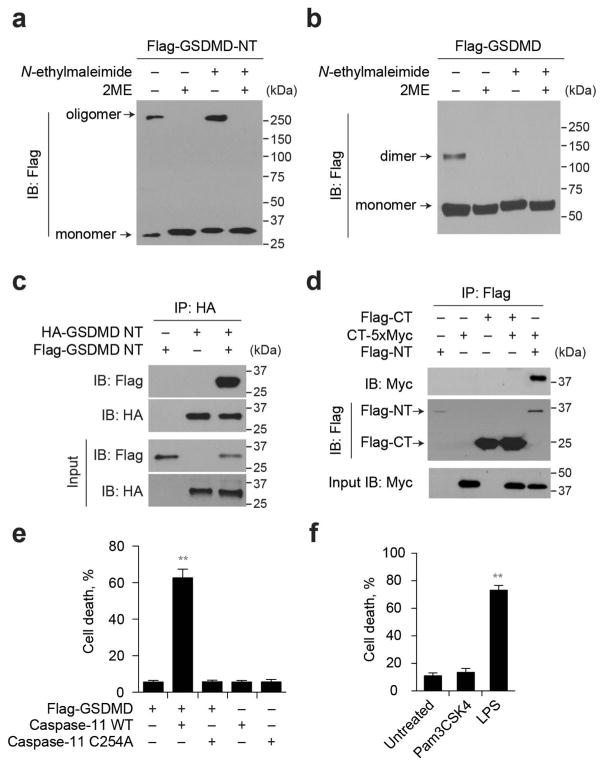
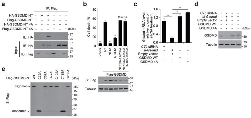
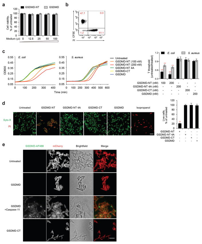
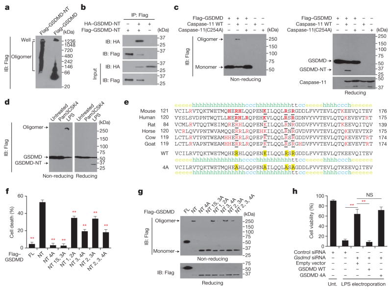
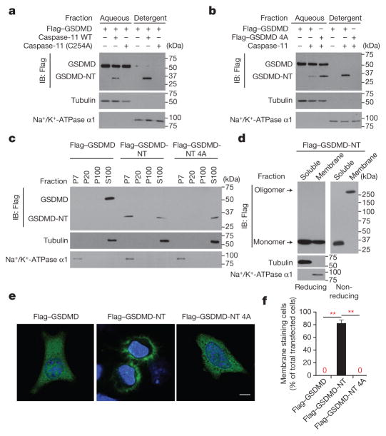
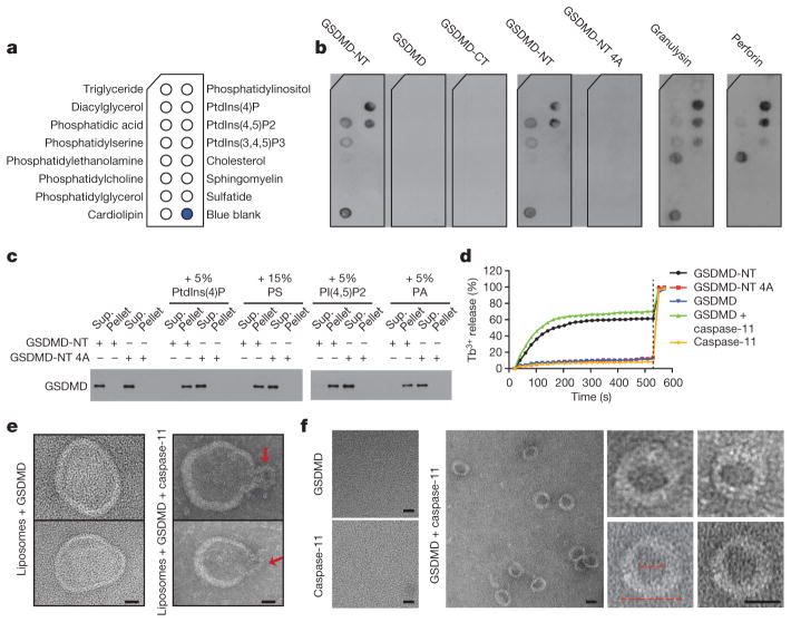
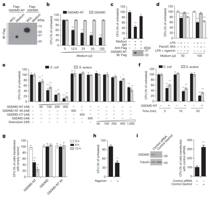
Inflammatory caspases (caspases 1, 4, 5 and 11) are activated in response to microbial infection and danger signals. When activated, they cleave mouse and human gasdermin D (GSDMD) after Asp276 and Asp275, respectively, to generate an N-terminal cleavage product (GSDMD-NT) that triggers inflammatory death (pyroptosis) and release of inflammatory cytokines such as interleukin-1β. Cleavage removes the C-terminal fragment (GSDMD-CT), which is thought to fold back on GSDMD-NT to inhibit its activation. However, how GSDMD-NT causes cell death is unknown. Here we show that GSDMD-NT oligomerizes in membranes to form pores that are visible by electron microscopy. GSDMD-NT binds to phosphatidylinositol phosphates and phosphatidylserine (restricted to the cell membrane inner leaflet) and cardiolipin (present in the inner and outer leaflets of bacterial membranes). Mutation of four evolutionarily conserved basic residues blocks GSDMD-NT oligomerization, membrane binding, pore formation and pyroptosis. Because of its lipid-binding preferences, GSDMD-NT kills from within the cell, but does not harm neighbouring mammalian cells when it is released during pyroptosis. GSDMD-NT also kills cell-free bacteria in vitro and may have a direct bactericidal effect within the cytosol of host cells, but the importance of direct bacterial killing in controlling in vivo infection remains to be determined.


Conflict of interest statement

The authors declare no competing financial interests.

Figures

Extended Data Figure 1
Extended Data Figure 1. GSDMD-NT oligomerizes and induces pyroptosis
a, b, HEK293T cells, transfected with Flag–GSDMD-NT (a) or Flag–GSDMD (b), were lysed with or without N-ethylmaleimide or 2ME, and analysed by SDS–PAGE and Flag immunoblot. c, Lysates of HEK293T cells, transfected with HA–GSDMD-NT and/or Flag–GSDMD-NT, were immunoprecipitated with anti-HA and analysed by immunoblot with the indicated antibodies. d, HEK293T cells were transfected with the indicated plasmids. Cell lysates were immunoprecipitated with anti-Flag and analysed by immunoblot with the indicated antibodies. Flag–GSDMD-NT (Flag-NT) was expressed at considerably lower levels than GSDMD-CT-MYC (CT-MYC) or Flag–GSDMD-CT (Flag-CT), which accounts for the relative weak intensity of the corresponding bands on the middle blot. e, HEK293T cells, transiently transfected with the indicated plasmids, were assessed 16 h after transfection for cell death by CytoTox96 assay. f, Immortalized iBMDMs expressing Flag–GSDMD were electroporated with PBS, ultra LPS or Pam3CSK4, as a negative control for pyroptosis. 2 h later, cell death was determined by CytoTox96 assay. Graphs show the mean ± s.d. of triplicate wells and data shown are representative of three independent experiments. **P < 0.01 (two-tailed t-test).
Extended Data Figure 2
Extended Data Figure 2. Mutation of four positively charged residues in GSDMD-NT or of two cysteine residues disrupts pyroptosis
a, Lysates of HEK293T cells, transfected with the indicated plasmids, were immunoprecipitated with anti-Flag and analysed by immunoblot with the indicated antibodies. The 4A mutant of GSDMD-NT does not self-associate in multimers. b, Mutations in other basic residues do not affect pyroptosis. The indicated wild-type or mutated Flag–GSDMD-NT constructs were transiently expressed in HEK293T cells. Medium was collected 18 h after transfection and cell death was measured by CytoTox96 assay. c, d, Knockdown in immortalized iBMDMs of Gsdmd and ectopic expression of wild-type or 4A Gsdmd mRNA (c, assessed by qRT–PCR relative to GAPDH) and protein (d, relative to tubulin). These data for the cells used in the rescue experiment in Fig. 1h show that the ectopic proteins are expressed at similar levels as the endogenous protein. e, Replacement of Cys37 or Cys192 by Ala in GSDMD-NT disrupts oligomerization. Mean ± s. d. of three technical replicates and data shown are representative of three independent experiments (b, c). Statistical differences are calculated by two-tailed t-test (in b, compared to samples transfected to express wild-type GSDMD-NT); **P < 0.01 (two-tailed t-test).
Extended Data Figure 3
Extended Data Figure 3. Treatment with GSDMD-NT reduces bacterial viability, but does not affect the viability of mammalian cells
a, Antibiotic-free culture supernatants (concentrated fivefold) from transfected HEK293T cells, collected 30 h after transfection, were added to iBMDMs, which were cultured at 37 °C in 200 μl final volume for 6 h before measuring viability by CellTiter-Glo. b, HEK293T cells, transfected with Flag–GSDMD-NT 6 h earlier, were mixed with an equal number of CFSE-labelled untransfected HEK293T cells and incubated for 18 h before assessing cell death by propidium iodide staining and flow cytometry. c, E. coli and S. aureus were untreated or treated with recombinant GSDMD, wild-type or 4A-mutant GSDMD-NT, or GSDMD-CT (200 nM or indicated concentrations) for 20 min before samples were collected and bacterial growth was assessed by monitoring turbidity by optical density (representative experiments, left). The time to reach OD600 of 0.05 above background, which is a quantitative measure of the lag in detectable growth because of fewer viable bacteria, was defined as Tthreshold (right). The right graph shows the mean ± s.d. of three technical replicates. d, Bacterial viability after 20 min incubation with indicated proteins (200 nM) or isopropanol. Syto-9 enters live and dead bacteria, PI only enters dead bacteria (representative images, left; percent live cells, right). e, Fluorescence microscopy of mCherry-expressing L. monocytogenes incubated with AlexaFluor 488-GSDMD (activated or not with caspase-11) or AlexaFluor488-GSDMD-CT for 30 min at 37 °C. Data shown are representative of results of three independent experiments. Statistical differences are relative to untreated samples; **P < 0.01 (two-tailed t-test). Scale bars, 5 μm.
Figure 1
Figure 1. GSDMD-NT forms oligomers, disrupted by mutation of four positively charged residues
a, HEK293T cells, transfected with indicated plasmids, lysed under non-reducing conditions, were resolved on a native gel, immunoblotted for Flag. b, Flag immunoprecipitation of lysates of HEK293T cells, transfected with HA–GSDMD-NT and/or Flag–GSDMD-NT, were analysed by immunoblot. c, HEK293T cells, transiently transfected with indicated plasmids, were assessed 16 h after transfection for oligomer formation by Flag immunoblot of non-reducing (left) or reducing (right) gels. The reducing gel was also blotted for caspase-11. d, iBMDMs expressing Flag–GSDMD were electroporated with phosphate-buffered solution (PBS), LPS or Pam3CSK4 and analysed 2 h later by Flag immunoblot. e, The cluster of four evolutionarily conserved, positively charged amino acids (red and underlined) in GSDMD-NT were mutated to Ala. Secondary structures of the wild-type and 4A-mutated mouse GSDMD fragment were predicted using the SOPMA algorithm. h, helix; e, sheet; t, turn; c, coil. f, g, Mutations of mouse GSDMD-NT block GSDMD-NT-mediated pyroptosis (f) and oligomerization (g). The first to fourth amino acids (R138/K146/R152/R154) were mutated to Ala. The mutated residues are indicated, i.e. in NT4A, all 4 residues are mutated; in NT 3, 4A, the 3rd and 4th residues are mutated. In NT1S3A, R138 was mutated to Ser, the other three residues were mutated to Ala. FL, full-length GSDMD; NT, GSDMD-NT. HEK293T cells, transfected with wild-type (WT) or mutated GSDMD-NT, were analysed 18 h later for cell death (CytoTox96 assay) and GSDMD oligomerization (Flag immunoblot). GSDMD-NT monomer and oligomer are indicated. h, iBMDMs, co-transfected with control or Gsdmd siRNA and the indicated siRNA-resistant Gsdmd expression plasmids, were electroporated with LPS. The number of surviving cells was determined by CellTiter-Glo assay 2.5 h later. Mean ±s.d. of three technical replicates from one of three independent experiments are shown (f, h). Statistical differences are relative to Flag–GSDMD-NT-expressing samples (f). **P <0.01 (two-tailed t-test). NS, not significant; unt., not transfected with LPS.
Figure 2
Figure 2. GSDMD-NT localizes to the plasma membrane
a, b, Lysates of HEK293T cells, transfected with indicated plasmids for 16 h, were separated into aqueous and detergent phases using Triton X-114, and analysed by immunoblot probed for Flag, tubulin, or Na+/K+-ATPase α1. c, HEK293T cells, transfected with indicated plasmids for 16 h, were separated into P7 (heavy membrane), P20 (light membrane), P100 (insoluble cytosol) and S100 (soluble cytosol) fractions and analysed by immunoblot with indicated antibodies. d, Soluble and crude membrane fractions of HEK293T cells, transfected to express Flag–GSDMD-NT, were analysed by immunoblot as indicated. e, f, Representative confocal microscopy images (e) and quantification (f) of distribution of ectopic Flag–GSDMD, Flag–GSDMD-NT and Flag–GSDMD-NT 4A (green) in HeLa cells co-stained with DAPI (blue). The ratio of cells with membrane versus cytosolic Flag staining was calculated by counting 10 high-power fields for each sample in 5 independent experiments (f). **P <0.0001 (paired t-test). Scale bar, 20 μm. Data are representative of three independent experiments (a–d).
Figure 3
Figure 3. N-terminal gasdermin D binds to phosphatidyl serine and cardiolipin and forms pores on liposomes
a, b, Membranes displaying lipids (a) were incubated with indicated proteins and binding was assessed by blotting for GSDMD, perforin or granulysin (b). c, Wild-type or 4A-mutant GSDMD-NT binding to PC–PE liposomes containing additional indicated phospholipids (molar proportion of added lipid indicated) was analysed by SDS–PAGE and GSDMD immunoblot. d, Liposome leakage was monitored by terbium (Tb3+) fluorescence after incubation with the indicated GSDMD protein ±caspase-11. Detergent was added after 9 min (dotted line). e, Negative staining electron microscopy images of PE–PC–PS liposomes treated with GSDMD (left) or caspase-11-activated GSDMD (right). Arrows indicate potential side views of GSDMD-NT pores. f, Negative staining electron microscopy images of GSDMD-NT pores formed in PS-containing liposomes and extracted by detergent. The left image of pores formed by GSDMD and caspase-11 shows a field with multiple rings, whereas the right images show enlarged single rings. The inner and outer diameters (red dotted lines) are approximately 15 nm and 32 nm, respectively. Data are representative of three independent experiments. Scale bars, 20 nm
Figure 4
Figure 4. GSDMD-NT kills bacteria
a, Culture supernatants and whole-cell lysates (WCL) of HEK293T cells, transiently expressing Flag–GSDMD-NT or Flag–GSDMD for 20 h, were analysed by Flag immunoblot of reducing gel. b, Antibiotic-free culture supernatants (concentrated fivefold) from transfected HEK293T cells, collected 30 h after transfection, were added to E. coli, which were cultured at 37 °C in 200 μl final volume for 30 min before measuring c.f.u. c, The concentrated culture medium from Flag–GSDMD-NT-expressing HEK293T cells was immunodepleted with anti-Flag or control IgG, before adding to E. coli, as in b. Lower panel, Flag immunoblot. d, L. monocytogenes were incubated at 37 °C for 30 min with antibiotic-free culture supernatants from iBMDMs, transfected with LPS or Pam3CSK4 or incubated with LPS and nigericin for 3 h, before assessing c.f.u. e, f, E. coli and S. aureus were treated with the indicated proteins for 20 min (e) or with 200 nM wild-type GSDMD-NT for the indicated times (f) before measuring c.f.u. g, HeLa cells, transfected for 6 h to express the indicated proteins, were infected with L. monocytogenes for the indicated times before cells were lysed to analyse intracellular c.f.u. h, LPS-primed-iBMDMs infected with L. monocytogenes, were treated or not with nigericin for 1 h before bacteria were collected and c.f.u. was analysed. Nigericin had no effect on cell-free bacteria (not shown). i, iBMDMs, transfected with control or Gsdmd siRNA, were infected with L. monocytogenes and assessed for intracellular c.f.u. 12 h later. GSDMD immunoblot, left. Shown are mean ± s.d. of triplicates of one experiment of three (b, d–f, h) or two (c, g, i) independent experiments. Statistical differences compared to untreated control samples (two-tailed t-test); *P <0.05, **P <0.01.

References

    1. Shi J, et al. Cleavage of GSDMD by inflammatory caspases determines pyroptotic cell death. Nature. 2015;526:660–665. - PubMed
    1. Kayagaki N, et al. Caspase-11 cleaves gasdermin D for non-canonical inflammasome signalling. Nature. 2015;526:666–671. - PubMed
    1. Rathinam VA, et al. TRIF licenses caspase-11-dependent NLRP3 inflammasome activation by gram-negative bacteria. Cell. 2012;150:606–619. - PMC - PubMed
    1. Hagar JA, Powell DA, Aachoui Y, Ernst RK, Miao EA. Cytoplasmic LPS activates caspase-11: implications in TLR4-independent endotoxic shock. Science. 2013;341:1250–1253. - PMC - PubMed
    1. Law RH, et al. The structural basis for membrane binding and pore formation by lymphocyte perforin. Nature. 2010;468:447–451. - PubMed

Publication types

MeSH terms