. 2016 Jul 7;535(7610):159-63.
doi: 10.1038/nature18631. Epub 2016 Jun 17.

Genetic dissection of Flaviviridae host factors through genome-scale CRISPR screens

Genetic dissection of Flaviviridae host factors through genome-scale CRISPR screens

Caleb D Marceau et al. Nature. .

Abstract

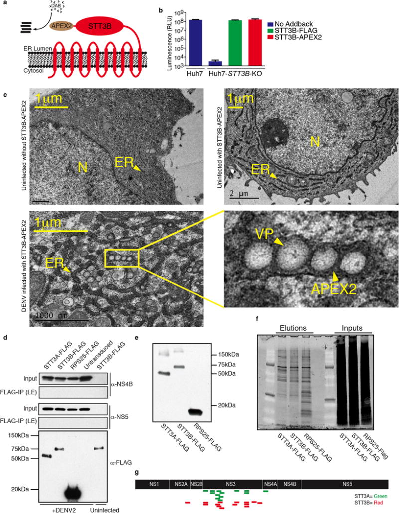
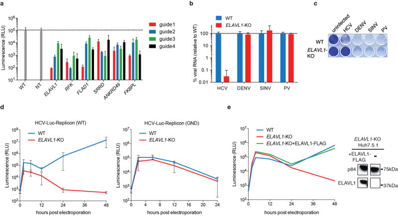
The Flaviviridae are a family of viruses that cause severe human diseases. For example, dengue virus (DENV) is a rapidly emerging pathogen causing an estimated 100 million symptomatic infections annually worldwide. No approved antivirals are available to date and clinical trials with a tetravalent dengue vaccine showed disappointingly low protection rates. Hepatitis C virus (HCV) also remains a major medical problem, with 160 million chronically infected patients worldwide and only expensive treatments available. Despite distinct differences in their pathogenesis and modes of transmission, the two viruses share common replication strategies. A detailed understanding of the host functions that determine viral infection is lacking. Here we use a pooled CRISPR genetic screening strategy to comprehensively dissect host factors required for these two highly important Flaviviridae members. For DENV, we identified endoplasmic-reticulum (ER)-associated multi-protein complexes involved in signal sequence recognition, N-linked glycosylation and ER-associated degradation. DENV replication was nearly completely abrogated in cells deficient in the oligosaccharyltransferase (OST) complex. Mechanistic studies pinpointed viral RNA replication and not entry or translation as the crucial step requiring the OST complex. Moreover, we show that viral non-structural proteins bind to the OST complex. The identified ER-associated protein complexes were also important for infection by other mosquito-borne flaviviruses including Zika virus, an emerging pathogen causing severe birth defects. By contrast, the most significant genes identified in the HCV screen were distinct and included viral receptors, RNA-binding proteins and enzymes involved in metabolism. We found an unexpected link between intracellular flavin adenine dinucleotide (FAD) levels and HCV replication. This study shows notable divergence in host-depenency factors between DENV and HCV, and illuminates new host targets for antiviral therapy.


Conflict of interest statement

The authors declare no competing financial interest.

Figures

Extended Data Figure 1
Extended Data Figure 1. Divergence of DENV and HCV host factors
(a) Gene ontology (GO) analysis for DENV and HCV CRISPR screens on the ranked gene lists. Curated (by redundancy) enriched GO terms are shown. A complete list of all enriched GO terms can be found in Supplementary Data Table 4. (b) Distribution of the subcellular location of the 30 most enriched host factors for DENV and HCV. (c) Cross-comparison of the effects of DENV or HCV host factor knockout in Huh7.5.1 cells on the replication of DENV or HCV using reporter viruses expressing luciferase. Data depict average with s.d. for triplicate infections.
Extended Data Figure 2
Extended Data Figure 2. Reproducibility of CRISPR screens
(a) Ranked lists of the 30 most enriched DENV and HCV host factors and their rankings in the individual replicate screens. The color code reflects in what percentile the gene scored in the replicate. (b) Gene enrichment based on RIGER score for the individual replicate screens. Red dots highlight where the 30 most significant host factors ranked in the individual replicates.
Extended Data Figure 3
Extended Data Figure 3. Genotyping of cell lines for DENV host factors
(a) The site of gene trap insertion in HAP1 cell lines was determined using a PCR based method. Bases depicted in red are the flanking sequences upstream of the genetrap insertion. Bases depicted in green are downstream gene-trap flanking sequences. (b) TALENs were used to edit the genomic region of AUP1 in HAP1 cells. Bases depicted in red are the left TALEN binding site, bases depicted in blue are the right TALEN binding site. Bases depicted in green are the TALEN target site. Arrow indicates site of 260bp insertion. (c) CRISPR Cas9 nuclease was targeted to bases depicted in red in HAP1 cells. Editing events are depicted at the gRNA target sites below the WT sequence. (d) CRISPR Cas9 nuclease was targeted to bases depicted in red in Huh7 cells. Editing events are depicted at the gRNA target sites below the WT sequence. (e) Immunoblots of wild type and KO cell lines.
Extended Data Figure 4
Extended Data Figure 4. Validation of DENV host factor genes
(a) Plaque-forming units (PFU) assay of DENV infection. ND indicates no plaques were detected (threshold of detection of the assay is 6 PFU/ml). (b) DENV luciferase levels in HAP1 isogenic knockout cells complemented using lentiviral stable expression of corresponding genes. (c) Crystal violet of complemented Huh7 knockout cells infected with DENV. (d) DENV luciferase levels in Raji DC-SIGN cells with KO in DENV host factors (lentiCRISPRv2). Empty denotes an empty vector control (expressing Cas9 but no guideRNA) and NT a cell line expressing a non-targeting guideRNA. (e) Time course of DENV and HCV expressing Renilla luciferase in Huh7 knockout cells. (f) Schematic diagram of the STT3A and STT3B isoforms. Gene names in red indicate OST subunits identified in the DENV screens. Data depict average with s.d. for triplicate infections.
Extended Data Figure 5
Extended Data Figure 5. Catalytic site mutations introduced in mammalian STT3A and STT3B
(a) Catalytic site amino acids highlighted in red as identified in the bacterial STT3 (C. lari pglb). Strong conservation allows their identification in other species. Alignments of STT3 isoforms across different species highlight the conserved catalytic sites that were mutated. The table specifies the amino acid position and the specific triple mutations that were made to abolish catalytic activity. (b) Huh7 STT3A and STT3B KO cells expressing FLAG tagged STT3A and STT3B WT and catalytic mutants.
Extended Data Figure 6
Extended Data Figure 6. Physical interaction between the OST complex and the replication complex of DENV
(a) APEX2, a protein tag for electron microscopy was fused to the C-terminus of STT3B enabling the imaging of subcellular protein localization by deposition of a polymer of 3,3′-diaminobenzidine (DAB). (b) Luminescence of Huh7 STT3B-KO cells complemented with STT3B-APEX2 and infected with DENV expressing Renilla luciferase. Data depict average with s.d. for triplicate infections. (c) STT3B localizes on ER membranes in the vicinity of DENV-induced vesicle packets as shown by transmission EM micrograph of DENV-infected or uninfected Huh7 cells expressing the STT3B-APEX2 construct. N represents the cell Nucleus and the arrowheads in samples transfected with STT3B-APEX2 represent APEX polymerized DAB staining in the lumen of the Endoplasmic Reticulum (ER) or around DENV-induced vesicle packets (VP). (d) Co-immunoprecipitations (IP) of STT3A-FLAG and STT3B-FLAG from DENV infected cell lysates. LE = long exposure. (e) Anti-FLAG Western blots of IP elutions of DENV infected cells stably expressing FLAG tagged STT3A, STT3B and RPS25. (f) SYPRO Ruby staining of elutions and inputs of IP of DENV infected cell lysates. (g) Co-IP elutions of DENV infected lysates were analyzed by mass spectrometry and DENV specific peptides aligned to DENV polyprotein.
Extended Data Figure 7
Extended Data Figure 7. Analysis of HCV host factor knockout cell lines
(a) Genotyping of CRISPR-induced KO Huh7.5.1 cells by Sanger sequencing showing the mutated locus and the WT reference. CRISPR/Cas9 induces mutations close to the PAM site resulting in frameshifts. CD81 and ELAVL1 KO cell lines are subclones whereas others are populations of cells mutagenized with lentiCRISPRv2. (b) Immunoblots of CRISPR-induced KO cells.
Extended Data Figure 8
Extended Data Figure 8. ELAVL1 is a critical host factor for HCV replication
(a) HCV luciferase infection in KO cell lines using 4 different guideRNAs per gene. NT = non-targeting guideRNA. (b) QPCR of viral RNA in WT or ELAVL1-KO Huh7.5.1 cells. (c) Crystal violet assay for different RNA virus infections. (d) HCV replicon assays using wild-type sgJFH1 (left) or GND sgJFH1 replicon. Note that one downward error bar for the right panel (6 hours, ELAVL1-KO) was not plotted because it would reach a negative value, which cannot be plotted on a logarithmic scale. (e) Transfection of ectopically expressed ELAVL1 restores HCV replication. Western blot of ELAVL1-FLAG transfected and untransfected Huh7.5.1 ELAVL1-KO cells. Data depict average with s.e.m. (QPCR) or s.d. (FFU, RLU) for triplicate infections, except e, which was a single infection.
Extended Data Figure 9
Extended Data Figure 9. Lumiflavin inhibits the replication of HCV but not other RNA viruses
(a) QPCR of HCV or DENV RNA replication in WT, RFK-KO or FLAD1-KO Huh7.5.1. (b) Immunofluorescence of HCV infection in WT, RFK-KO and FLAD1-KO Huh7.5.1 cells under treatment with lumiflavin, FMN or FAD. HCV core protein (green). Blue=DAPI. (c) Western blot for HCV core and DENV NS5 in untreated (UT) and lumiflavin-treated Huh7.5.1 cells. p84 and GAPDH served as loading controls. (d) QPCR of RNA viruses in untreated or lumiflavin-treated Huh7.5.1 cells. (e) MTT cell proliferation assay for lumiflavin-treated Huh7.5.1 cells. (f) Restoration of HCV replication in lumiflavin-treated cells by exogenous addition of FMN or FAD. Data depict average with s.e.m. (QPCR) or s.d. (MTT) for triplicate infections/treatments.
Extended Data Figure 10
Extended Data Figure 10. Comparison of knockout screen results to previous siRNA screens
(a) Venn diagram comparing the hits from the CRISPR and haploid screens for DENV host factors to previous siRNA screens from Sessions et al. (from Supplementary Table 2) and Krishnan et al. (from Supplementary Table 1). The top ten validated host factors (by strength of phenotype in the validation screen) for each screen are shown next to circle. (b) Venn diagram comparing the hits from the CRISPR screen for HCV host factors to previous siRNA screens from Tai et al. (from Table S2) and Li et al. (from Dataset S1).
Figure 1
Figure 1. Haploid and CRISPR genetic screens identify essential host factors of DENV and HCV infections
(a) Schematic for genome-wide screening approach. (b) Haploid genetic screen for DENV host factors. The y-axis represents significance of enrichment of gene-trap insertions in genes in DENV resistant population compared to unselected HAP1 cells. Each circle represents a specific gene and size corresponds to number of independent gene-trap insertions. All genes with p-value <0.05 were colored and grouped by function. The screen was performed once. (c) CRISPR genetic screen for DENV and (d) for HCV host factors in Huh7.5.1 cells. Significance of enrichment was calculated by RIGER analysis. The screens were performed in three replicates and the mean of the RIGER score is represented on the y-axis. The 30 most enriched genes were colored and grouped by function. (e) Comparison of the 30 most enriched genes from the DENV and HCV CRISPR screens and their position based on the mean RIGER score.
Figure 2
Figure 2. ER protein complexes play a crucial role in the replication phase of DENV and are also important for YFV, WNV and ZIKV infection
(a) QPCR of DENV16681, YFV17D, WNVKUNJIN and ZIKVUganda RNA in knockout HAP1 cells. (b) QPCR of prototypic strains of DENV serotypes 1–4 RNA in knockout Huh7 cells. (c) Confocal Microscopy of STT3B-KO Huh7 cells immunostained for DENV Envelope protein immediately or 30 minutes after DENV infection. (d) Luminescence of DENV replicon RNA expressing luciferase in knockout Huh7 cells. The DENV NS5-GDD mutant served as replication-deficient control. Data depict average with s.e.m. (QPCR) or s.d. (RLU) for triplicate infections.
Figure 3
Figure 3. DENV RNA replication requires a non-canonical function of OST, and DENV non-structural proteins interact with OST
(a) Viability of STT3A and STT3B double knockout cells complemented with wild-type (wt) or catalytic (cat) mutant cDNA. (b) Glycosylation of DENV protein NS1, SHBG, and pSAP in STT3A and STT3B knockout Huh7 cells. Different glycoforms are indicated by arrowheads. Tun. = Tunicamycin (c) Glycosylation state of pSAP and SHBG in STT3A- and STT3B-KO cells complemented with catalytic mutants. (d) DENV infection of knockout Huh7 cells complemented with WT or catalytic mutants of STT3A and STT3B. Data depict average with s.d. for triplicate infections. (e) Co-immunoprecipitations of STT3A-FLAG and STT3B-FLAG from DENV infected cell lysates. LE = long exposure.
Figure 4
Figure 4. FAD biosynthesis is required for HCV replication and can serve as antiviral target
(a) QPCR of HCV RNA in Huh7.5.1 cell lines. (b) HCV particle formation measured by focus-forming units (FFU) assay. ND indicates that no foci were detected (threshold of detection is 50 FFU/ml). (c) Biosynthesis pathway of FAD. Lumiflavin (LF) competitively inhibits uptake of riboflavin. (d) QPCR of HCV RNA in untreated, FMN- or FAD-treated RFK- and FLAD1-KO Huh7.5.1 cells. (e) QPCR of DENV or HCV RNA in lumiflavin-treated Huh7.5.1 cells. For each concentration the significance of the effect on HCV versus DENV was determined. (f) HCV replicon assay in untreated and lumiflavin-treated Huh7.5.1 cells using WT sgJFH1 replicon. (g) Model of identified DENV and HCV host factors. Data depict average with s.e.m. (QPCR) or s.d. (FFU, RLU) for triplicate infections. The p-values were determined using an unpaired, parametric, two-sided student t-test, with a Welch post-correction, where * – P <0.05, ** – P <0.01, *** – P <0.001. ns = non-significant

References

    1. Bhatt S, et al. The global distribution and burden of dengue. Nature. 2013;496:504–507. doi: 10.1038/nature12060. - DOI - PMC - PubMed
    1. Thomas SJ, Rothman AL. Trials and Tribulations on the Path to Developing a Dengue Vaccine. American journal of preventive medicine. 2015;49:S334–344. doi: 10.1016/j.amepre.2015.09.006. - DOI - PubMed
    1. Lavanchy D. Evolving epidemiology of hepatitis C virus. Clinical microbiology and infection : the official publication of the European Society of Clinical Microbiology and Infectious Diseases. 2011;17:107–115. doi: 10.1111/j.1469-0691.2010.03432.x. - DOI - PubMed
    1. Paul D, Bartenschlager R. Flaviviridae Replication Organelles: Oh, What a Tangled Web We Weave. Annu Rev Virol. 2015;2:289–310. doi: 10.1146/annurev-virology-100114-055007. - DOI - PubMed
    1. Shalem O, et al. Genome-scale CRISPR-Cas9 knockout screening in human cells. Science. 2014;343:84–87. doi: 10.1126/science.1247005. - DOI - PMC - PubMed

Publication types

MeSH terms

Substances