Review
. 2016 Jul;203(3):1027-67.
doi: 10.1534/genetics.115.186452.

Chromosome Duplication in Saccharomyces cerevisiae

Affiliations
Review

Chromosome Duplication in Saccharomyces cerevisiae

Stephen P Bell et al. Genetics. 2016 Jul.

Abstract

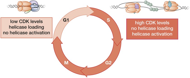
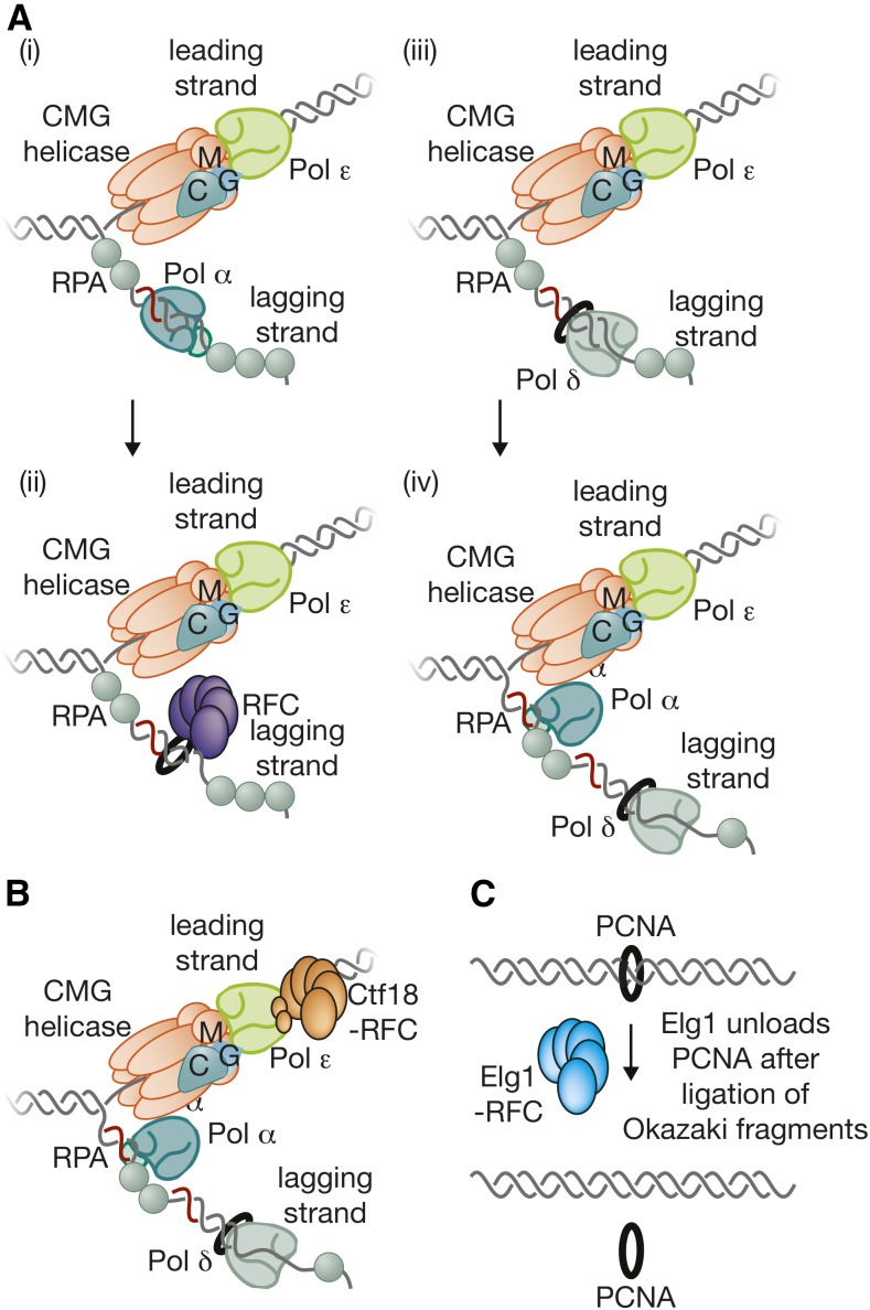
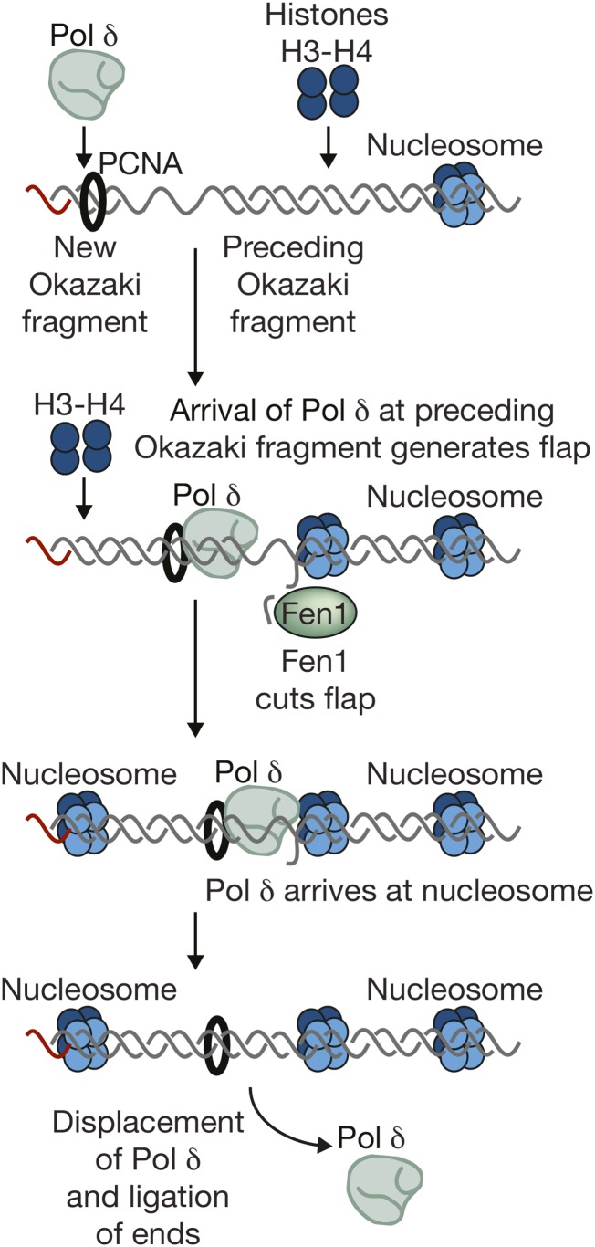
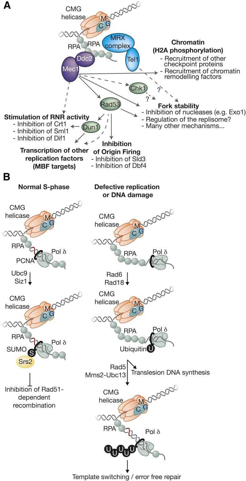
The accurate and complete replication of genomic DNA is essential for all life. In eukaryotic cells, the assembly of the multi-enzyme replisomes that perform replication is divided into stages that occur at distinct phases of the cell cycle. Replicative DNA helicases are loaded around origins of DNA replication exclusively during G1 phase. The loaded helicases are then activated during S phase and associate with the replicative DNA polymerases and other accessory proteins. The function of the resulting replisomes is monitored by checkpoint proteins that protect arrested replisomes and inhibit new initiation when replication is inhibited. The replisome also coordinates nucleosome disassembly, assembly, and the establishment of sister chromatid cohesion. Finally, when two replisomes converge they are disassembled. Studies in Saccharomyces cerevisiae have led the way in our understanding of these processes. Here, we review our increasingly molecular understanding of these events and their regulation.

Keywords: DNA replication; YeastBook; cell cycle; chromatin; chromosome duplication; genome stability.


Figures

Figure 1
Figure 1
Structure of S. cerevisiae replicator. The general structure of budding yeast replicators and the surrounding nucleosomes is illustrated. Although the precise nucleosome positions vary, the key elements of the replicator are located within a nucleosome-free region with the ORC binding site located asymmetrically within this region. The ORC-ACS consensus sequence shown is derived from Eaton et al. 2010.
Figure 2
Figure 2
Initial recruitment of Mcm2-7 to origin DNA. (A) The six Mcm subunits share a common structure and are arranged in a ring with a defined order of the subunits. (B) A model for the events during initial recruitment of the Mcm2-7 hexamer to origin DNA. The initial ORC-Cdc6 complex is proposed to form a second ring-shaped complex of AAA+-related subunits that encircle origin DNA. This complex is proposed to recruit one Mcm2-7/Cdt1 to the adjacent DNA to form the OCCM complex. The relative position of the N- and C-terminal domains of ORC/Cdc6 and Mcm2-7 in the OCCM are labeled.
Figure 3
Figure 3
Events of helicase loading after recruitment of the Mcm2-7 complex. Two models for the events of helicase loading after formation of the OCCM are shown. The one-ORC model is based on single-molecule studies of helicase loading and predicts that the second Mcm2-7 is recruited by interactions with first Mcm2-7. The two-ORC model is based on studies suggesting the first and second Mcm2-7 is loaded by the same mechanism as the first Mcm2-7. In this model, the time of binding of the second ORC and release of the first ORC is unclear. The color code for the Mcm subunits is the same as in Figure 2A.
Figure 4
Figure 4
Remodeling of the Mcm2-7 double hexamer and origin DNA during helicase activation. The loaded Mcm2-7 double hexamer encircles double-stranded origin DNA (top). In contrast, the active helicase (the Cdc45/Mcm2-7/GINS or CMG complex) contains one copy of the Mcm2-7 complex and encircles ssDNA (bottom). This transition requires dissolution of the interactions between the two Mcm2-7 hexamers, melting of the origin DNA, opening of each Mcm2-7 ring, extrusion of opposite ssDNAs from the two Mcm2-7 complexes, and reclosing of the Mcm2-7 rings. The relative order of these events during helicase activation is currently unknown.
Figure 5
Figure 5
A model for helicase activation during the initiation of DNA replication. (A) The model illustrates the first time that each factor is required. Although Sld2, Sld3, and Dpb11 are not thought to be part of the final replisome; it is unclear when these factors are released. Helicase activation is associated with the recruitment of many additional factors to form the replisome (see below). (B) A model for the mechanism of initial origin DNA melting by the Mcm2-7 double hexamer.
Figure 6
Figure 6
Helicase loading and activation are segregated during the cell cycle. The cell cycle can be split into two phases with respect to DNA replication. Helicase loading only occurs in G1 phase when CDK levels are low. The increased CDK levels present during S, G2, and M phases prevent helicase loading through multiple mechanisms. The same elevated CDK levels are required to activate CMG assembly and helicase activation, ensuring no helicase is activated during G1 phase. This regulation ensures no origin can initiate more than once per cell cycle.
Figure 7
Figure 7
Building the replisome. (A) The division of labor among DNA polymerases at the yeast replication fork. (B) The RPC assembles around the CMG helicase at replication forks. (C) The RPC is connected to Pol ε and Pol α at forks, but apparently not to Pol δ.
Figure 8
Figure 8
Multiple clamp loaders at the yeast replication fork. (A) Pol α detaches from the template after synthesizing an RNA-DNA primer (i), and Rfc1-RFC is then very effective at competing for access to the 3′ end of the primer bound to template, leading to loading of PCNA around dsDNA (ii). This in turn leads to recruitment of Pol δ (iii), which then extends the new Okazaki fragment (iv). (B) Ctf18-RFC associates with Pol ε and might contribute to loading of PCNA onto the leading-strand side of the fork. (C) Elg1-RFC is recruited to PCNA (aided by sumoylation) after ligation of Okazaki fragments, leading to removal of PCNA from the replicated DNA.
Figure 9
Figure 9
Processing of Okazaki fragments. The figure illustrates the model whereby nucleosome deposition plays a key role in completing the synthesis of Okazaki fragments. When Pol δ meets the 5′ end of the preceding Okazaki fragment, it displaces a short flap that is cut by Fen1 (or a longer flap that can be cut by Dna2). Strand displacement continues until Pol δ reaches the midpoint of the nucleosome deposited on the preceding fragment, at which point Pol δ detaches from the template, allowing ligation and thus completion of DNA synthesis.
Figure 10
Figure 10
Regeneration of chromatin during DNA replication. DNA unwinding by the CMG helicase displaces parental histones, but it is thought that a tetramer of H3-H4 is retained locally, probably by the histone-binding activity of replisome components including Mcm2 and FACT. This allows for the local redeposition of parental H3-H4 tetramers onto the nascent DNA, in parallel with the deposition of newly-synthesized histones H3-H4 by chaperones such as CAF1. Following addition of H2A-H2B, nucleosomes are regenerated, and in practice this whole process is extremely rapid. It is assumed that epigenetic modifications on parental histones are then copied to the neighboring newly-synthesized nucleosomes, thus restoring parental chromatin.
Figure 11
Figure 11
Surviving problems during chromosome replication. Replication defects expose more ssDNA at forks and thus lead to an accumulation of RPA. (A) This recruits Mec1-Ddc2 to initiate the S-phase checkpoint pathway and also (B) leads to ubiquitylation of PCNA, which activates translesion DNA synthesis and also an error-free repair pathway.
Figure 12
Figure 12
Disassembly of the CMG helicase is the final step in chromosome replication. When two forks converge, a poorly-characterized signal leads to ubiquitylation of the Mcm7 subunit of CMG, which is dependent upon the E3 ligase SCFDia2. The Cdc48 segregase is then required for disassembly of ubiquitylated CMG, by a mechanism that is not yet understood.

References

    1. Abid Ali F., Renault L., Gannon J., Gahlon H. L., Kotecha A., et al. 2016. Cryo-EM structures of the eukaryotic replicative helicase bound to a translocation substrate. Nat. Commun. 7: 10708. - PMC - PubMed
    1. Alabert C., Groth A., 2012. Chromatin replication and epigenome maintenance. Nat. Rev. Mol. Cell Biol. 13: 153–167. - PubMed
    1. Alabert C., Bianco J. N., Pasero P., 2009. Differential regulation of homologous recombination at DNA breaks and replication forks by the Mrc1 branch of the S-phase checkpoint. EMBO J. 28: 1131–1141. - PMC - PubMed
    1. Albert I., Mavrich T. N., Tomsho L. P., Qi J., Zanton S. J., et al. 2007. Translational and rotational settings of H2A.Z nucleosomes across the Saccharomyces cerevisiae genome. Nature 446: 572–576. - PubMed
    1. Alcasabas A. A., Osborn A. J., Bachant J., Hu F., Werler P. J., et al. 2001. Mrc1 transduces signals of DNA replication stress to activate Rad53. Nat. Cell Biol. 3: 958–965. - PubMed

MeSH terms

LinkOut - more resources