. 2016 Sep;26(9):1178-87.
doi: 10.1101/gr.204784.116. Epub 2016 Jul 6.

CENPT bridges adjacent CENPA nucleosomes on young human α-satellite dimers

Affiliations

CENPT bridges adjacent CENPA nucleosomes on young human α-satellite dimers

Jitendra Thakur et al. Genome Res. 2016 Sep.

Abstract

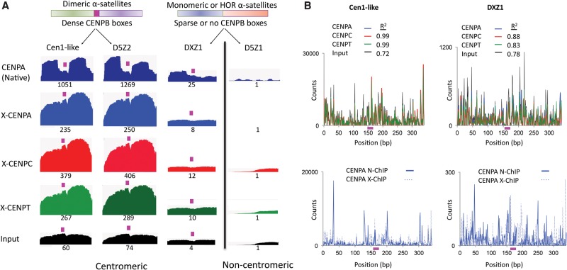
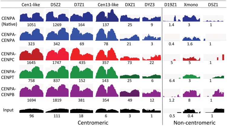
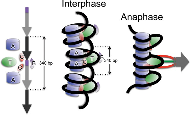
Nucleosomes containing the CenH3 (CENPA or CENP-A) histone variant replace H3 nucleosomes at centromeres to provide a foundation for kinetochore assembly. CENPA nucleosomes are part of the constitutive centromere associated network (CCAN) that forms the inner kinetochore on which outer kinetochore proteins assemble. Two components of the CCAN, CENPC and the histone-fold protein CENPT, provide independent connections from the ∼171-bp centromeric α-satellite repeat units to the outer kinetochore. However, the spatial relationship between CENPA nucleosomes and these two branches remains unclear. To address this issue, we use a base-pair resolution genomic readout of protein-protein interactions, comparative chromatin immunoprecipitation (ChIP) with sequencing, together with sequential ChIP, to infer the in vivo molecular architecture of the human CCAN. In contrast to the currently accepted model in which CENPT associates with H3 nucleosomes, we find that CENPT is centered over the CENPB box between two well-positioned CENPA nucleosomes on the most abundant centromeric young α-satellite dimers and interacts with the CENPB/CENPC complex. Upon cross-linking, the entire CENPA/CENPB/CENPC/CENPT complex is nuclease-protected over an α-satellite dimer that comprises the fundamental unit of centromeric chromatin. We conclude that CENPA/CENPC and CENPT pathways for kinetochore assembly are physically integrated over young α-satellite dimers.


Figures

Figure 1.
Figure 1.
Schematic representation of comparative ChIP methods. Under native conditions, partial MNase digestion produces insoluble chromatin arrays as the major population. Upon moderate MNase digestion under similar conditions, only nucleosomes or similar structures that wrap DNA are protected; and therefore, proteins associated with the linker DNA are lost from the chromatin. In X-ChIP and sequential ChIP, however, protein–protein interactions are stabilized by cross-linking, and the solubility of chromatin is enhanced by detergents, resulting in ChIP signals after both moderate and heavy MNase digestion. In sequential ChIP, false positives can arise from there being multiple complexes pulled down from longer arrays, in which the first antibody pulls down one complex and the second antibody pulls down a different complex: (DNA) black line; (nucleosomes) blue barrels; (DNA-binding proteins) colored balls.
Figure 2.
Figure 2.
CENPT is not retained at α-satellites by N-ChIP but is retained by X-ChIP. (A) Real-time PCR analysis of DNA obtained by native ChIP of CENPA, CENPC, and CENPT (using two different antibodies). The S1 primer pair was designed from Chromosome 21 alphoid arrays but is also present on Chromosome 13. Primer pair S2 was designed from the Cen1-like (SF1 family) sequences and is also present on Chromosomes 5 and 19 (Supplemental Table 1). Fold-enrichment over input was calculated relative to a control noncentromeric sequence from the 5S rDNA locus: Centromere (ChIP/input) ÷ 5S rDNA (ChIP/input). (B) CENPA and CENPT enrichment from native ChIP assays performed under 500 mM salt conditions. (C) CENPA and CENPT enrichment on DNA obtained from native ChIP assays performed on partially digested chromatin. (D) Enrichment of centromeric sequences in CENPA, CENPC, and CENPT cross-linking ChIP (X-ChIP). (E,F) Fragment length analysis of merged pairs obtained from (E) native or (F) X-ChIP data sets on D5Z2 (left), D7Z1 (middle), and DXZ1 (right) sequences. The sharp reduction in the size distribution above ∼160 bp and truncation at 185 bp is attributable to mapping of only paired-end reads in which the two 100-bp reads in a pair overlapped by 15 bp.
Figure 3.
Figure 3.
CENPA, CENPC, and CENPT cross-link in a single chromatin complex. (A) Close correspondence between X-ChIP of CENPA, CENPC, and CENPT data sets. X-ChIP fragments were clustered based on sequence identity, and frequency distributions of fragments within clusters between data sets were compared. Scatter plots show the regression of the number of distinct mapped merged pairs for the CENPC, CENPT, and input X-ChIP data sets on the CENPA X-ChIP data set. (B) Real-time PCR analysis of DNA obtained by sequential ChIP with indicated pairs of antibodies. The S1 primer pair was designed as described in the legend to Figure 2, and the S3 primer pair was designed from the Cen1-like (SF1 family) sequences and is also present on Chromosomes 1, 5, and 19 (Supplemental Table 1). Fold enrichment over input was calculated relative to a control noncentromeric sequence from the 5S rDNA locus.
Figure 4.
Figure 4.
Sequential ChIP-seq data sets of CCAN components are enriched for centromeric α-satellites and are highly correlated. (A) Sequential ChIP-seq data sets show that abundant centromeric α-satellite arrays (D5Z2 and Cen1-like) are enriched for CENPA, CENPT, CENPC, and CENPB, whereas representative noncentromeric satellites (Xmono and D19Z1) are not. The reads were mapped to previously characterized α-satellite arrays (Hayden et al. 2013; Henikoff et al. 2015), and the number of reads mapped to an array were calculated (using Trim Galore!) as a fraction of the total number of mapped reads processed. (B) Close correspondence between sequential ChIP-seq data sets: CENPA/CENPA, CENPA/CENPC, and CENPA/CENPT. Scatter plots show the regression of the number of mapped reads for the CENPA/CENPA, CENPA/CENPC, CENPA/CENPT, and CENPA/GFP sequential ChIP data sets on the CENPA X-ChIP data set.
Figure 5.
Figure 5.
CENPT and CENPC bridge the gap between adjacent CENPA nucleosomes over the CENPB box on young α-satellite dimers. (A) Mapping of X-ChIP merged pairs to previously characterized α-satellite arrays (Hayden et al. 2013; Henikoff et al. 2015). The profiles display normalized counts as described in Methods, in which entire fragments are “stacked,” and for each base-pair position the total number of counts is measured on the y-axis. A single α-satellite dimer from each array is shown. The CENPB box region is indicated by the magenta box. Cen1-like and D5Z2 arrays contain CENPB boxes in every α-satellite dimer (dense) (Henikoff et al. 2015). The DXZ1 tandem array, which is a 12-copy HOR, contains CENPB boxes only in a subset of dimeric units of the array (sparse), and a single CENPB box-containing dimer, which is embedded in α-satellite units that lack a CENPB box, is shown. The D5Z1 array does not contain CENPB boxes. The relative scale is the area of the indicated profile divided by the area of the D5Z1 profile, setting the D5Z1 value to 1, in which the numbers reflect the product of the total sequence abundance and enrichment. For example, Cen1-like is 60-fold enriched in the X-ChIP input, reflecting higher sequence abundance, and 235-fold enriched in CENPA ChIP, which implies that ChIP enrichment per copy is 235/60 or approximately fourfold. Biological replicates using different CENPT antibodies gave nearly identical patterns and relative scale values (Supplemental Fig. S2). (B) Fragment ends from native and X-ChIP data sets were mapped to the Cen1-like consensus and DXZ1 sequences. The CENPB box region is indicated by the magenta box. The top panel shows ends from CENPA, CENPC, CENPT X-ChIP, and input. The bottom panel compares ends of CENPA reads in N-ChIP (solid line) and X-ChIP (dotted line).
Figure 6.
Figure 6.
Sequential ChIP-seq profiles of CCAN components are nearly identical. Mapping of sequential ChIP-seq reads to α-satellite arrays (Hayden et al. 2013; Henikoff et al. 2015). A single α-satellite dimer from each array is shown, and the relative scale is the area of the indicated profile divided by the area of the D5Z1 profile, in which the numbers reflect the product of the total sequence abundance and enrichment. Because DXZ1, DYZ3, D19Z1, Xmono, and D5Z1 are not dimeric units, we chose pairs of tandem monomers as representatives.
Figure 7.
Figure 7.
A model for the CENPA/CENPB/CENPC/CENPT chromatin complex. (Left) Hypothesized arrangement of CENPA, B, C, and T along a single 340-bp Cen1-like dimeric unit. (Middle) During interphase, the CENPT complex occupies the CENPB box-containing linker on young 340-bp α-satellite dimers between tandem CENPA nucleosomes. Each CENPA particle binds a CENPC on one side and wraps ∼100-bp DNA with right-handed chirality. (Right) At anaphase, tension causes the positively supercoiled DNA to overwind and tighten up around the complex, favoring stacking of tandem dimers to form a stiff platform that spreads pulling forces.

Similar articles

Cited by

References

    1. Alexandrov I, Kazakov A, Tumeneva I, Shepelev V, Yurov Y. 2001. α-Satellite DNA of primates: old and new families. Chromosoma 110: 253–266. - PubMed
    1. Amano M, Suzuki A, Hori T, Backer C, Okawa K, Cheeseman IM, Fukagawa T. 2009. The CENP-S complex is essential for the stable assembly of outer kinetochore structure. J Cell Biol 186: 173–182. - PMC - PubMed
    1. Blower MD, Sullivan BA, Karpen GH. 2002. Conserved organization of centromeric chromatin in flies and humans. Dev Cell 2: 319–330. - PMC - PubMed
    1. Bodor DL, Mata JF, Sergeev M, David AF, Salimian KJ, Panchenko T, Cleveland DW, Black BE, Shah JV, Jansen LE. 2014. The quantitative architecture of centromeric chromatin. eLife 3: e02137. - PMC - PubMed
    1. Carroll CW, Milks KJ, Straight AF. 2010. Dual recognition of CENP-A nucleosomes is required for centromere assembly. J Cell Biol 189: 1143–1155. - PMC - PubMed

Publication types