. 2016 Aug 1;143(15):2753-9.
doi: 10.1242/dev.128348. Epub 2016 Jul 6.

IMP2 axonal localization, RNA interactome, and function in the development of axon trajectories

Affiliations

IMP2 axonal localization, RNA interactome, and function in the development of axon trajectories

Nicolas Preitner et al. Development. .

Abstract

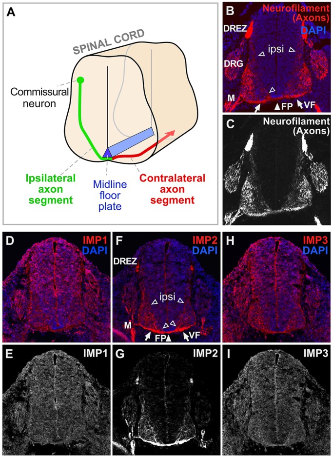
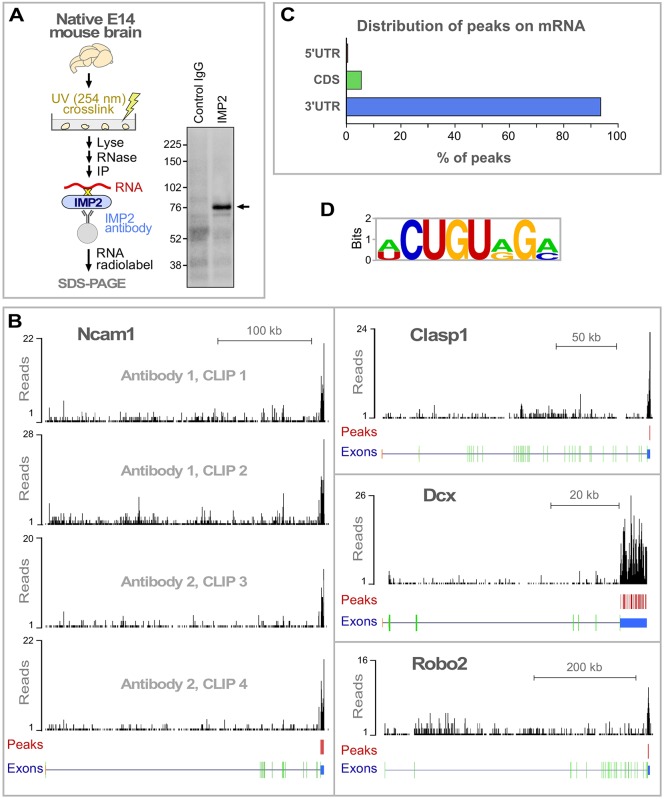
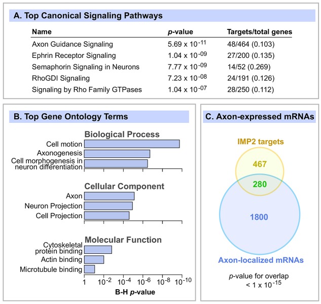
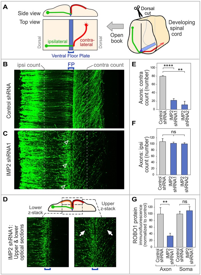
RNA-based regulatory mechanisms play important roles in the development and plasticity of neural circuits and neurological disease. Developing axons provide a model well suited to the study of RNA-based regulation, and contain specific subsets of mRNAs that are locally translated and have roles in axon pathfinding. However, the RNA-binding proteins involved in axon pathfinding, and their corresponding mRNA targets, are still largely unknown. Here we find that the RNA-binding protein IMP2 (Igf2bp2) is strikingly enriched in developing axon tracts, including in spinal commissural axons. We used the HITS-CLIP approach to perform a genome-wide identification of RNAs that interact directly with IMP2 in the native context of developing mouse brain. This IMP2 interactome was highly enriched for mRNA targets related to axon guidance. Accordingly, IMP2 knockdown in the developing spinal cord led to strong defects in commissural axon trajectories at the midline intermediate target. These results reveal a highly distinctive axonal enrichment of IMP2, show that it interacts with a network of axon guidance-related mRNAs, and reveal that it is required for normal axon pathfinding during vertebrate development.

Keywords: Axon guidance; IMP2; Igf2bp2; RNA interactome; RNA-binding protein.


Conflict of interest statement

The authors declare no competing or financial interests.

Figures

Fig. 1.
Fig. 1.
IMP2 is highly enriched in axon tracts. (A) Diagram of axons of dorsal commissural neurons navigating in developing spinal cord. Axon segments on the ipsilateral side (green) orient ventrally and medially from the cell bodies toward the midline floor plate (blue). After crossing the floor plate in the ventral commissure, most axons make a sharp anterior turn, then diverge away from the midline, before growing longitudinally in the ventral and lateral funiculi. (B-I) Transverse sections of E11.5 mouse spinal cord, with DAPI nuclear staining in blue and immunolabeling in red. (B,C) Neurofilament marker for axons. (D,E) Consistent with previous studies, IMP1 labeling was seen broadly in the nervous system as well as in other tissues. (F,G) IMP2 labeling was seen over the ventral funiculus, the ventral commissure, and ipsilateral axon segments oriented towards the floor plate. (H,I) IMP3 labeling, similar to that of IMP1, was seen broadly in the nervous system and other tissues. ipsi, ipsilateral axon segments (open arrowheads); FP, floor plate (white arrowhead); VF, ventral funiculus (arrows); DRG, dorsal root ganglion; DREZ, dorsal root entry zone; M, spinal motor axons.
Fig. 2.
Fig. 2.
HITS-CLIP identification of RNAs that interact with IMP2. (A) IMP2 HITS-CLIP. After UV-crosslinking protein-RNA complexes in native E14 mouse brain tissue, radiolabeled RNAs were co-immunoprecipitated with IMP2. CLIP results are shown on the right. Arrow marks the major band with increased intensity in the experimental lane, at the size expected for IMP2-RNA complexes, which was taken for high-throughput sequencing. Molecular mass markers in kDa. (B) Distribution of IMP2 HITS-CLIP sequence signals on representative target genes. Red vertical bars indicate high-confidence IMP2 binding peaks (see Fig. S1A and the supplementary Materials and Methods for details on peak identification). Structure of each gene is illustrated beneath: 5′UTR, orange; protein-coding, green; 3′UTR, blue. (C) Distribution of IMP2 binding peaks on target mRNAs. CDS, coding sequence. (D) A consensus sequence motif identified within the IMP2 binding regions using MEME software (see also Fig. S2).
Fig. 3.
Fig. 3.
IMP2 target mRNAs are highly enriched for functions related to axon development. (A) IMP2 target mRNAs were analyzed by IPA to identify enriched canonical signaling pathways, of which the top five are shown. Right-hand column shows target genes as a fraction of the total number of genes in the IPA category. (B) The top GO terms enriched among IMP2 target mRNAs, identified by the DAVID bioinformatic tools. P-values were adjusted for multiple comparisons by the Benjamini-Hochberg (B-H) method. (C) Venn diagram comparing IMP2 target mRNAs with a catalog of axon-localized mRNAs in embryonic mouse DRG axons (Gumy et al., 2011). Statistics used Fisher's exact test.
Fig. 4.
Fig. 4.
IMP2 knockdown causes defects in commissural axon pathfinding in vivo. (A) Diagram of spinal cord open-book preparation. The developing spinal cord is cut open along the dorsal midline, then mounted ventricular face downwards. A typical commissural axon ipsilateral segment (green), contralateral segment (red) and the floor plate (blue) are shown. (B-F) IMP2 or control shRNAs were introduced by in ovo electroporation into one side of the chick embryo spinal cord, with a Math1-GFP construct to trace commissural axons. Spinal cord was dissected 64 h later and imaged as an open-book. (B) Commissural axons in a confocal z-stack of open-book, viewed from the top. Electroporated cell bodies are visible on the left, and axons grow toward the right. (C) After IMP2 knockdown, in contrast to the control many growth cones are seen to have stalled at or near the ipsilateral side of the floor plate, showing an enlarged fusiform morphology typical of stalled growth cones (arrowheads). Correspondingly, a reduced number of axons was seen on the contralateral side, where they appeared to follow roughly normal trajectories without obvious signs of degeneration. (D) The lower confocal z-stack shows axon segments approaching the floor plate (green in diagram). The upper z-stack shows axon trajectories diverging away from the midline on both sides (contralateral, red in diagram; and ipsilateral, orange in diagram; examples of the approximately mirror image trajectories are arrowed). (E,F) Quantitation of ipsilateral and contralateral axon numbers, counted at the positions indicated by the white dashed lines in B and C. IMP2 knockdown did not significantly affect the number of axons emerging from the dorsal commissural neuron cell bodies and growing toward the floor plate, but strongly reduced the percentage of axons on the contralateral side. Each replicate was a separate embryo. (G) Effect of IMP2 RNAi on Robo1 expression in spinal commissural neurons. IMP2 or control shRNAs were introduced by in ovo electroporation into chick embryo spinal cord, with a Math1-GFP construct to identify transfected neurons; 64 h later, commissural neurons were dissociated and cultured for analysis of Robo1 immunofluorescence in axons and cell bodies. Immunolabeling was performed on permeabilized cells to detect both intracellular and cell surface Robo1. IMP2 shRNA reduced Robo1 expression in the axon, but not in the soma (see also Fig. S4B). Each replicate in this experiment was a different neuron (n=16 for control and n=28 for IMP2 shRNA), and the experiment was repeated three times to ensure reproducibility. Comparisons used Student's unpaired two-tailed t-test. Error bars show s.e.m. **P<0.01, ****P<0.0001; ns, not significant.

References

    1. Alther T. A., Domanitskaya E. and Stoeckli E. T. (2016). Calsyntenin 1-mediated trafficking of axon guidance receptors regulates the switch in axonal responsiveness at a choice point. Development 143, 994-1004. 10.1242/dev.127449 - DOI - PubMed
    1. Änkö M.-L. and Neugebauer K. M. (2012). RNA-protein interactions in vivo: global gets specific. Trends Biochem. Sci. 37, 255-262. 10.1016/j.tibs.2012.02.005 - DOI - PubMed
    1. Bell J. L., Wächter K., Mühleck B., Pazaitis N., Köhn M., Lederer M. and Hüttelmaier S. (2013). Insulin-like growth factor 2 mRNA-binding proteins (IGF2BPs): post-transcriptional drivers of cancer progression? Cell. Mol. Life Sci. 70, 2657-2675. 10.1007/s00018-012-1186-z - DOI - PMC - PubMed
    1. Brittis P. A., Lu Q. and Flanagan J. G. (2002). Axonal protein synthesis provides a mechanism for localized regulation at an intermediate target. Cell 110, 223-235. 10.1016/S0092-8674(02)00813-9 - DOI - PubMed
    1. Brose K., Bland K. S., Wang K. H., Arnott D., Henzel W., Goodman C. S., Tessier-Lavigne M. and Kidd T. (1999). Slit proteins bind robo receptors and have an evolutionarily conserved role in repulsive axon guidance. Cell 96, 795-806. 10.1016/S0092-8674(00)80590-5 - DOI - PubMed

Publication types