FNDC3B promotes cell migration and tumor metastasis in hepatocellular carcinoma
- PMID: 27385217
- PMCID: PMC5226524
- DOI: 10.18632/oncotarget.10374
FNDC3B promotes cell migration and tumor metastasis in hepatocellular carcinoma
Abstract
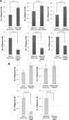
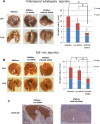
Recurrence and metastasis are common in hepatocellular carcinoma (HCC) and correlate with poor prognosis. We investigated the role of fibronectin type III domain containing 3B (FNDC3B) in HCC metastasis. Overexpression of FNDC3B in HCC cell lines enhanced cell migration and invasion. On the other hand, knockdown of FNDC3B using short-hairpin RNA reduced tumor nodule formation in both intra- and extra-hepatic metastasis. High levels of FNDC3B were observed in metastatic HCCs and correlated with poor patient survival and shorter recurrence time. Mutagenesis and LC-MS/MS analyses showed that FNDC3B promotes cell migration by cooperating with annexin A2 (ANXA2). Furthermore, FNDC3B and ANXA2 expression correlated negatively with patient survival. Our results indicate that FNDC3B behaves like an oncogene by promoting cell migration. This suggests FNDC3B could serve as a biomarker and therapeutic target for HCC metastasis.
Keywords: FNDC3B; hepatocellular carcinoma; metastasis.
Conflict of interest statement
No conflicts of interest to disclose.
Figures


Similar articles
-
Up-regulated microRNA-143 transcribed by nuclear factor kappa B enhances hepatocarcinoma metastasis by repressing fibronectin expression.Hepatology. 2009 Aug;50(2):490-9. doi: 10.1002/hep.23008. Hepatology. 2009. PMID: 19472311
-
Up-regulation of long non-coding RNA Sox2ot promotes hepatocellular carcinoma cell metastasis and correlates with poor prognosis.Int J Clin Exp Pathol. 2015 Apr 1;8(4):4008-14. eCollection 2015. Int J Clin Exp Pathol. 2015. PMID: 26097588 Free PMC article.
-
E2F1 promotes cell migration in hepatocellular carcinoma via FNDC3B.FEBS Open Bio. 2024 Apr;14(4):687-694. doi: 10.1002/2211-5463.13783. Epub 2024 Feb 25. FEBS Open Bio. 2024. PMID: 38403291 Free PMC article.
-
Overexpression of annexin A4 indicates poor prognosis and promotes tumor metastasis of hepatocellular carcinoma.Tumour Biol. 2016 Jul;37(7):9343-55. doi: 10.1007/s13277-016-4823-6. Epub 2016 Jan 16. Tumour Biol. 2016. PMID: 26779633 Clinical Trial.
-
Annexin A2 promotion of hepatocellular carcinoma tumorigenesis via the immune microenvironment.World J Gastroenterol. 2020 May 14;26(18):2126-2137. doi: 10.3748/wjg.v26.i18.2126. World J Gastroenterol. 2020. PMID: 32476780 Free PMC article. Review.
Cited by
-
Genomic association with pathogen carriage in bighorn sheep (Ovis canadensis).Ecol Evol. 2021 Mar 2;11(6):2488-2502. doi: 10.1002/ece3.7159. eCollection 2021 Mar. Ecol Evol. 2021. PMID: 33767816 Free PMC article.
-
Novel lncRNA XLOC_032768 protects against renal tubular epithelial cells apoptosis in renal ischemia-reperfusion injury by regulating FNDC3B/TGF-β1.Ren Fail. 2020 Nov;42(1):994-1003. doi: 10.1080/0886022X.2020.1818579. Ren Fail. 2020. PMID: 32972270 Free PMC article.
-
MiR-1225-5p acts as tumor suppressor in glioblastoma via targeting FNDC3B.Open Med (Wars). 2020 Sep 9;15(1):872-881. doi: 10.1515/med-2020-0156. eCollection 2020. Open Med (Wars). 2020. PMID: 33336045 Free PMC article.
-
FNDC3B is associated with ER stress and poor prognosis in cervical cancer.Oncol Lett. 2020 Jan;19(1):406-414. doi: 10.3892/ol.2019.11098. Epub 2019 Nov 14. Oncol Lett. 2020. PMID: 31897153 Free PMC article.
-
MicroRNA and cellular targets profiling reveal miR-217 and miR-576-3p as proviral factors during Oropouche infection.PLoS Negl Trop Dis. 2018 May 29;12(5):e0006508. doi: 10.1371/journal.pntd.0006508. eCollection 2018 May. PLoS Negl Trop Dis. 2018. PMID: 29813068 Free PMC article.
References
-
- El-Serag HB. Hepatocellular carcinoma. N Engl J Med. 2011;365:1118–1127. - PubMed
-
- Zimmerman MA, Ghobrial RM, Tong MJ, Hiatt JR, Cameron AM, Hong J, Busuttil RW. Recurrence of hepatocellular carcinoma following liver transplantation: a review of preoperative and postoperative prognostic indicators. Ann Surg. 2008;143:182–188. discussion 188. - PubMed
-
- Uchino K, Tateishi R, Shiina S, Kanda M, Masuzaki R, Kondo Y, Goto T, Omata M, Yoshida H, Koike K. Hepatocellular carcinoma with extrahepatic metastasis: clinical features and prognostic factors. Cancer. 2011;117:4475–4483. - PubMed
MeSH terms
Substances
LinkOut - more resources
Full Text Sources
Other Literature Sources
Medical
Miscellaneous

