. 2016 Oct;20(10):1908-19.
doi: 10.1111/jcmm.12883. Epub 2016 Jul 7.

Danshensu alleviates cardiac ischaemia/reperfusion injury by inhibiting autophagy and apoptosis via activation of mTOR signalling

Affiliations

Danshensu alleviates cardiac ischaemia/reperfusion injury by inhibiting autophagy and apoptosis via activation of mTOR signalling

Guanwei Fan et al. J Cell Mol Med. 2016 Oct.

Abstract

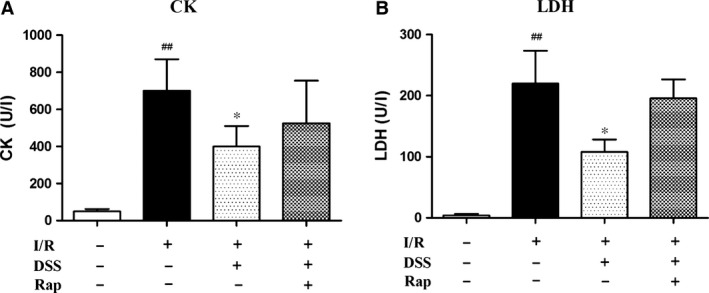
The traditional Chinese medicine Danshensu (DSS) has a protective effect on cardiac ischaemia/reperfusion (I/R) injury. However, the molecular mechanisms underlying the DSS action remain undefined. We investigated the potential role of DSS in autophagy and apoptosis using cardiac I/R injury models of cardiomyocytes and isolated rat hearts. Cultured neonatal rat cardiomyocytes were subjected to 6 hrs of hypoxia followed by 18 hrs of reoxygenation to induce cell damage. The isolated rat hearts were used to perform global ischaemia for 30 min., followed by 60 min. reperfusion. Ischaemia/reperfusion injury decreased the haemodynamic parameters on cardiac function, damaged cardiomyocytes or even caused cell death. Pre-treatment of DSS significantly improved cell survival and protected against I/R-induced deterioration of cardiac function. The improved cell survival upon DSS treatment was associated with activation of mammalian target of rapamycin (mTOR) (as manifested by increased phosphorylation of S6K and S6), which was accompanied with attenuated autophagy flux and decreased expression of autophagy- and apoptosis-related proteins (including p62, LC3-II, Beclin-1, Bax, and Caspase-3) at both protein and mRNA levels. These results suggest that alleviation of cardiac I/R injury by pre-treatment with DSS may be attributable to inhibiting excessive autophagy and apoptosis through mTOR activation.

Keywords: Danshensu; I/R injury; apoptosis; autophagy; mTOR.


Figures

Figure 1
Figure 1
Experimental protocol and effects of DSS treatment on cellular viability. (A) Hypoxia/reoxygenation (H/R) injury protocol. Control: cells were cultured constantly under normoxic condition. H/R: after normoxic condition cultured for 24 hrs, cells received 6 hrs of hypoxia and 18 hrs of reoxygenation. Drug treatments: after pre‐treatment with 10 μM DSS or 10 μM DSS + 100 nM Rap for 24 hrs, H/R was simulated as previously described. (B) Cell viability of cardiomyocytes. Data are expressed as the mean ± S.D., n = 4. ## P < 0.01 versus Control group. **P < 0.01 versus H/R group.
Figure 2
Figure 2
Effect of DSS on Autophagy flux in cardiomyocytes. (A) Representative immunofluorescence images of NRCMs expressing mRFPGFPLC3 adenovirus for 36 hrs. (B) Quantitative analysis of autophagosomes (AP, white bars), autolysosomes (AL, black bars), and both (AP+AL, grey bars) in NRCMs. Representative of n = 3 experiments. Data are expressed as the mean ± S.D. ## P < 0.01 versus Control group. **P < 0.01 versus H/R group.
Figure 3
Figure 3
Effects of DSS on autophagy and apoptosis related gene expression in cardiomyocytes. (A) Western blotting results of inhibition of autophagy‐related gene expression by DSS. (B) Western blotting results of inhibition of apoptosis‐related gene expression by DSS. Data are expressed as mean ± S.D., n = 3. # P < 0.05, ## P < 0.01 versus Control group. *P < 0.05, **P < 0.01 versus I/R group.
Figure 4
Figure 4
Effects of DSS on mTOR‐mediated autophagy and apoptosis in isolated rat hearts. (A) Western blot results of inhibition of autophagy by DSS. (B) Western blotting results of inhibition of apoptosis by DSS. Data are expressed as mean ± S.D., n = 3. ## P < 0.01 versus Control group. *P < 0.05, **P < 0.01 versus I/R group.
Figure 5
Figure 5
Effects of DSS on mTOR‐mediated autophagy and apoptosis in isolated rat hearts. (A) Evaluation of autophagy‐related gene expression after 60 min. reperfusion by RTPCR analysis. (B) Evaluation of apoptosis‐related gene expression after 60 min. reperfusion by RTPCR analysis. Data are expressed as mean ± S.D., n = 6. ## P < 0.01 versus Control group. *P < 0.05, **P < 0.01 versus I/R group.
Figure 6
Figure 6
Effects of DSS on cardiac function in isolated rat hearts. The hearts underwent 20 min. of stabilization, 30 min. of ischaemia, and 60 min. of reperfusion. Drugs were administered before ischaemia for 10 min. (A) Coronary flow (CF). (B) Heart rate (HR). (C) Left ventricular developed pressure (LVDP). (D and E) Maximal and minimum rate of pressure development (±dp/dt max), respectively. All parameters were measured at the end of the reperfusion period. Data are expressed as mean ± S.D., n = 6. ## P < 0.01 versus Control group. *P < 0.05, **P < 0.01 versus I/R group.
Figure 7
Figure 7
Effects of DSS on injury biomarkers in coronary effluents. (A) Creatine kinase (CK). (B) Lactate dehydrogenase (LDH). Data are expressed as mean ± S.D., n = 6. ## P < 0.01 versus Control group. *P < 0.05 versus I/R group.

References

    1. Yellon DM, Hausenloy DJ. Myocardial reperfusion injury. N Engl J Med. 2007; 357: 1121–35. - PubMed
    1. Magro M, Garg S, Serruys PW. Revascularization treatment of stable coronary artery disease. Expert Opin Pharmacother. 2011; 12: 195–212. - PubMed
    1. Minamino T. Cardioprotection from ischemia/reperfusion injury: basic and translational research. Circ J. 2012; 76: 1074–82. - PubMed
    1. Nishida K, Kyoi S, Yamaguchi O, et al The role of autophagy in the heart. Cell Death Differ. 2009; 16: 31–8. - PubMed
    1. Takemura G, Miyata S, Kawase Y, et al Autophagic degeneration and death of cardiomyocytes in heart failure. Autophagy. 2006; 2: 212–4. - PubMed

MeSH terms