. 2016 Sep 1;27(17):2757-70.
doi: 10.1091/mbc.E16-01-0048. Epub 2016 Jul 6.

Protein kinase A can block EphA2 receptor-mediated cell repulsion by increasing EphA2 S897 phosphorylation

Affiliations

Protein kinase A can block EphA2 receptor-mediated cell repulsion by increasing EphA2 S897 phosphorylation

Antonio Barquilla et al. Mol Biol Cell. .

Abstract

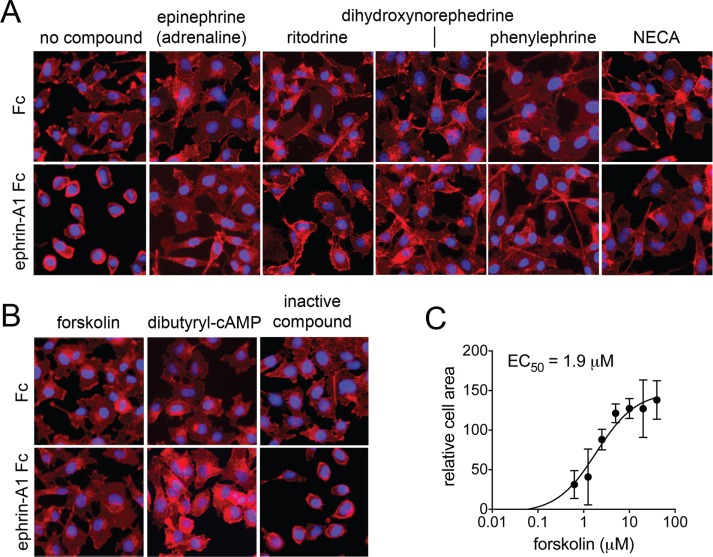
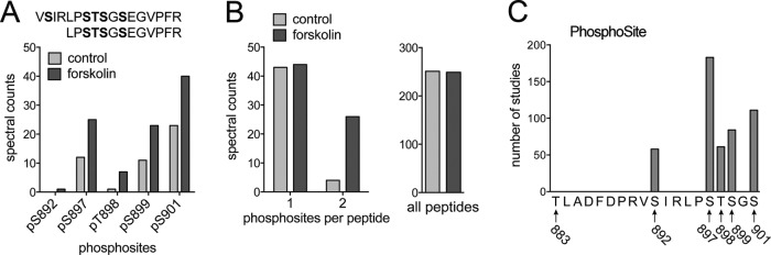
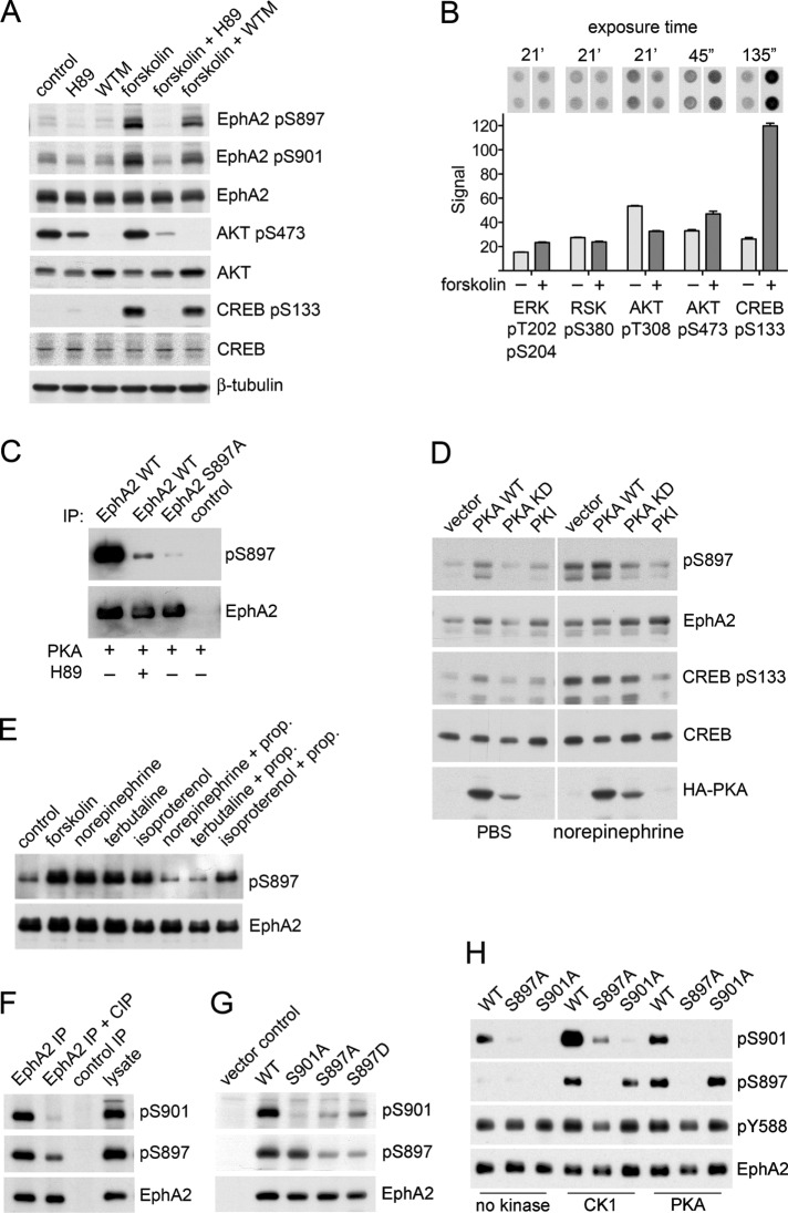
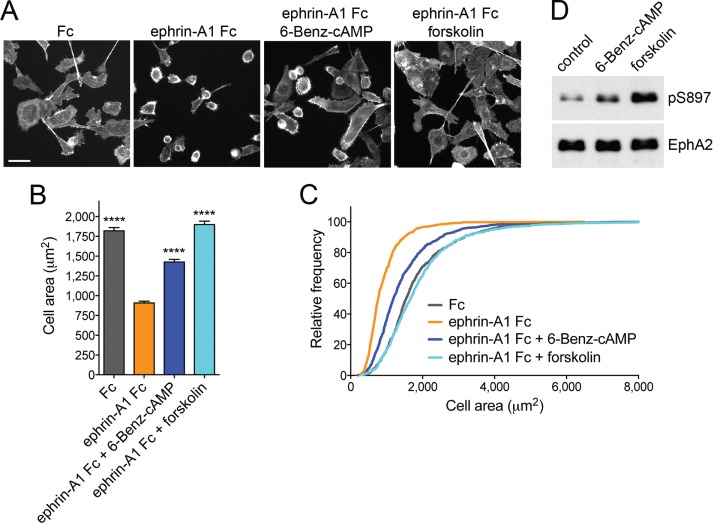
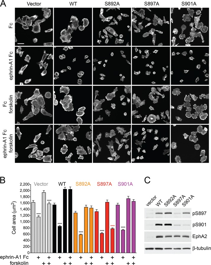
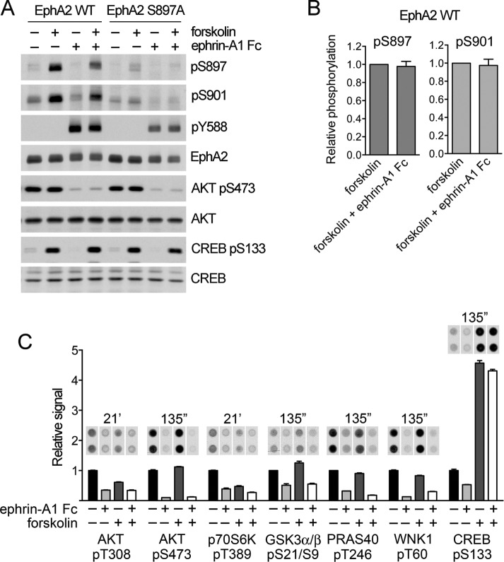
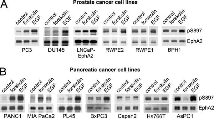
The EphA2 receptor tyrosine kinase plays key roles in tissue homeostasis and disease processes such as cancer, pathological angiogenesis, and inflammation through two distinct signaling mechanisms. EphA2 "canonical" signaling involves ephrin-A ligand binding, tyrosine autophosphorylation, and kinase activity; EphA2 "noncanonical" signaling involves phosphorylation of serine 897 (S897) by AKT and RSK kinases. To identify small molecules counteracting EphA2 canonical signaling, we developed a high-content screening platform measuring inhibition of ephrin-A1-induced PC3 prostate cancer cell retraction. Surprisingly, most hits from a screened collection of pharmacologically active compounds are agents that elevate intracellular cAMP by activating G protein-coupled receptors such as the β2-adrenoceptor. We found that cAMP promotes phosphorylation of S897 by protein kinase A (PKA) as well as increases the phosphorylation of several nearby serine/threonine residues, which constitute a phosphorylation hotspot. Whereas EphA2 canonical and noncanonical signaling have been viewed as mutually exclusive, we show that S897 phosphorylation by PKA can coexist with EphA2 tyrosine phosphorylation and block cell retraction induced by EphA2 kinase activity. Our findings reveal a novel paradigm in EphA2 function involving the interplay of canonical and noncanonical signaling and highlight the ability of the β2-adrenoceptor/cAMP/PKA axis to rewire EphA2 signaling in a subset of cancer cells.


Figures

FIGURE 1:
FIGURE 1:
Inhibition of EphA2-dependent cell retraction by representative hit compounds. (A, B) “No compound” refers to cells treated with Fc control or ephrin-A1 Fc and DMSO. Other cells were pretreated for 40 min with the indicated compounds and then for 10 min with Fc or ephrin-A1 Fc in the presence of the compounds. Forskolin was used at 20 μM and dibutyryl cyclical AMP (dbcAMP) at 0.5 mM. The other compounds were used at 5 μM. NECA, 5′-(N-ethylcarboxamido) adenosine. Cells were stained for F-actin with fluorescent phalloidin (red), and nuclei were labeled with DAPI (blue). The inactive compound (O100, oxotremorine methiodide) did not inhibit retraction and rounding of the cells induced by ephrin-A1 Fc, similar to the control. All other compounds shown inhibited retraction, and the cells remained flat. (C) Dose–response curve for forskolin-mediated inhibition of cell retraction analyzed by automated image analysis in the 384-well format. Averages and SEs from quadruplicate measurements from two experiments carried out on different days.
FIGURE 2:
FIGURE 2:
Forskolin increases phosphorylation of S897 and other hotspot sites in the EphA2 kinase–SAM domain linker segment. (A) Number of peptides (spectral counts) from the EphA2 kinase–SAM linker segment identified by mass spectrometry as containing the indicated phosphosite in control and forskolin-treated PC3 cells. The sequence of the two peptides containing hotspot residues is shown at the top, with the assigned phosphorylated residues in bold. (B) Left, number of peptides from A with one or two phosphosites, illustrating the dramatic increase in doubly phosphorylated peptides. Right, total number of EphA2 peptides detected, which was similar in control and forskolin-treated cells, indicating that similar total EphA2 levels were analyzed. (C) Number of studies in which each hotspot phosphosite was identified, which provides an indication of the abundance of the phosphosite.
FIGURE 3:
FIGURE 3:
Forskolin and β2-adrenoceptor agonists increase EphA2 phosphorylation on S897 by PKA as well as phosphorylation on S901. (A) PC3 cells were treated with the PKA inhibitor H89 or the PI3 kinase inhibitor wortmannin (WTM) for 1 h and with forskolin (FSK) for the last 40 min of inhibitor treatment. Cell lysates were probed by immunoblotting with the indicated antibodies. (B) Phosphokinase array signals show the effects of 40-min forskolin treatment on the indicated phosphosites. Images of the duplicate spots on the arrays and exposure times for the autoradiographs are shown at the top. The histogram shows averages from quantification of the spots, with the error bars representing SDs. (C) In vitro kinase reactions with immunoprecipitated EphA2 WT (with and without the H89 PKA inhibitor) or S897A mutant. A control immunoprecipitate is also included. The immunoblot was probed with a phospho-S897–specific antibody and reprobed for EphA2. (D) PC3 cells transfected with empty vector or with constructs encoding PKA WT, the kinase-inactive PKA K72H mutant (PKA KD), or the PKA inhibitor peptide PKI were treated for 30 min with PBS as a control or 10 μM norepinephrine, and lysates were probed with the indicated antibodies. (E) PC3 cells were treated with vehicle DMSO as a control, 20 μM forskolin, or 10 μM indicated β-adrenoceptor agonists for 1 h. Where indicated, cells were treated with 1 μM β-adrenoceptor antagonist propanolol (prop.) for 1 h before the 1-h treatment with β-adrenoceptor agonists. EphA2 immunoprecipitates were probed by immunoblotting for phospho-S897 and reprobed for EphA2. (F) EphA2 WT immunoprecipitated from transiently transfected HEK293 cells was either left untreated or treated with calf-intestinal alkaline phosphatase (CIP) to dephosphorylate the receptor. The control immunoprecipitate was obtained with nonimmune rabbit immunoglobulin Gs. The immunoprecipitates and a cell lysate for comparison were probed with the phospho-S901–specific antibody, a phospho-S897–specific antibody, or an EphA2 antibody. (G) Lysates of HEK293 cells transiently transfected constructs encoding EphA2 WT and the indicated EphA2 mutants or empty vector as a control were probed with the phospho-S901–specific antibody, a phospho-S897–specific antibody, or an EphA2 antibody. (H) In vitro kinase reactions measuring the phosphorylation of immunoprecipitated EphA2 WT and the indicated phosphosite mutants incubated with recombinant CK1 or PKA. EphA2 immunoprecipitates incubated only with ATP without any recombinant kinase served as a control. The EphA2 immunoprecipitates were probed by immunoblotting for phospho-S901 and phospho-S897 and then reprobed for EphA2.
FIGURE 4:
FIGURE 4:
PKA activation inhibits EphA2-dependent cell retraction. (A) Representative images of phalloidin-labeled PC3 cells stimulated for 12 min with 0.5 μg/ml control Fc or ephrin-A1 Fc or pretreated for 40 min with 20 μM forskolin or 200 μM of the PKA agonist 6-Benz-cAMP before ephrin-A1 Fc stimulation. Scale bar, 50 μm. (B) Histogram showing average cell areas ± SE under the different conditions (693 cells/condition from three experiments in each of which 77 cells/well from three wells were counted). ****p < 0.0001 for the comparison with the ephrin-A1 Fc condition by one-way analysis of variance (ANOVA) followed by Tukey’s multiple comparisons test. (C) Cumulative distribution showing the relative frequencies of cells with areas smaller than indicated on the x-axis. (D) Immunoblot of PC3 cells treated with 6-Benz-cAMP or forskolin to assess the levels of EphA2 phosphorylation on S897.
FIGURE 5:
FIGURE 5:
Inhibition of EphA2-dependent cell retraction by cAMP requires S897 phosphorylation. (A) Representative images of phalloidin-labeled, EphA2-knockdown PC3 cell populations infected with pLVX-IRES-Neo lentiviral vector (Vector) or lentivirus encoding WT EphA2 (WT) or the indicated EphA2 mutants. The cells, pretreated or not for 40 min with 20 μM forskolin, were stimulated for 12 min with 0.5 μg/ml control Fc or ephrin-A1 Fc. (B) Histogram showing average cell areas ± SE for the different conditions (160 cells/condition from an experiment in which 80 cells/well from two wells were measured). ****p < 0.0001 for the comparison of ephrin-A1 Fc–stimulated cells with the corresponding Fc-stimulated cells by one-way ANOVA followed by Sidak’s multiple comparisons test. (C) Immunoblot of PC3 cells transduced with empty lentiviral vector control and cells expressing the different EphA2 mutants to assess the levels of EphA2 expression and phosphorylation on S897 and S901.
FIGURE 6:
FIGURE 6:
EphA2 canonical signaling rapidly inhibits AKT but not PKA. (A) PC3 cells stably expressing EphA2 WT or the S897A mutant were treated with 20 μM forskolin for 40 min and/or 0.5 μg/ml ephrin-A1 Fc for 12 min. Lysates were probed by immunoblotting with the indicated antibodies. (B) Quantification of pS897 and pS901 phosphorylation relative to total EphA2 levels and normalized to the value in forskolin-treated cells. Averages ± SE from four independent experiments. The pS897 and pS901 levels in cells stimulated with forskolin and ephrin-A1 Fc are not significantly different from those in cells stimulated only with forskolin by one-sample t test. (C) Normalized phosphokinase array signals show the effects of ephrin-A1 Fc stimulation, with or without forskolin treatment, on the indicated phosphosites. Images of the duplicate spots on the arrays and exposure times for the autoradiographs are shown at the top. The histogram shows averages from quantification of the spots, normalized to the control condition for each phosphosite, with the error bars representing SDs.
FIGURE 7:
FIGURE 7:
PKA phosphorylates EphA2 on S897 in a subset of cancer cells. (A) Prostate cancer and immortalized prostate epithelial cell lines and (B) pancreatic cancer cell lines were treated with vehicle DMSO as a control, 20 μM forskolin, or 10 ng/ml EGF for 20 min. Cell lysates were probed by immunoblotting with the indicated antibodies. pAKT indicates AKT phosphorylated on S473; pCREB indicates CREB phosphorylated on S133. A vertical gap indicates that irrelevant intervening lanes from the blot were removed. Additional control lanes and immunoblots with additional control antibodies are shown in Supplemental Figure S4.

References

    1. Astin JW, Batson J, Kadir S, Charlet J, Persad RA, Gillatt D, Oxley JD, Nobes CD. Competition amongst Eph receptors regulates contact inhibition of locomotion and invasiveness in prostate cancer cells. Nat Cell Biol. 2010;12:1194–1204. - PubMed
    1. Baell J, Walters MA. Chemistry: chemical con artists foil drug discovery. Nature. 2014;513:481–483. - PubMed
    1. Baell JB, Holloway GA. New substructure filters for removal of pan assay interference compounds (PAINS) from screening libraries and for their exclusion in bioassays. J Med Chem. 2010;53:2719–2740. - PubMed
    1. Barquilla A, Pasquale EB. Eph receptors and ephrins: therapeutic opportunities. Annu Rev Pharmacol Toxicol. 2015;55:465–487. - PMC - PubMed
    1. Batson J, Maccarthy-Morrogh L, Archer A, Tanton H, Nobes CD. EphA receptors regulate prostate cancer cell dissemination through Vav2-RhoA mediated cell-cell repulsion. Biol Open. 2014;3:453–462. - PMC - PubMed

Publication types