. 2016 Aug 15;197(4):1489-97.
doi: 10.4049/jimmunol.1600052. Epub 2016 Jul 6.

A Critical Role for CD200R Signaling in Limiting the Growth and Metastasis of CD200+ Melanoma

Affiliations

A Critical Role for CD200R Signaling in Limiting the Growth and Metastasis of CD200+ Melanoma

Jin-Qing Liu et al. J Immunol. .

Abstract

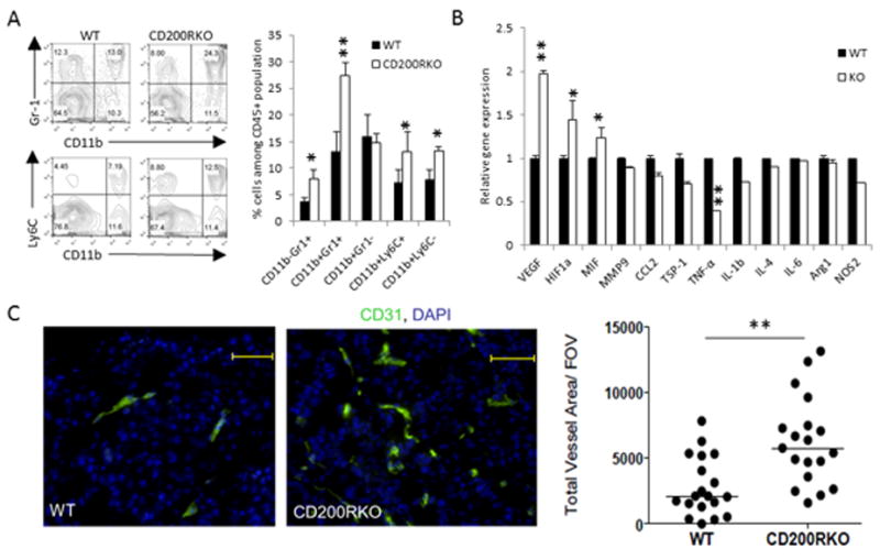
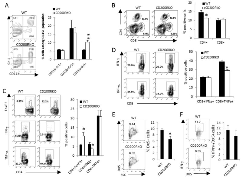
CD200 is a cell surface glycoprotein that functions through engaging CD200R on cells of the myeloid lineage and inhibits their functions. Expression of CD200 was implicated in a variety of human cancer cells, including melanoma cells; however, its roles in tumor growth and immunity are not clearly understood. In this study, we used CD200R-deficient mice and the B16 tumor model to evaluate this issue. We found that CD200R-deficient mice exhibited accelerated growth of CD200(+), but not CD200(-), B16 tumors. Strikingly, CD200R-deficient mice receiving CD200(+) B16 cells i.v. exhibited massive tumor growth in multiple organs, including liver, lung, kidney, and peritoneal cavity, whereas the growth of the same tumors in wild-type mice was limited. CD200(+) tumors grown in CD200R-deficient mice contained higher numbers of CD11b(+)Ly6C(+) myeloid cells, exhibited increased expression of VEGF and HIF1α genes with increased angiogenesis, and showed significantly reduced infiltration of CD4(+) and CD8(+) T cells, presumably as the result of reduced expression of T cell chemokines, such as CXCL9 and CXCL16. The liver from CD200R-deficient mice, under metastatic growth of CD200(+) tumors, contained significantly increased numbers of CD11b(+)Gr1(-) myeloid cells and Foxp3(+) regulatory T cells and reduced numbers of NK cells. Liver T cells also had a reduced capacity to produce IFN-γ or TNF-α. Taken together, we revealed a critical role for CD200R signaling in limiting the growth and metastasis of CD200(+) tumors. Thus, targeting CD200R signaling may potentially interfere with the metastatic growth of CD200(+) tumors, like melanoma.


Figures

Fig.1
Fig.1
IHC analyses of CD200 expression in human melanoma samples. Left image (A) shows a melanoma sample with overexpressed CD200 in tumor cells while the image in the right (B) shows a melanoma sample whose tumor cells do not express CD200. Frozen sections were used for the IHC staining. Anti-human CD200 mAb (OX-104) was purchased from eBioscience. The original images were 200 X.
Fig.2
Fig.2
Melanoma cell expression of CD200 inhibits tumor growth and alters tumor microenvironment. (A) B16.F10 melanoma cells were transfected with a CD200 expression plasmid or a control plasmid to generate CD200-positive (B16-CD200) or CD200-negative (B16-Ctrl) cells. Flow cytometery was used to analyze CD200 expression on B16-Ctrl (dotted line) or B16-CD200 (solid line) cells. (B) 1 × 10⁵ of B16-Ctrl or B16-CD200 cells were injected into each C57BL6 mouse subcutaneously. The tumor growth was observed over time. *P<0.05 by student’s t test. (C) Flow cytometry analysis of tumor infiltrating leukocytes was performed on disassociated tumor cells. (D) T cell subsets and activation status were analyzed by flow cytometry. Five mice per group were included in each group and data represents three experiments with similar results. **P<0.01 and ***P<0.001 by student’s t test.
Fig.3
Fig.3
Generation of CD200R-/- mice. (A) Diagram shows the targeting strategy for the generation of CD200R-/- mice. (B) CD200R expression on myeloid cells and T cells from different organs were analyzed by flow cytometry. Spleen CD4+ and CD8+ T cells were analyzed for the expression of CD200R.
Fig.4
Fig.4
CD200R-deficient mice had accelerated in situ and metastatic growth of B16-CD200 tumors. 1 × 10⁵ B16-CD200 cells (A) or B16-Ctrl cells (B) were injected into each CD200R-/- mice and their wild type breeding littermates (WT) s.c. and tumor growth was observed. (C) B16-ctrl and B16-CD200 tumors grew similarly in CD200R-/- mice. 1 × 10⁵ B16-CD200 cells or B16-Ctrl cells were injected into each CD200R-/- mice s.c. to observe tumor growth. (D) 1 × 10⁵ B16-CD200 cells were injected into each CD200R-/- mice and control WT littermates i.v. Nineteen days after melanoma cell injection mice were sacrificed. Representative mice and melanoma metastasis to the lungs, livers and kidneys are shown. The weights of lungs, livers and kidneys were quantified. *: p<0.05 and **p<0.01 by student’s t test. Data shown represents three (A-C) and five (D) experiments with similar results. Five mice per group were used for the experiments shown in A-C.
Fig.5
Fig.5
Increased tumor angiogenesis in CD200R-deficient tumors. B16-CD200 cells (1 × 10⁵/mouse) were injected into cohorts (n=5) of WT or CD200R-/- mice s.c. (A) Flow cytometry analyses of myeloid cells from B16-CD200 tumors grown in C57BL6 or CD200R-/- mice. Representative flow cytometry analyses of myeloid cells in tumors and summary of their different subsets are shown. Established tumors were also analyzed for the expression of a number of genes by qRT-PCR (B) and histological immunofluorescence staining for CD31 (C). CD31+ cells were quantified from images captured under a fluorescence microscope and quantified using the ImageJ software. Five tumors from each group were analyzed. *P<0.05; **P<0.01 by student’s t test.
Fig.6
Fig.6
Flow cytometry analyses of tumor infiltrating lymphocytes (TIL) from subcutaneously grown B16-CD200 tumors in CD200R-/- and WT mice. B16-CD200 cells (1 × 10⁵/mouse) were injected into cohorts (n=5) of WT and CD200R-/- mice s.c. Established tumors with a tumor size about 1 cm in diameter were disassociated followed by flow cytometry analyses. (A) Disassociated tumors were stained for CD45, CD4 and CD8 followed by flow cytometry analysis. Data shown were gated on CD45+ cells and summary of data of five tumors from each group are shown. *P<0.05 and **P<0.01 by student’s t test. Intracellular staining and flow cytometry were used to analyze CD8+ (B) and CD4+ (C) T cells for the expression of IFN-γ, TNF-α, and FoxP3, and (D) dada shown are plotted from 5 tumors in each group. (E) Expression of chemokine genes in B16-CD200 tumors from WT and CD200R-/- mice were quantified by qPCR. Five tumors from each group were analyzed. *P<0.05 and **P<0.01 by the student’s t test.
Fig.7
Fig.7
Flow cytometry analyses of leukocytes from the livers of CD200-deficient and WT mice underwent metastatic growth of B16-CD200 tumors. B16-CD200 cells (1 × 10⁵/mouse) were injected into cohorts (n=5) of WT and CD200R-/- mice i.v. Nineteen days after tumor cell injection mice were sacrificed, and liver leukocytes were isolated followed by flow cytometry analyses. Leukocytes from livers were stained for CD45, CD11b and Gr1 (A) or CD45, CD4 and CD8 (B) or DX5 (E) followed by flow cytometry analysis. Data shown were gated on CD45+ cells and summary of data of five samples from each group are shown. Intracellular staining and flow cytometry were used to analyze the liver CD4+ (C), CD8+ (D) T cells and DX5+ NK cells (F) for the expression of IFN-γ, TNF-α, or FoxP3. Data shown are summary of 5 samples from each group and representing two experiments with similar results. *P<0.05; **P<0.01 by student’s t test.

References

    1. Mantovani A, Allavena P, Sica A, Balkwill F. Cancer-related inflammation. Nature. 2008;454:436–444. - PubMed
    1. Yang L, DeBusk LM, Fukuda K, Fingleton B, Green-Jarvis B, Shyr Y, Matrisian LM, Carbone DP, Lin PC. Expansion of myeloid immune suppressor Gr+CD11b+ cells in tumor-bearing host directly promotes tumor angiogenesis. Cancer Cell. 2004;6:409–421. - PubMed
    1. Murdoch C, Muthana M, Coffelt SB, Lewis CE. The role of myeloid cells in the promotion of tumour angiogenesis. Nat Rev Cancer. 2008;8:618–631. - PubMed
    1. Qian BZ, Pollard JW. Macrophage diversity enhances tumor progression and metastasis. Cell. 2010;141:39–51. - PMC - PubMed
    1. Mantovani A, Schioppa T, Porta C, Allavena P, Sica A. Role of tumor-associated macrophages in tumor progression and invasion. Cancer Metastasis Rev. 2006;25:315–322. - PubMed

MeSH terms