

A case of sarcoidosis diagnosed by positron emission tomography/computed tomography
- PMID: 27385890
- PMCID: PMC4918483
- DOI: 10.4103/0972-3919.183608
A case of sarcoidosis diagnosed by positron emission tomography/computed tomography
Abstract
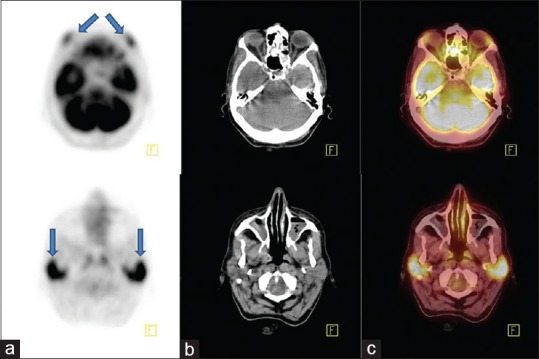
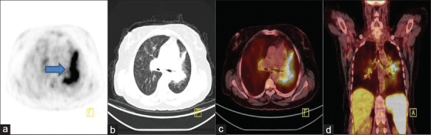
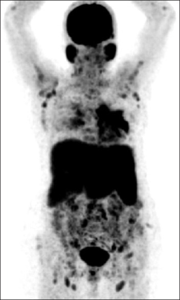
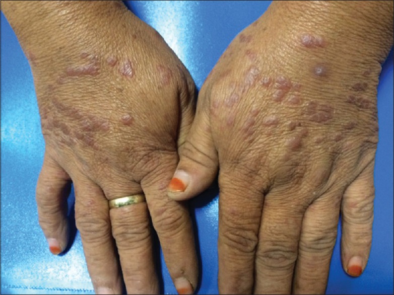
Sarcoidosis is a multisystem granulomatous disorder of unknown cause which may affect any organ or system but primarily involve the lungs and the lymphatic system. Extrapulmonary sarcoidosis represents approximately 30-50% of patients. We report the case of a 51-year-old female who presented with increasing complaints of a cough, weakness, weight loss, and chest pain and who was found to have a suspicious lesion on thorax computed tomography(CT). Fluorodeoxyglucose (FDG) positron emission tomography/CT performed for diagnostic purposes demonstrated increased FDG accumulation at the bilateral enlarged parotid and lacrimal gland and in the reticulonodular infiltration area located in the left lung as well as multiple lymphadenopathies with increased FDG accumulation. There were also hepatosplenomegaly and splenic uptake. Skin biopsy showed noncaseating granulomas, and the patient was diagnosed as stage 2 sarcoidosis.
Keywords: FDG; Fluorodeoxyglucose positron emission tomography/computed tomography; sarcoidosis.
Figures
Similar articles
-
Differentiation of sarcoidosis-lymphoma syndrome lesions: a case report on the use of two different positron emission tomography tracers.BMC Med Imaging. 2016 Jan 8;16:1. doi: 10.1186/s12880-015-0104-x. BMC Med Imaging. 2016. PMID: 26746426 Free PMC article.
-
[Usefulness of fluorodeoxyglucose-positron emission tomography in a case of pleural sarcoidosis].Nihon Kokyuki Gakkai Zasshi. 2009 Jul;47(7):658-62. Nihon Kokyuki Gakkai Zasshi. 2009. PMID: 19637812 Japanese.
-
Scar arcoidosis on 18F-FDG PET/CT.Asia Ocean J Nucl Med Biol. 2019 Spring;7(2):185-187. doi: 10.22038/AOJNMB.2019.37888.1253. Asia Ocean J Nucl Med Biol. 2019. PMID: 31380459 Free PMC article.
-
Sarcoidosis Stage II: a Case Report and Review of the Literature.Clin Lab. 2021 Feb 1;67(2). doi: 10.7754/Clin.Lab.2020.200632. Clin Lab. 2021. PMID: 33616320 Review.
-
Functional imaging in extrapulmonary sarcoidosis: FDG-PET/CT and MR features.Clin Nucl Med. 2014 Feb;39(2):e146-59. doi: 10.1097/RLU.0b013e318279f264. Clin Nucl Med. 2014. PMID: 23579973 Review.
Cited by
-
Looking beyond the cosmetic tattoo lesion near the eyebrow: Screening the lungs.J Postgrad Med. 2017 Apr-Jun;63(2):132-134. doi: 10.4103/0022-3859.201421. J Postgrad Med. 2017. PMID: 28272073 Free PMC article.
-
Liver: glucose metabolism and 18F-fluorodeoxyglucose PET findings in normal parenchyma and diseases.Am J Nucl Med Mol Imaging. 2021 Aug 15;11(4):233-249. eCollection 2021. Am J Nucl Med Mol Imaging. 2021. PMID: 34513277 Free PMC article. Review.
References
-
- Hunninghake GW, Costabel U, Ando M, Baughman R, Cordier JF, du Bois R, et al. ATS/ERS/WASOG statement on sarcoidosis. American Thoracic Society/European Respiratory Society/World Association of Sarcoidosis and other Granulomatous Disorders. Sarcoidosis Vasc Diffuse Lung Dis. 1999;16:149–73. - PubMed
-
- Iannuzzi MC, Rybicki BA, Teirstein AS. Sarcoidosis. N Engl J Med. 2007;357:2153–65. - PubMed
-
- Malaisamy S, Dalal B, Bimenyuy C, Soubani AO. The clinical and radiologic features of nodular pulmonary sarcoidosis. Lung. 2009;187:9–15. - PubMed
-
- Akira M, Kozuka T, Inoue Y, Sakatani M. Long-term follow-up CT scan evaluation in patients with pulmonary sarcoidosis. Chest. 2005;127:185–91. - PubMed
-
- Lebtahi R, Crestani B, Belmatoug N, Daou D, Genin R, Dombret MC, et al. Somatostatin receptor scintigraphy and gallium scintigraphy in patients with sarcoidosis. J Nucl Med. 2001;42:21–6. - PubMed
Publication types
LinkOut - more resources
Full Text Sources
Other Literature Sources
