. 2016 Jun 10;2(6):e1600502.
doi: 10.1126/sciadv.1600502. eCollection 2016 Jun.

Matrix-bound nanovesicles within ECM bioscaffolds

Affiliations

Matrix-bound nanovesicles within ECM bioscaffolds

Luai Huleihel et al. Sci Adv. .

Abstract

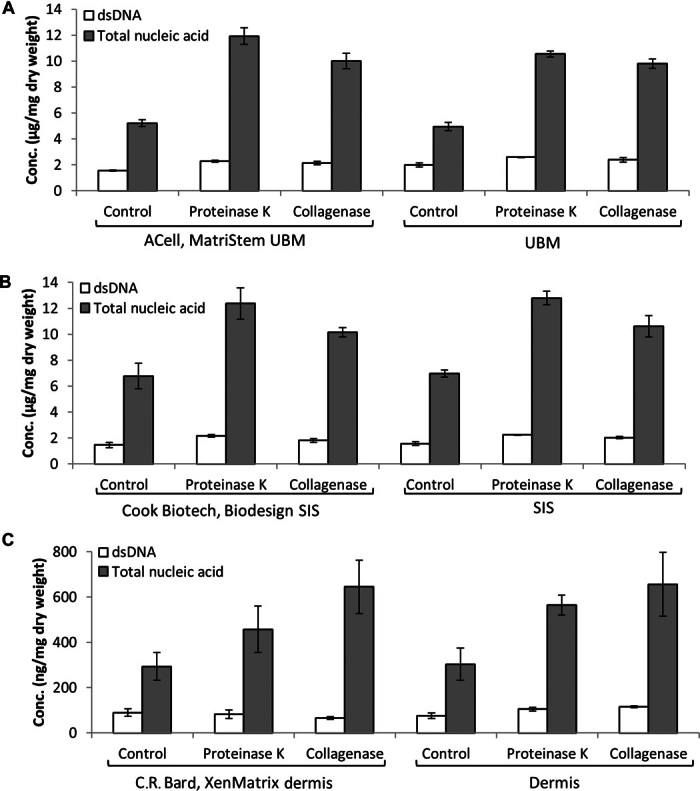
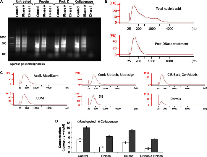
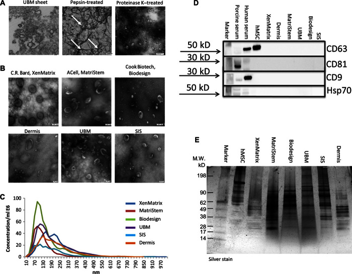
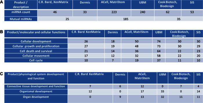
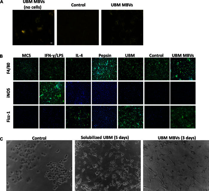
Biologic scaffold materials composed of extracellular matrix (ECM) have been used in a variety of surgical and tissue engineering/regenerative medicine applications and are associated with favorable constructive remodeling properties including angiogenesis, stem cell recruitment, and modulation of macrophage phenotype toward an anti-inflammatory effector cell type. However, the mechanisms by which these events are mediated are largely unknown. Matrix-bound nanovesicles (MBVs) are identified as an integral and functional component of ECM bioscaffolds. Extracellular vesicles (EVs) are potent vehicles of intercellular communication due to their ability to transfer RNA, proteins, enzymes, and lipids, thereby affecting physiologic and pathologic processes. Formerly identified exclusively in biologic fluids, the presence of EVs within the ECM of connective tissue has not been reported. In both laboratory-produced and commercially available biologic scaffolds, MBVs can be separated from the matrix only after enzymatic digestion of the ECM scaffold material, a temporal sequence similar to the functional activity attributed to implanted bioscaffolds during and following their degradation when used in clinical applications. The present study shows that MBVs contain microRNA capable of exerting phenotypical and functional effects on macrophage activation and neuroblastoma cell differentiation. The identification of MBVs embedded within the ECM of biologic scaffolds provides mechanistic insights not only into the inductive properties of ECM bioscaffolds but also into the regulation of tissue homeostasis.

Keywords: Exosomes; Extracellular Matrix (ECM); Extracellular vesicles (EV); Matrix Bound Nano Vesicles (MBV); Microvesicles (MV).


Figures

Fig. 1
Fig. 1. Comparison of nucleic acid concentration from UBM, SIS, or dermis and their commercially available equivalents.
(A to C) Concentration of total nucleic acid and dsDNA per milligram dry weight of ECM scaffold from untreated (control) and proteinase K– or collagenase-treated samples of (A) UBM and ACell MatriStem (porcine UBM), (B) SIS and Cook Biotech Biodesign (porcine SIS), and (C) dermis and C.R. Bard XenMatrix (porcine dermis). Total nucleic acid concentration was assessed by UV absorbance at 260 nm. dsDNA concentration was assessed using PicoGreen dsDNA quantification reagent. Variability from isolation to isolation is depicted by SD. Data are means ± SD; n = 3 isolations per sample.
Fig. 2
Fig. 2. Enzymatic digestion of decellularized ECM scaffolds releases small RNA molecules.
(A) Nucleic acid extracted from untreated UBM (no digest) and pepsin-, proteinase K–, or collagenase-treated UBM was exposed to RNase A, DNase I, or no-nuclease treatment (control). (B) Electropherogram depicting the small RNA pattern of nucleic acid in fluorescence units (FU) before (top panel) and after (bottom panel) DNase I treatment. (C) Electropherogram depicting small RNA pattern from the indicated samples in FU. (D) A subset of nucleic molecules in biologic scaffolds is protected from nuclease degradation.
Fig. 3
Fig. 3. Identification of ECM-embedded MBVs.
(A) TEM imaging of MBVs identified in a UBM sheet stained positive with osmium (left panel), pepsin-treated UBM (middle panel), or proteinase K–treated UBM (right panel). (B) TEM imaging of MBVs identified in proteinase K–treated ECM from three commercial and three laboratory-produced scaffolds. Scale bars, 100 nm. (C) Validation of MBV size was measured with NanoSight. (D) Western blot analysis was performed on four exosomal surface markers: CD63, CD81, CD9, and Hsp70. Expression levels were not detectable as compared to porcine serum, human serum, and human bone marrow–derived mesenchymal stem cell controls. (E) MBV protein cargo signature was different between MBVs and hMSCs as evaluated using SDS-PAGE and silver stain imaging.
Fig. 4
Fig. 4. Identification of miRNA packaged within MBVs.
MBV small RNA sequencing analysis reveals specific miRNA signature between commercial products and comparable in-house products (n = 1). (A) Numbers in each box represents different miRNAs within each sample. (B and C) Molecular and cellular functions (B) and physiological system development and function pathways (C) associated with identified miRNAs were generated using IPA. Each box represents the numbers of different miRNAs involved in each pathway.
Fig. 5
Fig. 5. MBVs are biologically active.
MBVs isolated from UBM were labeled with Exo-Glow. (A) C2C12 cells were exposed to labeled MBVs for 4 hours. The left panel shows a representative image of successful labeling of MBVs before exposure to cell culture. The right panel represents exposure of labeled MBVs in C2C12 compared to the middle panel image (control). Green fluorescence represents DNA, whereas red fluorescence represents RNA MBV cargo that is successfully integrated with target cells. (B) Bone marrow was isolated from C57bl/6 mice and cultured in medium supplemented with macrophage colony-stimulating factor (M-CSF) to derive macrophages. Macrophages were treated with IFN-γ (20 ng/ml) and LPS (100 ng/ml) to derive M1 macrophages, IL-4 (20 ng/ml) to derive M2 macrophages, and isolated MBVs (5 μg/ml) from a UBM source. Macrophages were fixed and immunolabeled for the pan-macrophage marker (F4/80) and markers associated with the M1 (iNOS) and M2 (Fizz-1) phenotype. MBV-treated macrophages are predominantly F4/80 + Fizz-1 + macrophages, indicating an M2-like phenotype. Experiment was conducted with n = 2 samples with four technical replicates. (C) N1E-115 neuroblastoma cells were exposed to pepsin-solubilized UBM and MBVs. Five days (solubilized UBM) and three days (MBVs) after exposure, neurite extensions were visible in treated cells compared to control.

References

    1. Alicuben E. T., DeMeester S. R., Onlay ventral hernia repairs using porcine non-cross-linked dermal biologic mesh. Hernia 18, 705–712 (2014). - PMC - PubMed
    1. Sicari B. M., Rubin J. P., Dearth C. L., Wolf M. T., Ambrosio F., Boninger M., Turner N. J., Weber D. J., Simpson T. W., Wyse A., Brown E. H., Dziki J. L., Fisher L. E., Brown S., Badylak S. F., An acellular biologic scaffold promotes skeletal muscle formation in mice and humans with volumetric muscle loss. Sci. Transl. Med. 6, 234ra258 (2014). - PMC - PubMed
    1. Badylak S. F., Hoppo T., Nieponice A., Gilbert T. W., Davison J. M., Jobe B. A., Esophageal preservation in five male patients after endoscopic inner-layer circumferential resection in the setting of superficial cancer: A regenerative medicine approach with a biologic scaffold. Tissue Eng. Part A 17, 1643–1650 (2011). - PMC - PubMed
    1. Bejjani G. K., Zabramski J. Durasis Study Group , Safety and efficacy of the porcine small intestinal submucosa dural substitute: Results of a prospective multicenter study and literature review. J. Neurosurg. 106, 1028–1033 (2007). - PubMed
    1. Longo U. G., Lamberti A., Petrillo S., Maffulli N., Denaro V., Scaffolds in tendon tissue engineering. Stem Cells Int. 2012, 517165 (2012). - PMC - PubMed