. 2016 Aug;35(4):217-26.
doi: 10.1089/mab.2016.0014. Epub 2016 Jul 7.

Novel Monoclonal Antibodies for Studies of Human and Rhesus Macaque Secretory Component and Human J-Chain

Affiliations

Novel Monoclonal Antibodies for Studies of Human and Rhesus Macaque Secretory Component and Human J-Chain

Ruijun Zhang et al. Monoclon Antib Immunodiagn Immunother. 2016 Aug.

Abstract

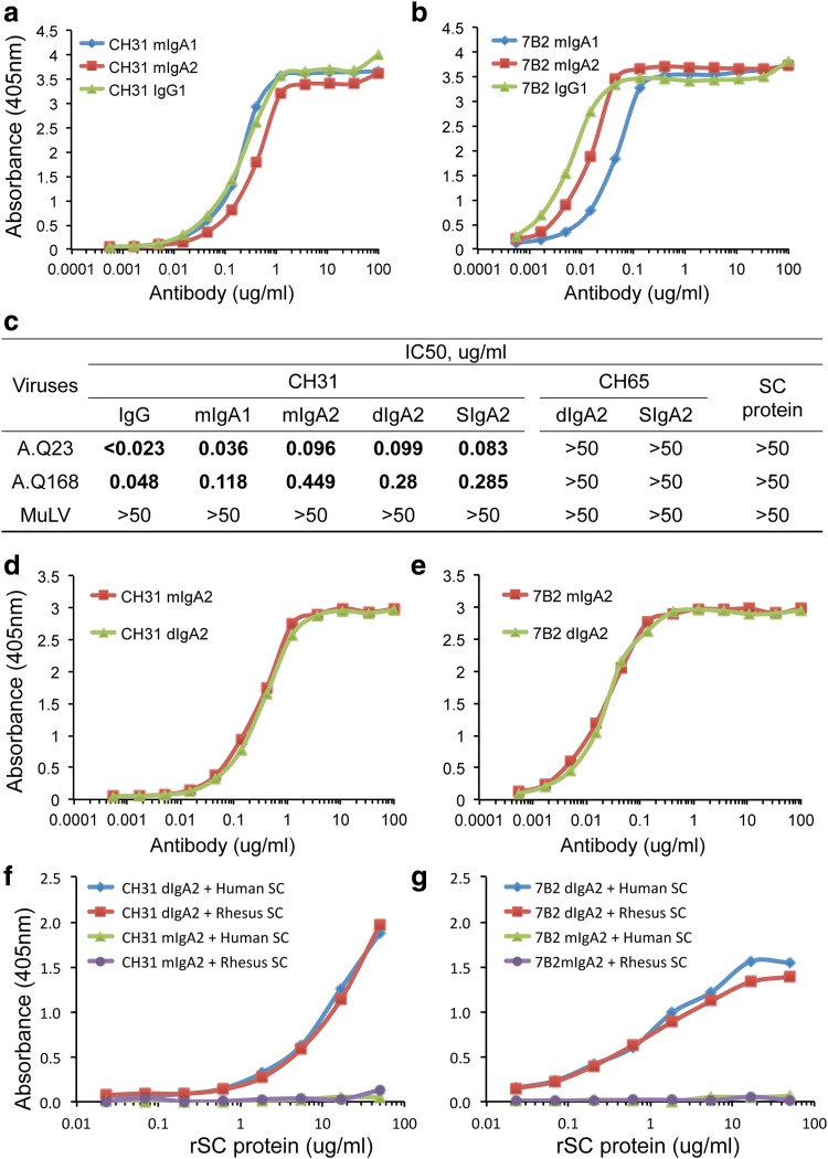
Immunoglobulin A (IgA) antibodies exist in monomeric, dimeric, and secretory forms. Dimerization of IgA depends on a 15-kD polypeptide termed "joining (J) chain," which is also part of the binding site for an epithelial glycoprotein called "secretory component (SC)," whether this after apical cleavage on secretory epithelia is ligand bound in secretory IgA (SIgA) or in a free form. Uncleaved membrane SC, also called the "polymeric Ig receptor," is thus crucial for transcytotic export of SIgA to mucosal surfaces, where it interacts with and modulates commensal bacteria and mediates protective immune responses against exogenous pathogens. To evaluate different forms of IgA, we have produced mouse monoclonal antibodies (MAbs) against human J-chain and free SC. We found that J-chain MAb 9A8 and SC MAb 9H7 identified human dimeric IgA and SIgA in enzyme-linked immunoassay and western blot analysis, as well as functioning in immunohistochemistry to identify cytoplasmic IgA of intestinal lamina propria plasmablasts/plasma cells and crypt epithelium of distal human intestine. Finally, we demonstrated that SC MAb 9H7 cross-reacted with rhesus macaque SIgA. These novel reagents should be of use in the study of the biology of various forms of IgA in humans and SIgA in macaques, as well as in monitoring the production and/or isolation of these forms of IgA.


Figures

<b>FIG. 1.</b>
FIG. 1.
Comparison of binding and neutralizing activity of mIgA1, mIgA2, IgG, dIgA2, and SIgA antibodies. Serial dilutions from 100 μg/mL to 0.001 μg/mL of purified CH31 (a, d, and f) and 7B2 (b, e, and g) in forms of mIgA1, mIgA2, dIgA2, SIgA2, and IgG (as control) were tested in ELISA for binding to CON-S gp120 (a and d) or HIV-1 gp41 immunodominant region peptide (b and e) by coating on 384-well ELISA plates at saturated amounts (2 μg/mL and 30 ng/well). Serial dilutions of recombinant human or rhesus free rSC at concentrations ranging from 50 μg to 0.023 μg/mL were tested in ELISA for binding to CH31 (f) and 7B2 (g) dIgA2 coated in the wells of the 384-well ELISA plates at saturated amounts (2 μg/mL and 30 ng/well). The binding of free SC to CH31 and 7B2 IgA was then detected by SC MAb 9H7 and expressed as absorbance at OD450 nm (vertical axis). (c) Neutralization activity of CH31 IgG, mIgA1, mIgA2, dIgA2, and SIgA against two clade A HIV-1 tier 2 isolates (A.Q23 and A.Q168) and MuLV as control in TZM-bl cell-based HIV-1 neutralization assays. ELISA, enzyme-linked immunoassay; MAb, monoclonal antibodies; MuLV, murine leukemia virus.
<b>FIG. 2.</b>
FIG. 2.
Reactivity of J-chain antibodies to recombinant J-chain, CH31 dIgA2, and human IgA MAbs derived from human B cell lines. Recombinant J-chain and purified CH31 dIgA2 were fractionated in 4%–20% gradient SDS-PAGE under nonreducing (a) and reducing (b) conditions. Human IgA MAbs HG129 and HG130 (c and d) were purified from EBV-transformed B cell lines and fractionated in 4%–20% gradient blue native-PAGE (c and d). Western blots were carried out by incubation with J-chain MAbs 4G10 and 9A8 2 at 2 μg/mL or our rabbit anti-human J-chain R-282 as a positive control, followed by incubation with either AP-labeled anti-mouse or anti-rabbit secondary antibody. J-chain MAb 4G10 and 9A8 at concentrations ranging from 100 μg to 0.01 μg/mL were also tested in ELISA for binding to recombinant J-chain (e) and CH31 dIgA2 antibody (f) coated in the wells of the 384-well ELISA plates at saturated amounts (2 μg/mL and 30 ng/well). AP, alkaline phosphatase; SDS-PAGE, sodium dodecyl sulfate–polyacrylamide gel electrophoresis.
<b>FIG. 3.</b>
FIG. 3.
Detection of SIgA by western blots with anti-SC MAb 9H7. CH31 SIgA antibody was produced by either in vitro formation by incubation of CH31 dIgA with free SC (a–d) or in vivo production in CHO cell line stably transfected with the human J-chain and SC genes (e–h). Purified CH31 dIgA (a) and in vitro-formed CH31 SIgA (b) were shown as a single major peak in size exclusion column chromatography, fractionated in 4%–20% gradient SDS-PAGE under nonreducing conditions (c), and detected in western blots (d) by anti-SC MAb9H7. Recombinant human free SC was used as a positive control. CH31 dIgA and SIgA produced in CHO cell lines stably transfected with human J-chain alone, or with both human J-chain and SC genes, were also analyzed by 4%–20% gradient SDS-PAGE under nonreducing (N) and reducing (R) conditions (c). Gels were stained with Coomassie blue (e) or western blotted with J-chain MAbs 9A8 (f), 9H7 (g), or mouse anti-human IgA antibody (h), followed by AP-conjugated goat anti-mouse IgG antibody (1/2000) conjugated with alkaline phosphatase.
<b>FIG. 4.</b>
FIG. 4.
Detection of IgA derived from human BM and human or rhesus plasma. IgA purified from human BM and human (CH1700) and rhesus plasma was fractionated by 4%–20% SDS-PAGE under nonreducing condition. Recombinant human and rhesus free SC was used as control. Gels were stained with Coomassie blue (a) or blotted with anti-human SC MAb 9H7 (b). BM, breast milk.
<b>FIG. 5.</b>
FIG. 5.
Immunostaining of two key proteins in mucosal immunity, SC and J-chain, on human tissue sections from the distal small intestine. (a) Immunoperoxidase staining with MAb 9H7 (0.5 μg/mL) shows normal distribution of SC in the crypts, with decreasing intensity toward the tips of the villi. The goblet cells appear negative. (b–e) Immunostaining of J-chain distribution comparing results obtained with MAb 9A8 (10 μg/mL) and our rabbit antibody R-282 (1/800). (b) Numerous lamina propria plasmablasts/plasma cells with abundant cytoplasmic J-chain are detected with the MAb. In the crypts, J-chain is seen particularly not only at the luminal side of the epithelium but also basolaterally on the epithelial cells, signifying dIgA (and probably much less pentameric IgM) transported externally by the polymeric Ig receptor (membrane SC). (c) The middle part of the field in (b) shown at higher magnification. (d) Double immunofluorescence staining with the monoclonal (Alexa Fluor 488-conjugated anti-mouse IgG, green) and polyclonal (Alexa Fluor 555-conjugated anti-rabbit IgG, red) antibodies in mixture. The two stainings overlap (merge, yellow) in most of the lamina propria PCs, while epithelial staining in crypts to the left and right seems to be dominated by green color, which, however, may depend on the concentration of the reagents. (e) Immunoperoxidase staining with our rabbit R-282 shows a distribution of J-chain in PCs and crypts that is virtually identical to that obtained with the MAb in (b) and (c).

References

    1. Brandtzaeg P: Secretory IgA: Designed for anti-microbial defense. Front Immunol 2013;4:222. - PMC - PubMed
    1. Brandtzaeg P, Kiyono H, Pabst R, and Russell MW: Terminology: Nomenclature of mucosa-associated lymphoid tissue. Mucosal Immunol 2008;1:31–37 - PubMed
    1. Fagarasan S, and Honjo T: Intestinal IgA synthesis: Regulation of front-line body defences. Nat Rev Immunol 2003;3:63–72 - PubMed
    1. Fernandez MI, Pedron T, Tournebize R, Olivo-Marin JC, Sansonetti PJ, and Phalipon A: Anti-inflammatory role for intracellular dimeric immunoglobulin A by neutralization of lipopolysaccharide in epithelial cells. Immunity 2003;18:739–749 - PubMed
    1. Macpherson AJ, and Uhr T: Induction of protective IgA by intestinal dendritic cells carrying commensal bacteria. Science 2004;303:1662–1665 - PubMed

MeSH terms

Substances

LinkOut - more resources