. 2016 Jul 8;17(1):145.
doi: 10.1186/s13059-016-0995-z.

Widespread parainflammation in human cancer

Affiliations

Widespread parainflammation in human cancer

Dvir Aran et al. Genome Biol. .

Abstract

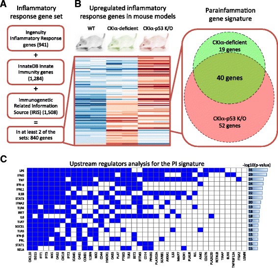
Background: Chronic inflammation has been recognized as one of the hallmarks of cancer. We recently showed that parainflammation, a unique variant of inflammation between homeostasis and chronic inflammation, strongly promotes mouse gut tumorigenesis upon p53 loss. Here we explore the prevalence of parainflammation in human cancer and determine its relationship to certain molecular and clinical parameters affecting treatment and prognosis.

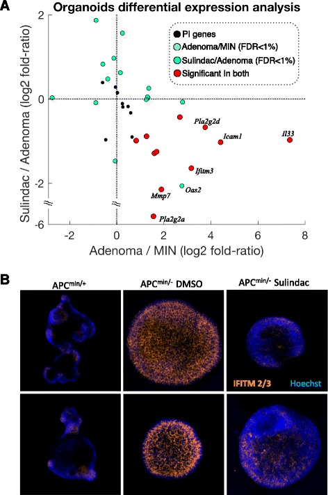
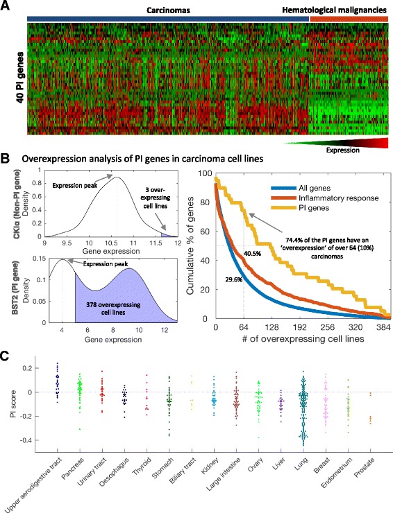
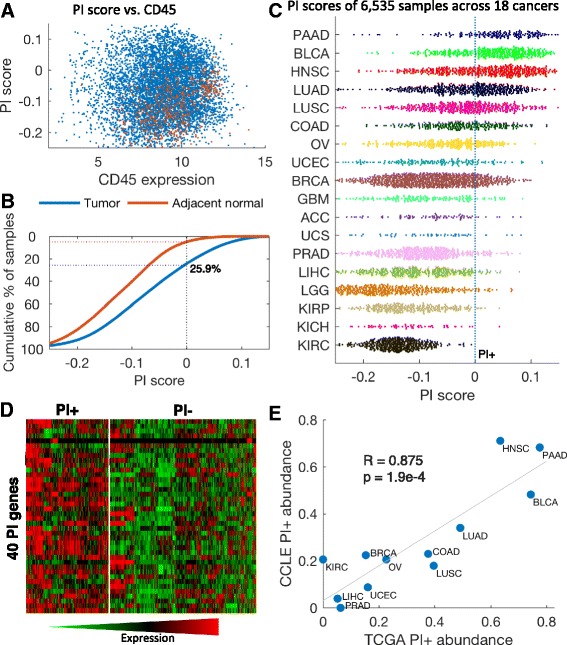
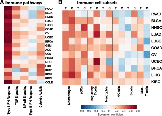
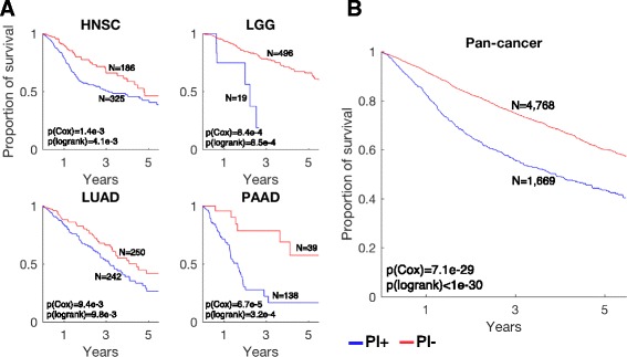
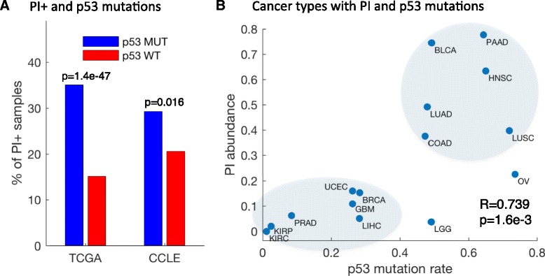
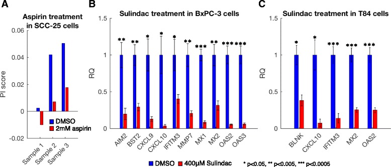
Results: We generated a transcriptome signature to identify parainflammation in many primary human tumors and carcinoma cell lines as distinct from their normal tissue counterparts and the tumor microenvironment and show that parainflammation-positive tumors are enriched for p53 mutations and associated with poor prognosis. Non-steroidal anti-inflammatory drug (NSAID) treatment suppresses parainflammation in both murine and human cancers, possibly explaining a protective effect of NSAIDs against cancer.

Conclusions: We conclude that parainflammation, a low-grade form of inflammation, is widely prevalent in human cancer, particularly in cancer types commonly harboring p53 mutations. Our data suggest that parainflammation may be a driver for p53 mutagenesis and a guide for cancer prevention by NSAID treatment.

Keywords: Cancer prevention; Genomics; Inflammation; NSAID treatment; Parainflammation; p53 mutations.


Figures

Fig. 1
Fig. 1
The parainflammation signature. a An inflammatory response gene set was generated by combining genes that are found in at least two of three manually curated databases: the Ingenuity inflammatory response gene list, InnateDB innate immunity genes, and the Immunogenetic Related Information Source (IRIS). b Differential expression analysis of CKIα-deficient and CKIα-p53-deficient mice against wild type (WT) revealed 59 and 92 upregulated inflammatory response genes in the single and double knock outs (K/O), respectively, and 40 genes upregulated in both. The 40 genes were designated as the parainflammation (PI) gene signature. The heatmap shows the expression levels of the 111 upregulated genes (red, high expression; blue, low expression). c Upstream regulatory analysis revealed a strong enrichment for genes regulated by lipopolysaccharide and immune pathways that are activated by it. The rows show the top enriched, non-chemical regulators, the columns are the 40 PI genes, and a blue cell represents regulation
Fig. 2
Fig. 2
Parainflammation decreases in response to NSAID treatment in mouse organoids. a The adenoma/MIN log2 expression fold ratio of PI genes (x-axis) against the sulindac-treated adenomas/adenoma log2 expression fold ratio of PI genes (y-axis). Colors represent significance in the differential expression analyses (false discovery rate (FDR) <1 %). Along the x-axis it can be observed that 17 of 19 significant genes in adenoma/MIN are upregulated in the adenomas. Of those, 11 are significantly downregulated in the sulindac-treated samples and none are significantly upregulated. b IFITM2/3 immunofluorescence in APCmin/+, adenoma and Sulindac-treated adenoma organoids. Both rows show different fields of same experiment.
Fig. 3
Fig. 3
Parainflammation genes are overexpressed in carcinoma cell lines. a Heatmap of the expression of 39 PI genes in 634 carcinoma cell lines and 180 hematopoietic and lymphoid cancer cell lines from CCLE. One PI gene, IFITM3, is not represented in CCLE. Of the PI genes, 19 show significantly higher expression in carcinomas compared with cancers originated from immune cell types; 10 PI genes are more abundant in immune cancers. b Left: the distribution of expression of two representative genes across 634 carcinoma cell lines from CCLE. We detected the expression peak and counted the number of samples that express the gene twofold (1 in log2 scale) more than the peak. The top example, the gene CSNK1A1 (CKIα), which here represents a housekeeping gene, is a typical normally distributed gene with low “overexpression” rate and the bottom example, BST2, which is part of the PI signature, shows a gene with a bimodal expression pattern, corresponding to a high “overexpression” rate. Right: the cumulative overexpression rates for the PI genes, all inflammatory response genes, and all genes. Of the PI signature genes, 29 (74.4 %) are overexpressed in at least 10 % of the carcinoma cell lines (≥64 samples) compared with 29.6 % and 40.5 % of all and inflammatory response genes, respectively. The yellow curve (PI genes) shows remarkably higher levels of overexpression along the whole graph. c Spread plot of tThe PI score in 634 carcinoma cell lines grouped by tissue types. The dashed blue line differentiates PI+ and PI− samples as defined by the proportion of PI+ of tumor samples
Fig. 4
Fig. 4
Pan-cancer parainflammation in human cancers. a PI scores (y-axis) of all 6535 tumor samples (blue) and 582 adjacent normal samples (red) versus the CD45 expression level (x-axis). No correlation is observed between the PI score and CD45 after the adjustment. b PI scores in the tumor samples (blue) and the adjacent normal samples (red). The y-axis is the cumulative percentage of samples over the score in x; 25.9 % of the tumor samples (dashed blue line) are over a threshold which only 5 % of the adjacent normal tissues pass (dashed red line). The PI score is shifted accordingly, so PI+ samples have positive scores. c Spread plot of the PI score in 6535 tumor samples from 18 cancer types. The dashed blue line differentiates PI+ and PI− samples. PAAD pancreatic adenocarcinoma, BLCA bladder carcinoma, HNSC head and neck squamous cell carcinoma, LUAD lung adenocarcinoma, LUSC lung squamous cell carcinoma, COAD colon adenocarcinoma, OV ovarian serous cystadenocarcinoma, UCEC uterine corpus endometrial carcinoma, BRCA breast carcinoma, GBM glioblastoma multiforme, ACC adrenocortical carcinoma, UCS uterine carcinosarcoma, PRAD prostate adenocarcinoma, LIHC liver hepatocellular carcinoma, LGG lower grade glioma, KIRP kidney renal papillary cell carcinoma, KICH kidney chromophobe, KIRC kidney renal clear cell carcinoma. d Heatmap of expression profiles of PI genes across TCGA samples: left, PI+ samples; right, PI− samples. Different subsets of PI genes are expressed in PI+ samples. The expression levels presented are after adjustment to immune infiltrations and are standardized across all samples. e Correlation of the fraction of PI+ samples in each tumor type from TCGA with corresponding tissue origin in CCLE. The same types of cancers have low or high PI+ levels in tumors and in cell lines. The Pearson coefficient is presented
Fig. 5
Fig. 5
Cancer parainflammation resembles macrophage infiltration. a Heatmap of the Spearman correlations between the PI score and immune functional gene sets across different cancer types. Correlations in CCLE are shown as well (Additional file 3: Tables S5 and S6). b Heatmap of the Spearman correlations between the PI score and the immune subset enrichments calculated using gene sets (Additional files 6 and 9) across different cancer types derived from both TCGA (T) and CCLE (C). Similar correlation trends are observed for a cancer type whether the data were derived from TCGA or CCLE, suggesting that the correlation is due not to association of PI with immune subset presence but to shared functionality with the gene signatures. PAAD pancreatic adenocarcinoma, BLCA bladder carcinoma, HNSC head and neck squamous cell carcinoma, LUAD lung adenocarcinoma, LUSC lung squamous cell carcinoma, COAD colon adenocarcinoma, OV ovarian serous cystadenocarcinoma, UCEC uterine corpus endometrial carcinoma, BRCA breast carcinoma, GBM glioblastoma multiforme, ACC adrenocortical carcinoma, UCS uterine carcinosarcoma, PRAD prostate adenocarcinoma, LIHC liver hepatocellular carcinoma, LGG lower grade glioma, KIRP kidney renal papillary cell carcinoma, KICH kidney chromophobe, KIRC kidney renal clear cell carcinoma
Fig. 6
Fig. 6
Parainflammation is associated with worse prognosis. a Kaplan-Meier plots for PI+ samples (blue) versus PI− samples (red) in head and neck squamous cell carcinoma (HNSC), lower grade glioma (LGG), lung adenocarcinoma (LUAD), and pancreatic adenocarcinoma (PAAD). The p values were calculated using Cox proportional hazard regression of the PI scores and the log-rank method between PI+ and PI− samples. b Pan-cancer survival analysis of 6437 patients with survival data and PI scores
Fig. 7
Fig. 7
Parainflammation is associated with p53 mutations. a Percentage of p53 nonsense mutated PI+ samples (blue) versus percentage of p53 WT PI+ samples (red) in TCGA and in CCLE. b Abundance of p53 missense mutations (x-axis) versus the abundance of PI+ samples (y-axis) in 15 cancer types with sufficient information. Cancer types with higher rates of p53 nonsense mutations tend to have higher rates of PI. The Spearman coefficient is presented
Fig. 8
Fig. 8
Parainflammation decreases in response to NSAID treatment in human cell lines. a average of the differential expression (in log2 scale) of three replicates each of control and aspirin-treated SCC-25 cells for the PI genes. A kernel smoothed regression curve for all genes is presented as reference (red). The p value presented was calculated using a Wilcoxon rank-sum test. b qPCR analysis of highly expressed PI genes in control and sulindac-treated BxPC3 cells. Error bars show the standard error of the mean (SEM) of triplicates. c qPCR analysis of highly expressed PI genes in control and sulindac-treated T84 cells. Error bars show the SEM of triplicates

References

    1. Hanahan D, Weinberg RA. Hallmarks of cancer: the next generation. Cell. 2011;144:646–74. doi: 10.1016/j.cell.2011.02.013. - DOI - PubMed
    1. Aggarwal BB, Vijayalekshmi RV, Sung B. Targeting inflammatory pathways for prevention and therapy of cancer: short-term friend, long-term foe. Clin Cancer Res. 2009;15:425–30. doi: 10.1158/1078-0432.CCR-08-0149. - DOI - PubMed
    1. Balkwill FR, Mantovani A. Cancer-related inflammation: common themes and therapeutic opportunities. Semin Cancer Biol. 2012;22:33–40. doi: 10.1016/j.semcancer.2011.12.005. - DOI - PubMed
    1. Grivennikov SI, Greten FR, Karin M. Immunity, inflammation, and cancer. Cell. 2010;140:883–99. doi: 10.1016/j.cell.2010.01.025. - DOI - PMC - PubMed
    1. Elyada E, Pribluda A, Goldstein RE, Morgenstern Y, Brachya G, Cojocaru G, Snir-Alkalay I, Burstain I, Haffner-Krausz R, Jung S, et al. CKIalpha ablation highlights a critical role for p53 in invasiveness control. Nature. 2011;470:409–13. - PubMed

Publication types

Substances