. 2016 Jul 7;11(7):e0158657.
doi: 10.1371/journal.pone.0158657. eCollection 2016.

Activation of the miR-34a/SIRT1/p53 Signaling Pathway Contributes to the Progress of Liver Fibrosis via Inducing Apoptosis in Hepatocytes but Not in HSCs

Affiliations

Activation of the miR-34a/SIRT1/p53 Signaling Pathway Contributes to the Progress of Liver Fibrosis via Inducing Apoptosis in Hepatocytes but Not in HSCs

Xiao-Feng Tian et al. PLoS One. .

Abstract

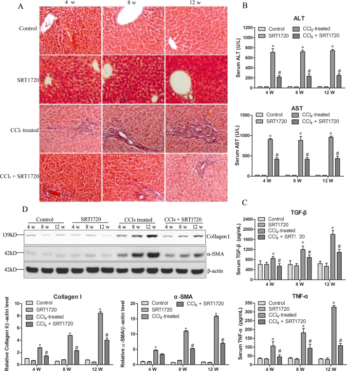
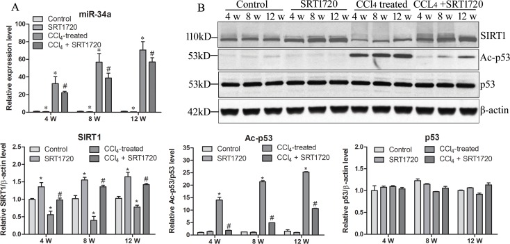
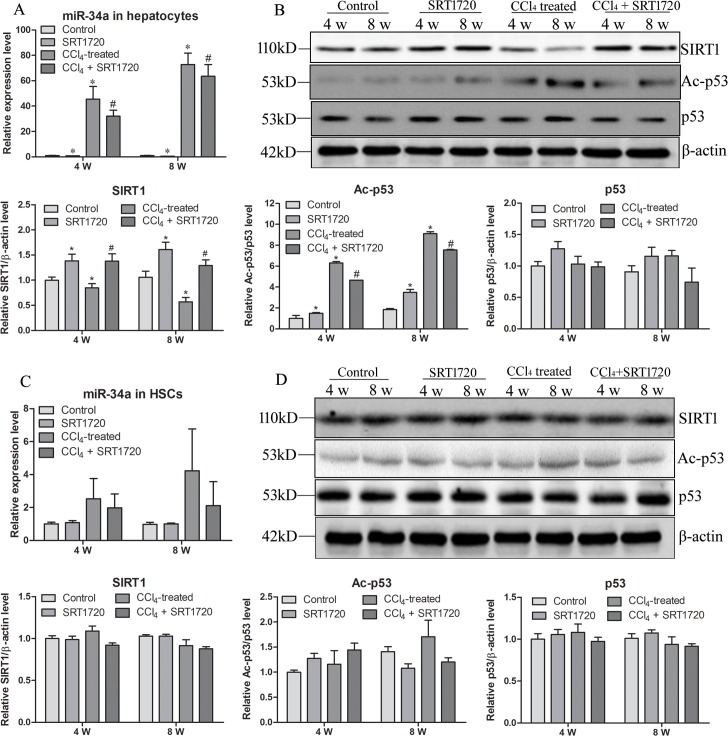
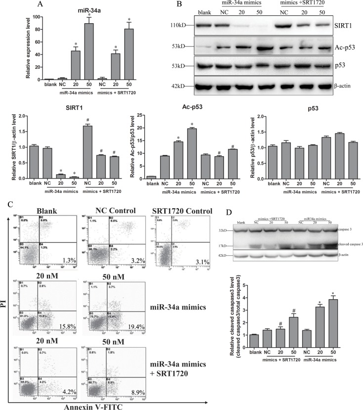
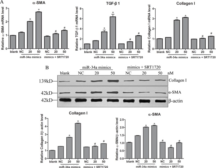
Liver fibrosis results from a sustained wound healing response to chronic liver injury, and the activation of nonparenchymal hepatic stellate cells (HSCs) is the pivotal process. MicroRNA-34a (miR-34a) is the direct target gene of p53 and activates p53 through sirtuin 1 (SIRT1) simultaneously. The miR-34a/SIRT1/p53 signaling pathway thus forms a positive feedback loop wherein p53 induces miR-34a and miR-34a activates p53 by inhibiting SIRT1, playing an important role in cell proliferation and apoptosis. miR-34a expression has been found to be increased in animal models or in human patients with different liver diseases, including liver fibrosis. However, the exact role of this classical miR-34a/SIRT1/p53 signaling pathway in liver fibrosis remains unclear. In the present study, using a CCl4-induced rat liver fibrosis model, we found that the miR-34a/SIRT1/p53 signaling pathway was activated and could be inhibited by SIRT1 activator SRT1720. Further studies showed that the miR-34a/SIRT1/p53 signaling pathway was activated in hepatocytes but not in HSCs. The activation of this pathway in hepatocytes resulted in the apoptosis of hepatocytes and thus activated HSCs. Our data indicate that the miR-34a/SIRT1/p53 signaling pathway might be a promising therapeutic target for liver fibrosis.


Conflict of interest statement

Competing Interests: The authors have declared that no competing interests exist.

Figures

Fig 1
Fig 1. Upregulation of SIRT1 inhibits liver fibrosis.
Rats were divided into four groups: Control, SIRT1 activator SRT1720-treated group, liver fibrosis model (CCl4 treated) and SRT1720-treated liver fibrosis model (CCl4 + SRT1720). The liver fibrosis model was established through intraperitoneally injecting rats with 0.2 mL/kg of CCl4 (mixed in 1:1 vegetable oil) twice a week for 4 weeks and then once a week for the next 12 weeks. For the CCl4 + SRT1720 group, in addition to CCl4, rats were treated every other day with an intraperitoneal injection of SRT1720 at doses of 50 mg/kg. Rats were sacrificed at 4, 8 and 12 w. The liver fibrosis of the model was confirmed by hematoxylin and eosin staining, showing the histopathology of liver (A), serum levels of alanine aminotransferase (ALT), aspartate aminotransferase (AST) (B), tumor necrosis factor (TNF)-α and tumor growth factor (TGF)-β (C) showing the liver function and immunology response, and expression of α-SMA (D). * P<0.05 compared to control. # P<0.05 compared to CCl4-treated group.
Fig 2
Fig 2. miR-34a/SIRT1/p53 signaling pathway is activated during liver fibrosis.
Expression of miR-34a (A), SIRT1, p53, acylated p53 (Ac-p53) and β-actin (B) were detected in four rat groups at three time points. * P<0.05 compared to control. # P<0.05 compared to CCl4-treated group.
Fig 3
Fig 3. miR-34a/SIRT1/p53 signaling pathway is activated in hepatocytes but not in HSCs.
Primary hepatocytes and HSCs were isolated from rats at 4 and 8 w. Expression of miR-34a (A, C), SIRT1, p53, Ac-p53 and β-actin (B, D) were detected in the two types of primary cells. A, B, primary hepatocytes. C, D, primary HSCs. * P<0.05 compared to control. # P<0.05 compared to CCl4-treated group.
Fig 4
Fig 4. Upregulation of miR-34a-induced apoptosis of hepatocytes.
Human normal hepatocytes cell line L-02 cells were transfected with miR-34a mimics (20, 50 nM) or negative miRNA control (NC) with or without SRT1720 for 48 h. Expression of miR-34a was confirmed (A) at 24 h after transfection. Changes in the expression of SIRT1, p53 and Ac-p53 were determined (B) after 48 h. The apoptosis of hepatocytes was detected using an Annexin V-FITC/PI Apoptosis Detection Kit in a flow cytometry system (C) or by detecting the cleavage of caspase3 (D). Blank, L-02 cells; NC, negative control (L-02 cells transfected with negative mimics). * P<0.05 compared to NC. # P<0.05 compared to mimics transfection.
Fig 5
Fig 5. miR-34a-induced apoptosis of hepatocytes activates HSCs.
HSCs LX-2 cells were co-cultured with pre-treated L-02 cells without SRT1720 for 24 h. L-02 cell were transfected with miR-34a mimics (20, 50 nM) in the presence/absence of SRT1720 for 24 h. The mRNA levels of α-SMA, TGF-β1 and collagen I (A) and the protein expression ofα-SMA and collagen I (B) in LX-2 cells were detected. * P<0.05 compared to NC and to miR-34a mimic-transfected cells. Blank, HSC LX-2 co-cultured with L -02 cells; NC, negative control (HSC LX-2 co-cultured with L -02 cells transfected with negative mimics). * P<0.05 compared to NC. # P<0.05 compared to mimics transfection.

References

    1. Friedman SL. Liver fibrosis—from bench to bedside. Journal of hepatology. 2003;38 Suppl 1:S38–53. . - PubMed
    1. Moreira RK. Hepatic stellate cells and liver fibrosis. Arch Pathol Lab Med. 2007;131(11):1728–34. Epub 2007/11/06. doi: 2006-0688-RSR-1 [pii] 10.1043/1543-2165(2007)131[1728:HSCALF]2.0.CO;2 . - DOI - PubMed
    1. Duval F, Moreno-Cuevas JE, Gonzalez-Garza MT, Rodriguez-Montalvo C, Cruz-Vega DE. Liver fibrosis and protection mechanisms action of medicinal plants targeting apoptosis of hepatocytes and hepatic stellate cells. Advances in pharmacological sciences. 2014;2014:373295 10.1155/2014/373295 - DOI - PMC - PubMed
    1. Mallat A, Lodder J, Teixeira-Clerc F, Moreau R, Codogno P, Lotersztajn S. Autophagy: a multifaceted partner in liver fibrosis. BioMed research international. 2014;2014:869390 10.1155/2014/869390 - DOI - PMC - PubMed
    1. Bartel DP. MicroRNAs: genomics, biogenesis, mechanism, and function. Cell. 2004;116(2):281–97. Epub 2004/01/28. doi: S0092867404000455 [pii]. . - PubMed