. 2016 Jul 8:6:29256.
doi: 10.1038/srep29256.

Cathepsin S attenuates endosomal EGFR signalling: A mechanical rationale for the combination of cathepsin S and EGFR tyrosine kinase inhibitors

Affiliations

Cathepsin S attenuates endosomal EGFR signalling: A mechanical rationale for the combination of cathepsin S and EGFR tyrosine kinase inhibitors

Chien-Chang Huang et al. Sci Rep. .

Abstract

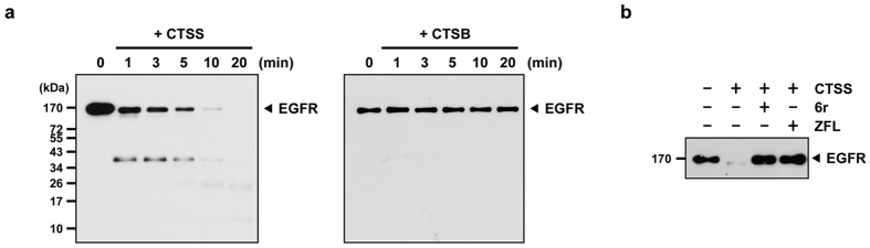
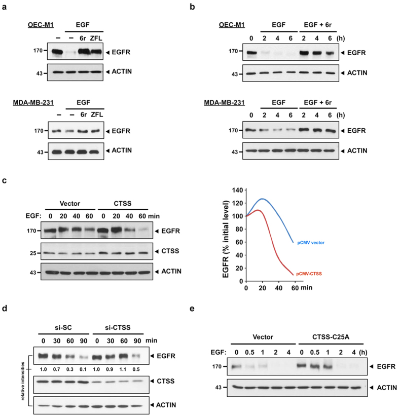
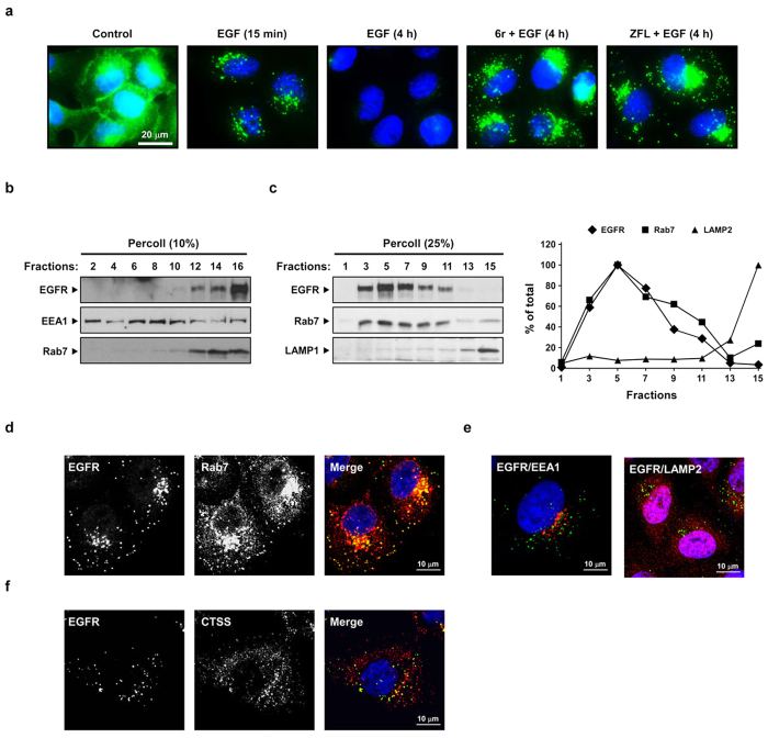
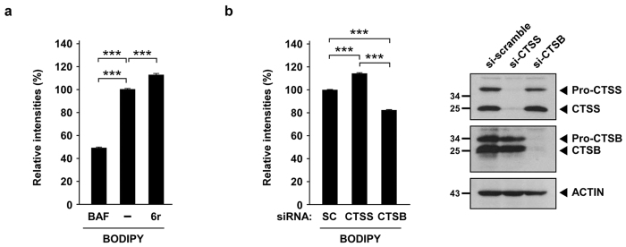
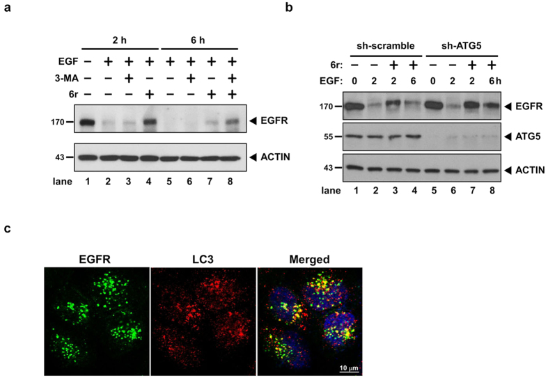
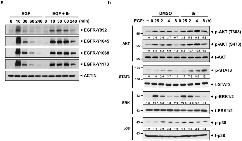
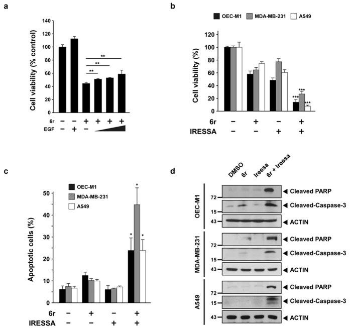
EGF-mediated EGFR endocytosis plays a crucial role in the attenuation of EGFR activation by sorting from early endosomes to late endosomes and transporting them into lysosomes for the final proteolytic degradation. We previously observed that cathepsin S (CTSS) inhibition induces tumour cell autophagy through the EGFR-mediated signalling pathway. In this study, we further clarified the relationship between CTSS activities and EGFR signalling regulation. Our results revealed that CTSS can regulate EGFR signalling by facilitating EGF-mediated EGFR degradation. CTSS inhibition delayed the EGFR degradation process and caused EGFR accumulation in the late endosomes at the perinuclear region, which provides spatial compartments for prolonged EGFR and sustained downstream signal transducer and activator of transcription 3 and AKT signalling. Notably, cellular apoptosis was markedly enhanced by combining treatment with the EGFR inhibitor Iressa and CTSS inhibitor 6r. The data not only reveal a biological role of CTSS in EGFR signalling regulation but also evidence a rationale for its clinical evaluation in the combination of CTSS and EGFR tyrosine kinase inhibitors.


Figures

Figure 1
Figure 1. Proteolytic cleavage of EGFR by CTSS.
(a) Recombinant EGFR was incubated with CTSS (left panel) and CTSB (right panel) at 37 °C for the indicated durations. The reaction was stopped by adding a sample buffer, and the reaction mixtures were subjected to SDS-PAGE, followed by Western blotting with an EGFR antibody. (b) Recombinant EGFR was incubated with CTSS in the presence of a CTSS inhibitor 6r or ZFL. The reaction mixtures were incubated at 37 °C for 20 min and then subjected to SDS-PAGE.
Figure 2
Figure 2. CTSS attenuates EGF-mediated EGFR degradation.
(a) OEC-M1 and MDA-MB-231 cells were pretreated with 20 μM 6r or ZFL for 1 h and subsequently incubated with 100 ng/mL EGF for an additional 2 h. The total cell lysates were analysed using EGFR-specific antibodies. ACTIN was used as the internal control for semiquantitative loading in each lane. (b) The cells were stimulated with EGF (100 ng/mL) with or without the pretreatment of 20 μM 6r for the indicated durations. EGFR degradation was examined through immunostaining by using an anti-EGFR antibody. Notably, a substantial amount of EGFR was detectable even after 6 h of EGF stimulation in 6r-treated cells. (c) The OEC-M1 cells were transiently transfected with plasmids (pCMV) that encoded wild-type CTSS. After 24 h of transfection, the cells were treated with 100 ng/mL EGF for the indicated durations and the cellular EGFR, CTSS, and ACTIN signals were determined through Western blotting. The lifespan of EGF-mediated EGFR degradation was calculated by normalising the signal intensity of EGFR with that of ACTIN. (d) The MDA-MB-231 cells were transfected with specific 50 nM CTSS siRNA (si-CTSS) for 24 h and subsequently incubated with 100 ng/mL EGF for the indicated durations. The nontargeting scramble siRNA (si-SC) was used as the scramble control. (e) The MDA-MB-231 cells were transiently transfected with plasmids encoding the CTSS-C25A mutant. After 24 h of transfection, the cells were incubated with 100 ng/mL of EGF for the indicated durations. Furthermore, the cells were harvested and subjected to SDS-PAGE and Western blotting. EGFR degradation was determined using an antibody against EGFR. ACTIN signalling was included as the loading control.
Figure 3
Figure 3. CTSS inhibition causes EGFR accumulation in late endosomes.
(a) After the pretreatment of 20 μM 6r or ZFL for 1 h, the cells were stimulated with 100 ng/mL of EGF for the indicated durations. The cells were fixed, permeabilised, and immunostained for EGFR (green), as described in Methods. Nuclei were stained with DAPI. Note that CTSS inhibition caused EGFR accumulation in punctate intracellular vesicles. (b,c) The cells were pretreated with 20 μM 6r for 1 h and subsequently incubated with 100 ng/mL EGF for 1 h further. The cells were then homogenised and fractionated into gradients by using 10% Percoll (b) and 25% Percoll (c), as described in Methods. The Percoll gradient fractions were then subjected to SDS-PAGE, followed by Western blotting with EGFR, EEA1, Rab7, and LAMP1 antibodies. (d–f) The cells were pretreated with 20 μM 6r for 1 h and subsequently incubated with 100 ng/mL EGF for 2 h further and fixed, permeabilised, and stained with antibodies to EGFR (green; d–f) and Rab7 (red; d), EEA1 (red, e), LAMP2 (red, e), or CTSS (red; f). The cells were imaged through confocal microscopy. Scale bars, 10 μm. (d) Confocal images showed the colocalisation between accumulated EGFR and Rab7. (e) Confocal images showed that a very small amount of accumulated EGFR was colocalised with EEA1 or LAMP2. (f) Confocal images showing colocalisation between CTSS and accumulated EGFR.
Figure 4
Figure 4. CTSS inhibition does not impair lysosomal activities.
(a) OEC-M1 cells were pretreated with vesicle or 100 nM BAF for 1 h and then incubated with 20 μM 6r for 1 h further. Lysosomal proteolytic activities were determined using BODIPY–BSA and quantified through flow cytometry. (b) After 48 h of siRNA knockdown of CTSS and CTSB, the relative expression of CTSS and CTSB were determined through Western blotting (right panel). Lysosomal proteolytic activities were determined using BODIPY–BSA and quantified through flow cytometry (right panel). Data represent the mean ± SD of three independent experiments. Differences were found to be statistically significant at ***P < 0.001.
Figure 5
Figure 5. Autophagy is involved in the clearance of accumulated EGFR.
(a) The cells were pretreated with 10 mM 3-MA or 20 μM 6r for 1 h and then stimulated with 100 ng/mL EGF for additional periods of 2 and 6 h. Subsequently, the cells were harvested and subjected to SDS-PAGE and Western blotting. EGFR degradation was examined through immunostaining by using an anti-EGFR antibody. ACTIN signalling was considered the loading control. (b) After 24 h of shRNA knockdown of CTSS, the cells were treated with 100 ng/mL EGF for the indicated durations. The harvested cells were subjected to Western blotting. (c) The cells were pretreated with 20 μM 6r for 1 h and then treated with 100 ng/mL EGF for 30 min. The cells were immediately fixed, permeabilised, stained with EGFR and LC3 antibodies, as described in Methods.
Figure 6
Figure 6. CTSS inhibition results in a prolonged EGFR activation and sustained downstream signaling.
(a) OEC-M1 cells with and without the treatment of 20 μM 6r were stimulated with 100 ng/mL EGF for the indicated durations. Cell lysates were separated through SDS-PAGE and then probed with the indicated phospho-EGFR antibodies. (b) After 1 h of pretreatment of 20 μM 6r, the cells were stimulated with 100 ng/mL EGF for the indicated durations. The harvested cells were then subjected to Western blotting analysis and probed with the indicated antibodies. The relative band intensities were shown.
Figure 7
Figure 7. Cotreatment with 6r and Iressa enhances cancer cells apoptosis.
(a) OEC-M1 cells were pretreated with or without 20 μM 6r for 1 h and subsequently incubated with 100 ng/mL EGF for an additional 72 h. Cell viabilities were determined by a methylene blue dye assay. The bars represent the relative mean survival of four independent wells ± SD. Differences were considered statistically significant at *P < 0.05, **P < 0.01, and ***P < 0.001. (b) The cells were treated with 6r alone or in combination with Iressa for 72 h. Cell viabilities were determined by a methylene blue dye assay. In the OEC-M1 cells, 20 μM 6r and 10 μM Iressa were used. For the MDA-MB-231 cells, 15 μM 6r and 15 μM Iressa were used. Moreover, 40 μM 6r and 27 μM Iressa were used in the A549 cells. (c) After 24 h of treatment with 6r or Iressa or the 6r–Iressa combination, cell apoptotic rates were evaluated with annexin V/propidium iodide double staining. The data represent the mean ± SD of three independent experiments. Differences were considered statistically significant when *P < 0.05, **P < 0.01, and ***P < 0.001. (d) The cells were treated with 6r, Iressa, or the 6r–Iressa combination for 24 h; cleaved PARP and caspase-3 were assessed through Western blotting.

References

    1. Saranath D. et al.. Amplification and overexpression of epidermal growth factor receptor gene in human oropharyngeal cancer. Eur J Cancer B Oral Oncol 28B, 139–143 (1992). - PubMed
    1. Rush J. S., Quinalty L. M., Engelman L., Sherry D. M. & Ceresa B. P. Endosomal accumulation of the activated epidermal growth factor receptor (EGFR) induces apoptosis. J Biol Chem 287, 712–722 (2012). - PMC - PubMed
    1. Hyatt D. C. & Ceresa B. P. Cellular localization of the activated EGFR determines its effect on cell growth in MDA-MB-468 cells. Exp Cell Res 314, 3415–3425 (2008). - PMC - PubMed
    1. Sousa L. P. et al.. Suppression of EGFR endocytosis by dynamin depletion reveals that EGFR signaling occurs primarily at the plasma membrane. Proc Natl Acad Sci USA 109, 4419–4424 (2012). - PMC - PubMed
    1. Taub N., Teis D., Ebner H. L., Hess M. W. & Huber L. A. Late endosomal traffic of the epidermal growth factor receptor ensures spatial and temporal fidelity of mitogen-activated protein kinase signaling. Mol Biol Cell 18, 4698–4710 (2007). - PMC - PubMed

Publication types

MeSH terms