. 2016 Aug;47(8):2112-8.
doi: 10.1161/STROKEAHA.115.012578. Epub 2016 Jul 7.

Diabetes Mellitus Impairs Cognitive Function in Middle-Aged Rats and Neurological Recovery in Middle-Aged Rats After Stroke

Affiliations

Diabetes Mellitus Impairs Cognitive Function in Middle-Aged Rats and Neurological Recovery in Middle-Aged Rats After Stroke

Li Zhang et al. Stroke. 2016 Aug.

Abstract

Background and purpose: Diabetes mellitus (DM) is a common metabolic disease among the middle-aged and older population, which leads to an increase of stroke incidence and poor stroke recovery. The present study was designed to investigate the impact of DM on brain damage and on ischemic brain repair after stroke in aging animals.

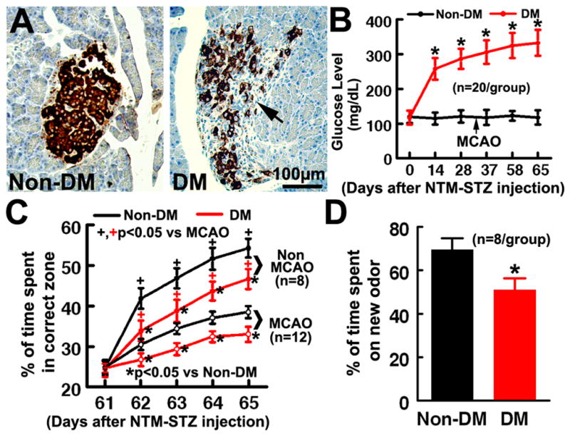
Methods: DM was induced in middle-aged rats (13 months) by administration of nicotinamide and streptozotocin. Rats with confirmed hyperglycemia status 30 days after nicotinamide-streptozotocin injection and age-matched non-DM rats were subjected to embolic middle cerebral artery occlusion.

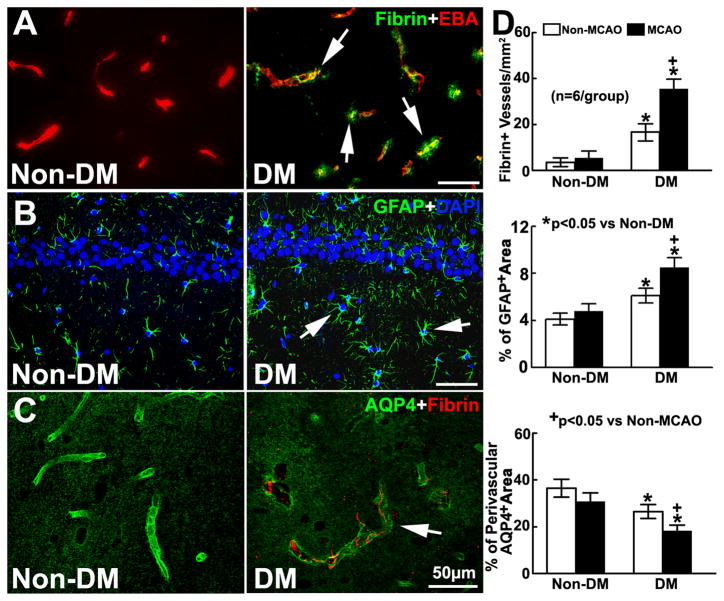
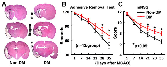
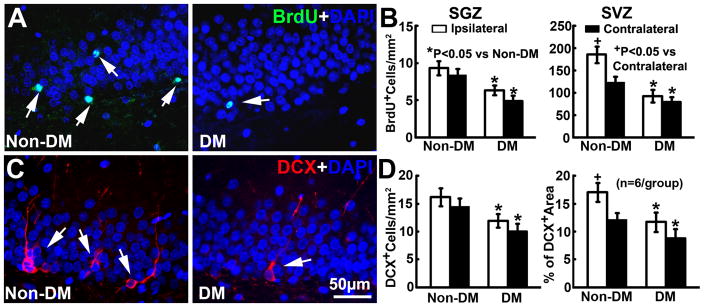
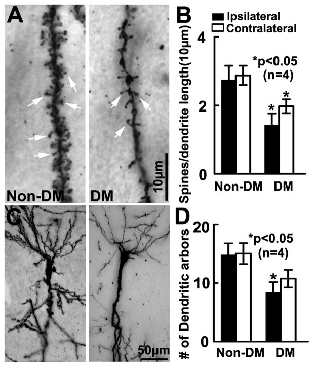
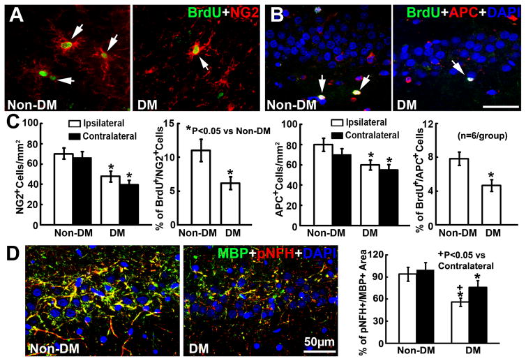
Results: Middle-aged rats subjected to nicotinamide-streptozotocin injection became hyperglycemic and developed cognitive deficits 2 months after induction of DM. Histopathologic analysis revealed that there was sporadic vascular disruption, including cerebral microvascular thrombosis, blood-brain barrier leakage, and loss of paravascular aquaporin-4 in the hippocampi. Importantly, middle-aged DM rats subjected to stroke had exacerbated sensorimotor and cognitive deficits compared with age-matched non-DM ischemic rats during stroke recovery. Compared with age-matched non-DM ischemic rats, DM ischemic rats exhibited aggravated neurovascular disruption in the bilateral hippocampi and white matter, suppressed stroke-induced neurogenesis and oligodendrogenesis, and impaired dendritic/spine plasticity. However, DM did not enlarge infarct volume.

Conclusions: Our data suggest that DM exacerbates neurovascular damage and hinders brain repair processes, which likely contribute to the impairment of stroke recovery.

Keywords: blood–brain barrier; diabetes mellitus; hyperglycemia; recovery; stroke.


Figures

Figure 1
Figure 1. Pancreatic β-cells islet morphology, blood glucose level, and cognitive deficits
Insulin immunoreactivity within the pancreas of middle-aged non-DM and DM rats (A). Insulin+ cells occupied most of the central region of the β-cell islets (brown) in the pancreas of middle-aged non-DM rats, whereas decreased insulin+ cells and distortion of islet shape (A, arrow) were evident in middle-aged rats after NTM-STZ administration. B shows glucose levels in rats with or without induction of DM. DM rats exhibited significant cognitive deficits measured by the Morris water maze test (C) and odor recognition test (D) 2 months after induction of DM. *P< 0.05 as compared with the Non-DM rats. ANCOVA for panels B and C. ANOVA/two-sample t-test for panel D.
Figure 2
Figure 2. Vascular damage in the DM brain
A shows the double immunofluorescent images of fibrin (green) with EBA (red) in the ipsilateral hippocampus in ischemic rats with and without induction of DM. B and C are the immunofluorescent images of GFAP+ astrocytes (green, B) and AQP4 immunoreactivity (green, C) within the ipsilateral hippocampus. Bar graphs in D show DM rats with and without MCAO exhibited significant increases of fibrin deposition and GFAP immunoreactivity, and reductions of paravascular AQP4 immunoreactivity in the hippocampus, respectively. ANOVA.
Figure 3
Figure 3. Infarction and neurological functional outcome
A shows H&E stained coronal brain sections from representative non-DM and DM rats after MCAO. a: anterior. p: posterior. B&C show the neurological functional outcome measured by adhesive removal test (B), and modified neurological severity score (C) in middle aged Non-DM and DM rats after MCAO. *P< 0.05 as compared with the Non-DM rats. ANCOVA.
Figure 4
Figure 4. Neurogenesis
A shows the immunofluorescent images of BrdU+(green) cells within the SGZ of the ipsilateral dentate gyrus. Ischemic DM rats exhibited significant reductions of BrdU+ cell density in the bilateral SGZ and the SVZ (B). C shows the immunofluorescent images of DCX+(red) neuroblasts within the SGZ of the ipsilateral dentate gyrus. Ischemic DM rats exhibited marked bilateral reductions of DCX+ neuroblasts (D). ANCOVA.
Figure 5
Figure 5. Dendritic spines and arborization
Golgi-Cox staining of dendritic spines (A, arrows) and dendrites (C) in the hippocampal CA1 of Non-DM and DM rats 35d after MCAO. DM rats exhibited significant bilateral reduction of spine density (B) and ipsilateral basal dendritic arborization (D) compared to Non DM rats. ANCOVA.
Figure 6
Figure 6. Oligodendrogenesis
The double immunofluorescent images of BrdU+(green)/NG2+(red) OPCs (A) and BrdU+(green)/APC+(red) oligodendrocytes (B) within the ipsilateral hippocampus. Bar graphs in C show that compared to ischemic non-DM rats, ischemic DM rats exhibited marked bilateral reductions of NG2+ OPCs and APC+oligodendrocytes, and the ipsilateral reduction of proliferating OPCs (BrdU+/NG2+ cells) and newly generated oligodendrocytes (BrdU+/APC+ cells). D shows the double immunofluorescent images of MBP+(green)/pNFH+(red) myelinated axon within the ipsilateral hippocampus acquired from the adjacent coronal sections from B. The bar graph in D shows that ischemic DM rats exhibited significant bilateral reductions of myelinated axon density in the hippocampus and subcortical white matter structures. ANCOVA.

References

    1. Barzilai N, Huffman DM, Muzumdar RH, Bartke A. The critical role of metabolic pathways in aging. Diabetes. 2012;61:1315–1322. - PMC - PubMed
    1. Lukovits TG, Mazzone TM, Gorelick TM. Diabetes mellitus and cerebrovascular disease. Neuroepidemiology. 1999;18:1–14. - PubMed
    1. Jorgensen H, Nakayama H, Raaschou HO, Olsen TS. Stroke in patients with diabetes. The Copenhagen stroke study. Stroke. 1994;25:1977–1984. - PubMed
    1. Newman GC, Bang H, Hussain SI, Toole JF. Association of diabetes, homocysteine, and hdl with cognition and disability after stroke. Neurology. 2007;69:2054–2062. - PubMed
    1. Mizrahi EH, Waitzman A, Blumstein T, Arad M, Adunsky A. Diabetes mellitus predicts cognitive impairment in patients with ischemic stroke. Am J Alzheimers Dis Other Demen. 2010;25:362–366. - PMC - PubMed

Publication types

MeSH terms