. 2016 Jul 7:16:415.
doi: 10.1186/s12885-016-2467-y.

Tumor-suppressive effects of atelocollagen-conjugated hsa-miR-520d-5p on un-differentiated cancer cells in a mouse xenograft model

Affiliations

Tumor-suppressive effects of atelocollagen-conjugated hsa-miR-520d-5p on un-differentiated cancer cells in a mouse xenograft model

Yoshitaka Ishihara et al. BMC Cancer. .

Erratum in

Abstract

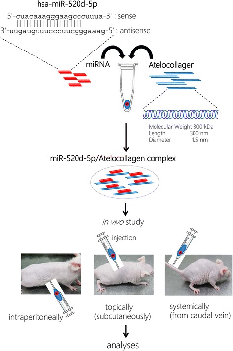
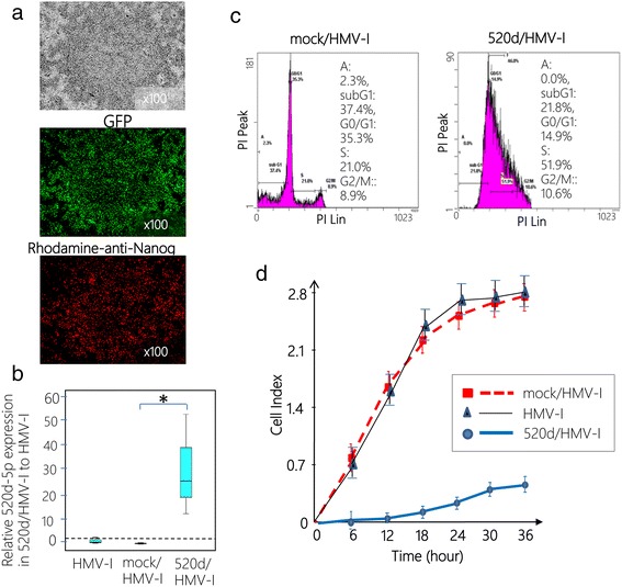
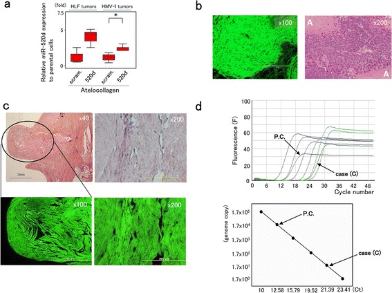
Background: We previously demonstrated that hsa-miR-520d-5p can convert cancer cells into induced pluripotent stem cells (iPSCs) or mesenchymal stem cells (MSCs) via a demethylation process and p53 upregulation in vivo. Additionally, we have reported the non-tumorigenic effect of miR-520d-5p on normal human cells, including fibroblasts.

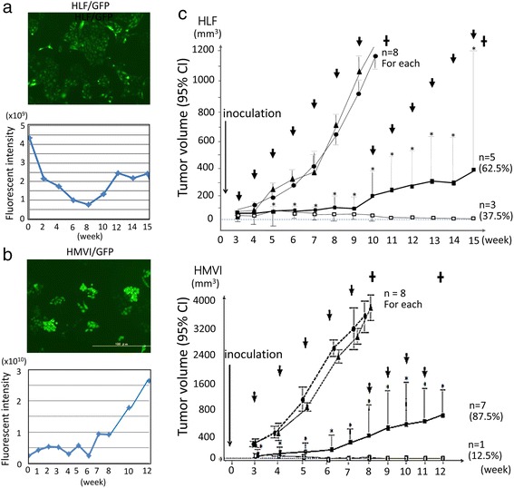
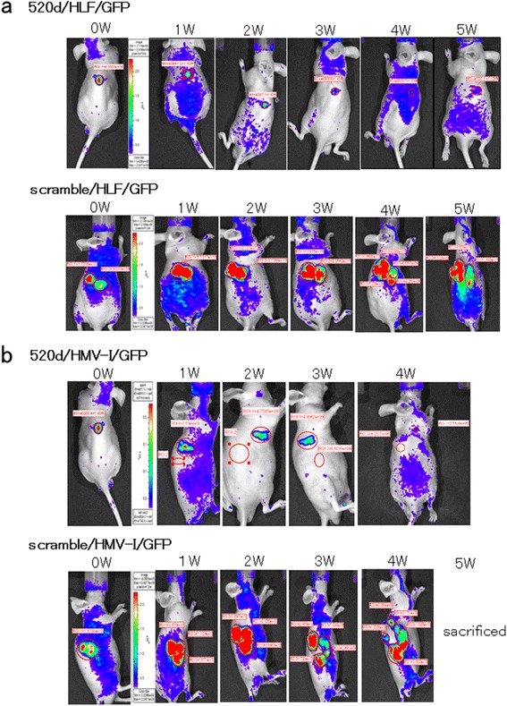
Methods: We used atelocollagen-conjugated miR-520d-5p (520d/atelocollagen) to confirm the possibility of a therapeutic effect on cancer cells. We traced the size and signal intensity of GFP-expressing tumors in mice each week, beginning 4 weeks after subcutaneous inoculation.

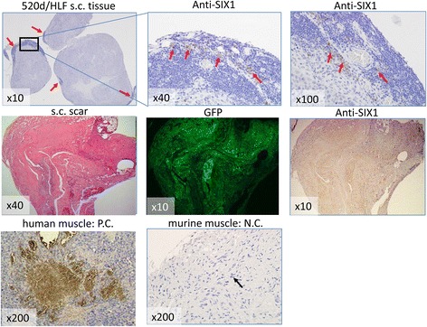
Results: 520d/atelocollagen treatment suppressed tumor growth by greater than 80 % each week relative to controls and resulted in an approximately 30 % disappearance of tumors. In mice whose tumors disappeared, the existence of human genomic material at the injection site was examined by quantitative Alu-PCR, and we confirmed the co-existence of both species-derived cells. In every site where a tumor disappeared in immunodeficient mice, GFP protein was expressed in the connective tissues, and approximately 0.1 % of the extracted DNA contained human genomic material. We could not identify any adverse effects in vivo.

Conclusions: This is the first report to confirm an inhibitory effect of 520d/atelocollagen on cancer cells in vivo. The development of optimized modifications of this carrier is expected to enhance the efficiency of entry into tumor cells and the induction of its inhibitory effect.

Keywords: Atelocollagen; Cancer; Therapeutic effect; Xenograft model; miR-520d-5p.


Figures

Fig. 1
Fig. 1
Schematic of the study design and processes. Atelocollagen and miR-520d-5p were conjugated following the manufacturer’s instructions, and the resulting complex was injected into immunodeficient mice that were inoculated with cancer cells. In addition, 520d-conjugated atelocollagen was administered to mice via three different methods (see Methods for further details)
Fig. 2
Fig. 2
Lenti-viral transfection of 520d-5p to HMV-I cells in vitro. a A representative phenotype of 520d/HMV-I is presented (x100 magnification). Spheroid-like cells comprising a small fraction of the cell population are presented (top). Analysis of GFP expression confirmed the effective induction (more than 99 %) of 520d/HMV-I cells (x100 magnification) (middle). miR-520d-5p induced the expression of Nanog in HMV-I cells (x100 magnification) (bottom). The phenotypic changes were not sufficient to assess whether they were malignant or benign. b Significant 520d-5p expression was confirmed in 520d/HMV-I cells (*, P < 0.01 by the Mann-Whitney U test). The relative expression of 520d-5p in 520d/HMV-I cells compared with mock/HMV-I cells is presented. All expression data are standardized to the β-actin expression level (n = 4). c Fluorescence activated cell sorting (FACS) analysis revealed that GFP-positive 520d/HMV-I cells (right) exhibited increased DNA content in the S phase compared with GFP-positive mock/HMV-I cells (left). d The invasive abilities of HMV-I, mock-transfected HMV-I (mock/HMV-I) and HMV-I cells treated with miR-520d-5p (520d/HMV-I) were estimated with a migration assay using a fibronectin membrane (10 μg/ml). Most of the 520d/HMV-I cells did not pass through the membrane
Fig. 3
Fig. 3
An in vivo study to assess the topical effects of 520d-5p-conjugated atelocollagen on cancer tissues. a, b After we generated GFP-expressing HLF (HLF/GFP) or HMV-I (HMV-I/GFP) cells using lentiviral constructs (top for each), the therapeutic effects of 520d/atelocollagen on the resulting GFP-expressing tumors in mice were examined. The average fluorescence intensity in tumors was measured, and representative data are presented through week 8 (bottom of each panel). GFP expression was maintained during the observation period in both studies. Dotted lines indicate tumor growth in controls. c Overviews of the in vivo study using 520d/atelocollagen in a mouse xenograft model (top, HLF; bottom, HMV-I). In the group that was administered the 520d/atelocollagen complex, tumor volume was significantly suppressed relative to the control group. *, P < 0.01 by the Mann-Whitney U test. The tumor volume of HLF cells from 11 to 15 weeks or of HMV-I cells from 9 to 12 weeks was analyzed and compared with control or scrambled data at 10 weeks or 8 weeks, respectively. The arrow indicates the timing of the doses that were administered to each group from 3 to 9 weeks (c) and from 3 to 7 weeks (d). : injection of complex (once a week); ▲: atelocollagen alone; ●: scramble/atelocollagen; ■: miR-520d-5p/atelocollagen (cases with suppressive growth); □: miR-520d-5p/atelocollagen (cases with tumor disappearance); ✝: mice were sacrificed; CI: confidence interval
Fig. 4
Fig. 4
The therapeutic effect of 520d-5p-conjugated atelocollagen on subcutaneously inoculated tumors as monitored by an in vivo imaging system. a A representative image of the therapeutic effect of 520d-5p/atelocollagen using an in vivo imaging system. At each inoculation site, 37.5 % (3/8) of the tumors disappeared. The growth of tumors that received 520d-5p was significantly suppressed, and metastatic ability was entirely inhibited in the remaining mice. A time course (top) of 520d/HLF/GFP cells (520d-5p-transfected and GFP-expressing HLF) is depicted. Representative sequential images (bottom) of scrambled/HLF/GFP cells (scrambled-transfected GFP-expressing HLF). b In HMV-I tumors, 12.5 % (1/8) of the tumors disappeared. After treatment with 520d-5p, the growth of other tumors was significantly suppressed, and their metastatic ability was entirely inhibited. 520d/HMV-I/GFP and scrambled/HMV-I/GFP indicate 520d-5p-transfected, GFP-expressing HLF (top) and scramble-transfected, GFP-expressing HMV-I (bottom), respectively
Fig. 5
Fig. 5
Evidence of human genomic DNA at the injection site. a miR-520d-5p expression was examined in HLF (n = 5) and HMV-I (n = 7) tumors resected after 520d/atelocollagen treatment. 520d/HMV-I tumors significantly expressed 520d-5p (*, P < 0.05), and 520d/HLF tumors tended to express miR-520d-5p (P = 0.051). scram.: scrambled. b Microscopic examination of HLF (left; HE stain, x100 magnification) and HMV-I tumors (right; HE stain, x200 magnification) that displayed suppressed tumor growth. HLF tumors treated with 520d/atelocollagen exhibited GFP-expressing muscle tissues, including undifferentiated tumor cells unaccompanied by necrosis. HMV-I tumors treated with 520d/atelocollagen showed undifferentiated neoplastic cells. This finding is not inconsistent, as malignant melanomas are observed to be accompanied by extensive necrosis, as shown around A. c Histological findings in the area where there should have been a tumor are presented (HE stain, x40 magnification). The area of the HLF tumors that disappeared is surrounded by an oval (left top). GFP expression was confirmed (left bottom). Histological findings in the disappearance site of HMV-I tumors showed no malignant cells. This finding was similar to that for 520d-treated HLF tumors (HE stain, x200 magnification) (right top), but we observed GFP even in the connective tissue (right bottom). We did not observe any malignant cells in the injection sites of mice in either case. d Fluorescence (F) (top) and a quantitative calibration line (bottom) in quantitative Alu-PCR. P.C. (left arrow) indicates a representative positive control (a subcutaneous tumor generated from mock/HLF cells). The reaction of interest was performed using DNA from the scar-like area of cases (C; right arrow) to confirm that human genomic material was present in the injection site. The average amount of human genomic DNA derived from human hepatoma (mock/HLF: n = 3) was approximately 50 ng. Case (C) showed 55.0 pg. The genome copy number was calculated based on the fact that 1 pg corresponds to approximately 0.333 human genome copies. Correlation coefficient, r = −0.996
Fig. 6
Fig. 6
Pathological findings of human muscle-derived gene expression at the injection site. Immunohistochemistry showing human muscle-derived gene expression (SIX1) in 3 normal subcutaneous regions (upper) and the scar-like, slightly protruding region (middle). The human muscular region in subcutaneous tissue (bottom left) and the murine subcutaneous region (bottom right), including muscle, were used as a positive control and a negative control, respectively. Scar-like formation (HE stain; middle left) induced by injection retained GFP expression (middle center) and weak human SIX1 expression in murine tissue (middle right). The representative region for immunostaining is denoted by a rectangle (top left), and arrows denote moderate human SIX1 expression. The arrow shows the murine muscle region without human SIX1 staining (bottom left)

Similar articles

Cited by

References

    1. Lagos-Quintana M, Rauhut R, Lendeckel W, Tuschl T. Identification of novel genes coding for small expressed RNAs. Science. 2001;294:853–858. doi: 10.1126/science.1064921. - DOI - PubMed
    1. Meltzer PS. Cancer genomics: small RNAs with big impacts. Nature. 2005;435:745–746. doi: 10.1038/435745a. - DOI - PubMed
    1. Chen CZ, Li L, Lodish HF, Bartel DP. MicroRNAs modulate hematopoietic lineage differentiation. Science. 2004;303:83–86. doi: 10.1126/science.1091903. - DOI - PubMed
    1. Thum T, Galuppo P, Wolf C, Fiedler J, Kneitz S, van Laake LW, et al. MicroRNAs in the human heart: a clue to fetal gene reprogramming in heart failure. Circulation. 2007;116:258–267. doi: 10.1161/CIRCULATIONAHA.107.687947. - DOI - PubMed
    1. Viswanathan SR, Daley GQ, Gregory RI. Selective blockade of microRNA processing by Lin28. Science. 2008;320:97–100. doi: 10.1126/science.1154040. - DOI - PMC - PubMed

Publication types