

Dcsbis (PA2771) from Pseudomonas aeruginosa is a highly active diguanylate cyclase with unique activity regulation
- PMID: 27388857
- PMCID: PMC4937426
- DOI: 10.1038/srep29499
Dcsbis (PA2771) from Pseudomonas aeruginosa is a highly active diguanylate cyclase with unique activity regulation
Abstract
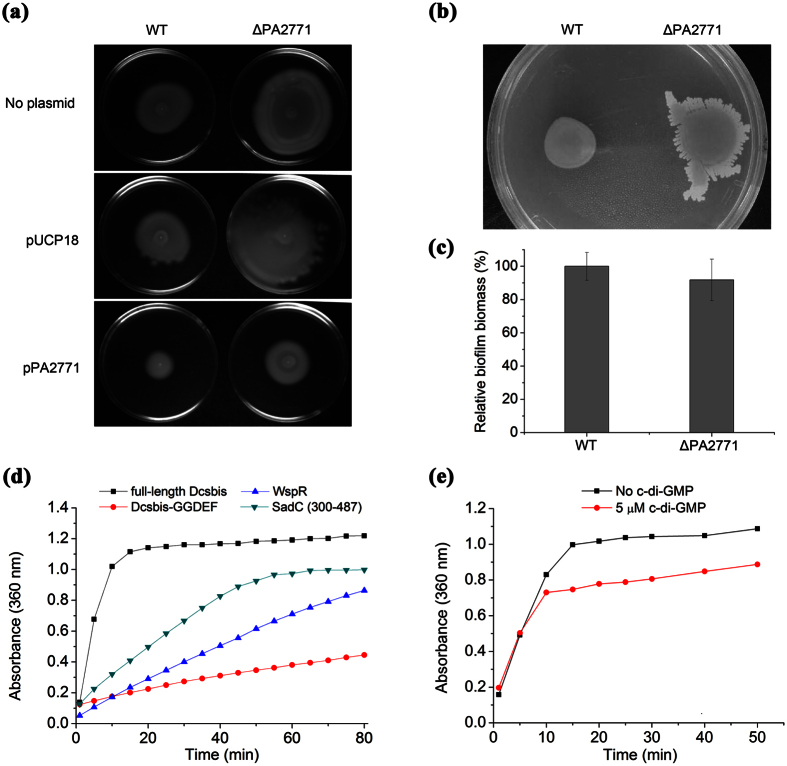
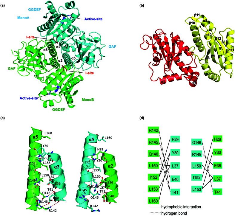
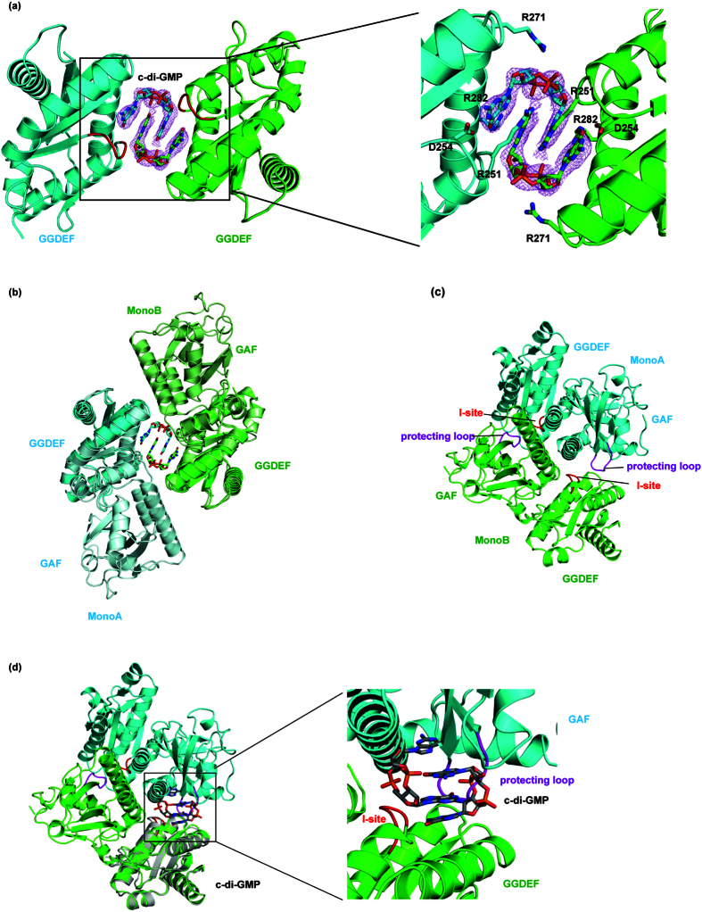
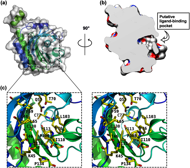
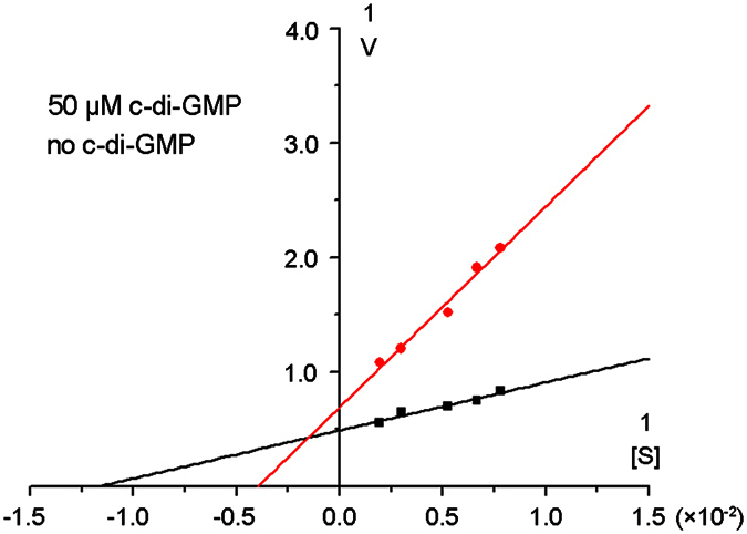
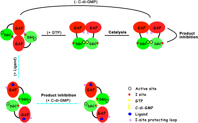
C-di-GMP (3',5' -Cyclic diguanylic acid) is an important second messenger in bacteria that influences virulence, motility, biofilm formation, and cell division. The level of c-di-GMP in cells is controlled by diguanyl cyclases (DGCs) and phosphodiesterases (PDEs). Here, we report the biochemical functions and crystal structure of the potential diguanylase Dcsbis (PA2771, a diguanylate cyclase with a self-blocked I-site) from Pseudomonas aeruginosa PAO1. The full-length Dcsbis protein contains an N-terminal GAF domain and a C-terminal GGDEF domain. We showed that Dcsbis tightly coordinates cell motility without markedly affecting biofilm formation and is a diguanylate cyclase with a catalytic activity much higher than those of many other DGCs. Unexpectedly, we found that a peptide loop (protecting loop) extending from the GAF domain occupies the conserved inhibition site, thereby largely relieving the product-inhibition effect. A large hydrophobic pocket was observed in the GAF domain, thus suggesting that an unknown upstream signaling molecule may bind to the GAF domain, moving the protecting loop from the I-site and thereby turning off the enzymatic activity.
Conflict of interest statement
The authors declare no competing financial interests.
Figures
Similar articles
-
More than Enzymes That Make or Break Cyclic Di-GMP-Local Signaling in the Interactome of GGDEF/EAL Domain Proteins of Escherichia coli.mBio. 2017 Oct 10;8(5):e01639-17. doi: 10.1128/mBio.01639-17. mBio. 2017. PMID: 29018125 Free PMC article.
-
Structure of the active GGEEF domain of a diguanylate cyclase from Vibrio cholerae.Biochem Biophys Res Commun. 2020 Mar 5;523(2):287-292. doi: 10.1016/j.bbrc.2019.11.179. Epub 2019 Dec 18. Biochem Biophys Res Commun. 2020. PMID: 31862141
-
Analysis of Pseudomonas aeruginosa diguanylate cyclases and phosphodiesterases reveals a role for bis-(3'-5')-cyclic-GMP in virulence.Proc Natl Acad Sci U S A. 2006 Feb 21;103(8):2839-44. doi: 10.1073/pnas.0511090103. Epub 2006 Feb 13. Proc Natl Acad Sci U S A. 2006. PMID: 16477007 Free PMC article.
-
C-di-GMP Synthesis: Structural Aspects of Evolution, Catalysis and Regulation.J Mol Biol. 2016 Sep 25;428(19):3683-701. doi: 10.1016/j.jmb.2016.07.023. Epub 2016 Aug 4. J Mol Biol. 2016. PMID: 27498163 Review.
-
Bacterial diguanylate cyclases: structure, function and mechanism in exopolysaccharide biofilm development.Biotechnol Adv. 2015 Jan-Feb;33(1):124-141. doi: 10.1016/j.biotechadv.2014.11.010. Epub 2014 Dec 10. Biotechnol Adv. 2015. PMID: 25499693 Review.
Cited by
-
Pleiotropic Effects of c-di-GMP Content in Pseudomonas syringae.Appl Environ Microbiol. 2019 May 2;85(10):e00152-19. doi: 10.1128/AEM.00152-19. Print 2019 May 15. Appl Environ Microbiol. 2019. PMID: 30850427 Free PMC article.
-
c-di-GMP Regulates Various Phenotypes and Insecticidal Activity of Gram-Positive Bacillus thuringiensis.Front Microbiol. 2018 Feb 13;9:45. doi: 10.3389/fmicb.2018.00045. eCollection 2018. Front Microbiol. 2018. PMID: 29487570 Free PMC article.
-
Structural studies of the periplasmic portion of the diguanylate cyclase CdgH from Vibrio cholerae.Sci Rep. 2017 May 12;7(1):1861. doi: 10.1038/s41598-017-01989-6. Sci Rep. 2017. PMID: 28500346 Free PMC article.
-
Structural basis for activation of a diguanylate cyclase required for bacterial predation in Bdellovibrio.Nat Commun. 2019 Sep 9;10(1):4086. doi: 10.1038/s41467-019-12051-6. Nat Commun. 2019. PMID: 31501441 Free PMC article.
-
Negative feedback of cyclic di-GMP levels optimizes switching between sessile and motile lifestyles in Vibrio cholerae.bioRxiv [Preprint]. 2024 Sep 1:2024.09.01.610008. doi: 10.1101/2024.09.01.610008. bioRxiv. 2024. PMID: 39257796 Free PMC article. Preprint.
References
-
- Ross P. et al. Regulation of cellulose synthesis in Acetobacter xylinum by cyclic diguanylic acid. Nature. 325, 279–281 (1987). - PubMed
-
- Kalia D. et al. c-di-GMP, c-di-AMP, cGMP, cAMP, (p)ppGpp signaling in bacteria and implications in pathogenesis. Chem Soc Rev. 42, 305–341 (2013). - PubMed
-
- Jenal U. & Malone J. Mechanisms of cyclic-di-GMP signaling in bacteria. Annu Rev Genet. 40, 385–407 (2006). - PubMed
-
- Romling U., Gomelsky M. & Galperin M. Y. C-di-GMP: the dawning of a novel bacterial signalling system. Mol Microbiol. 57, 629–639 (2005). - PubMed
Publication types
MeSH terms
Substances
LinkOut - more resources
Full Text Sources
Other Literature Sources
Molecular Biology Databases
