. 2016 Nov;136(11):2251-2259.
doi: 10.1016/j.jid.2016.06.618. Epub 2016 Jul 5.

AP1S3 Mutations Cause Skin Autoinflammation by Disrupting Keratinocyte Autophagy and Up-Regulating IL-36 Production

Affiliations

AP1S3 Mutations Cause Skin Autoinflammation by Disrupting Keratinocyte Autophagy and Up-Regulating IL-36 Production

Satveer K Mahil et al. J Invest Dermatol. 2016 Nov.

Abstract

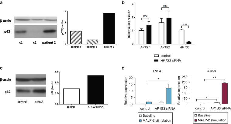
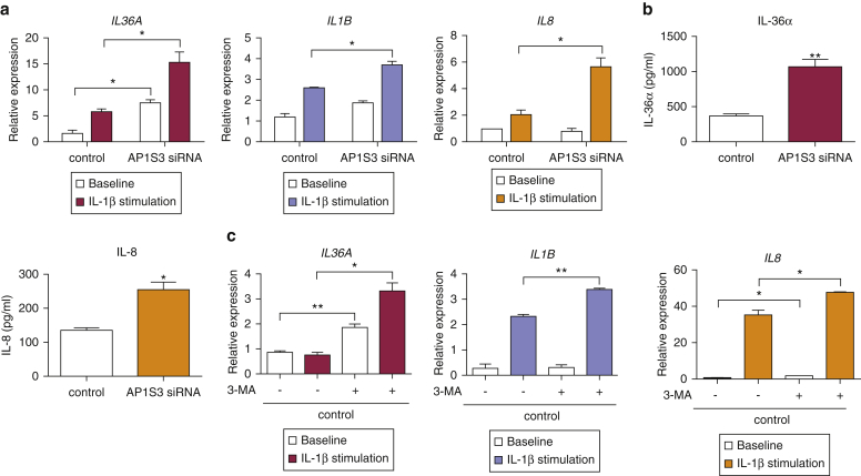
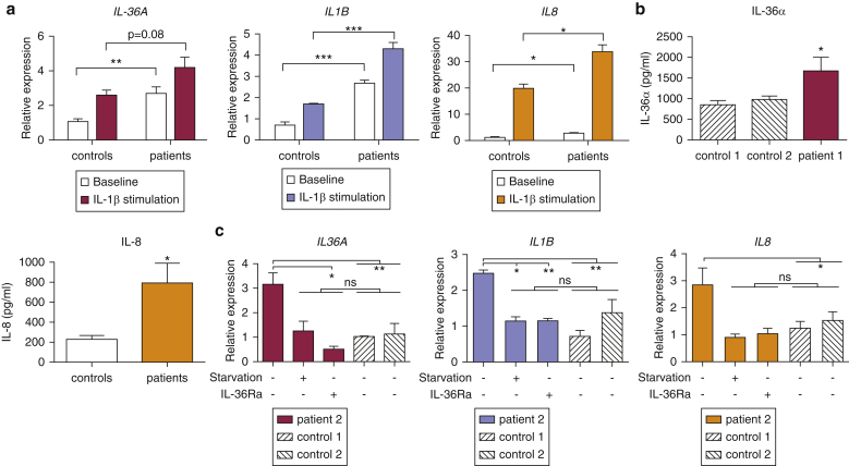
Prominent skin involvement is a defining characteristic of autoinflammatory disorders caused by abnormal IL-1 signaling. However, the pathways and cell types that drive cutaneous autoinflammatory features remain poorly understood. We sought to address this issue by investigating the pathogenesis of pustular psoriasis, a model of autoinflammatory disorders with predominant cutaneous manifestations. We specifically characterized the impact of mutations affecting AP1S3, a disease gene previously identified by our group and validated here in a newly ascertained patient resource. We first showed that AP1S3 expression is distinctively elevated in keratinocytes. Because AP1S3 encodes a protein implicated in autophagosome formation, we next investigated the effects of gene silencing on this pathway. We found that AP1S3 knockout disrupts keratinocyte autophagy, causing abnormal accumulation of p62, an adaptor protein mediating NF-κB activation. We showed that as a consequence, AP1S3-deficient cells up-regulate IL-1 signaling and overexpress IL-36α, a cytokine that is emerging as an important mediator of skin inflammation. These abnormal immune profiles were recapitulated by pharmacological inhibition of autophagy and verified in patient keratinocytes, where they were reversed by IL-36 blockade. These findings show that keratinocytes play a key role in skin autoinflammation and identify autophagy modulation of IL-36 signaling as a therapeutic target.


Figures

Figure 1
Figure 1
AP1S3 loss-of-function mutations are most likely to affect skin keratinocytes. (a) Schematic representation of AP-1 structure. (b) HEK293 cells were transfected with wild-type and mutant AP1S3 constructs. Lysates were subjected to the indicated temperature gradient, and soluble (nondenatured) proteins were analyzed by Western blotting. The densitometry shows the fraction of nondenatured protein (mean ± standard error of the mean of the results obtained in two experiments). (c) HEK293 cells were transfected with myc-tagged AP1S3 and FLAG-tagged AP1M1 constructs. Lysates were subjected to immune precipitation (IP) and immune blotting (IB) as indicated. The image is representative of results obtained in two experiments. (d) Real-time PCR analysis showing abundant AP1S3 expression in keratinocytes. The data show the mean ± standard error of the mean of measurements obtained in two donors. P ≤ 0.05. IB, immune blotting; IP, immune precipitation; WCE, whole cell extracts; wt, wild type.
Figure 2
Figure 2
AP1S3 deficiency results in impaired autophagy. (a) After the generation of a HaCaT AP1S3 knockdown cell line, gene silencing was measured by real-time PCR, because of cross-reactivity of the anti-AP1σ1c antibody with the proteins encoded by AP1S1 and AP1S2. (b) Starvation-induced LC3-II accumulation was measured by Western blotting and densitometry. The data are presented as mean ± standard error of the mean of measurements obtained in four independent experiments. (c) HEK293 AP1S3 knockout cell lines harboring a c.124delC change (highlighted by a red asterisk in the chromatogram) were generated by CRISPR/Cas-9 editing. (d) Cells were starved to induce autophagy, and LC3-II accumulation was measured by Western blotting. The data are presented as described. (e) Control and AP1S3 KO HEK293 cells were transfected with GFP-LC3 and either an empty vector (control and KO panels) or a rescue construct (wild-type AP1S3 in KO/wt panel and p.Arg33Trp AP1S3 in KO/mut). Starvation-induced LC3 punctae were visualized by confocal fluorescence microscopy. The data are presented as mean ± standard error of the mean of measurements obtained in at least 15 cells per experiment. Scale bar = 5 μm. P ≤ 0.05, ∗∗∗∗P ≤ 0.0001. Cas9, CRISPR-associated protein-9; CRISPR, clustered regularly interspaced short palindromic repeats; GFP, green fluorescent protein; KD, knockdown; KO, knockout; mut, mutated; ns, not significant; wt, wild-type.
Figure 3
Figure 3
Abnormal p62 accumulation and enhanced TLR-2/6 signaling in AP1S3-deficient keratinocytes. (a) p62 levels were measured in patient and control subject keratinocytes by Western blotting and densitometry. (b) After the transfection of silencing (AP1S3 siRNA) and nonsilencing (control) siRNA pools into primary keratinocytes, (c) baseline p62 levels were measured by Western blotting and densitometry. (d) Alternatively, cells were stimulated with MALP-2 in triplicate, and the induction of TLR2/6-dependent genes was measured by real-time PCR. The data are representative of results obtained in at least two independent experiments and are presented as mean ± standard error of the mean of duplicate stimulations. P ≤ 0.05, ∗∗P ≤ 0.01, ∗∗∗P ≤ 0.001. c, control; ns, not significant; siRNA, small interfering RNA; TLR, Toll-like receptor.
Figure 4
Figure 4
AP1S3-deficient primary keratinocytes exhibit an abnormal immune profile, which can be recapitulated by autophagy inhibition. (a) After siRNA-mediated AP1S3 silencing, primary keratinocytes were stimulated with IL-1β, and gene expression was determined by real-time PCR. (b) Alternatively, cells were cultured for a further 48 hours in the absence of stimuli, and cytokine production was measured by ELISA. (c) Normal primary keratinocytes were cultured in the presence or absence of 3-MA and subsequently stimulated with IL-1β. Gene expression was determined by real-time PCR. All data are representative of results obtained in two independent experiments and are presented as mean ± standard error of the mean of (a) duplicate or (b, c) triplicate measurements. P ≤ 0.05, ∗∗P ≤ 0.01. 3-MA, 3-methyladenine; siRNA, small interfering RNA.
Figure 5
Figure 5
Abnormal cytokine expression in the keratinocytes of patients harboring AP1S3 mutations. (a) Primary keratinocytes were stimulated with IL-1β, and cytokine induction was measured by real-time PCR. The data are presented as mean ± standard error of the mean of duplicate stimulations carried out in the cells of two unrelated patients and two healthy control subjects. (b) IL-36α and IL-8 production was measured in culture supernatants by ELISA. Data are presented as mean ± standard error of the mean of triplicate measurements. (c) Primary keratinocytes were starved to induce autophagy or cultured in the presence of IL-36Ra. Gene expression was measured by real-time PCR. The data are presented as mean ± standard error of the mean of triplicate measurements, obtained in one patient and two healthy control subjects. P < 0.05, ∗∗P < 0.01, ∗∗∗P < 0.001. ns, not significant.

References

    1. Aasen T., Izpisua Belmonte J.C. Isolation and cultivation of human keratinocytes from skin or plucked hair for the generation of induced pluripotent stem cells. Nat Protoc. 2010;5:371–382. - PubMed
    1. Aksentijevich I., Kastner D.L. Genetics of monogenic autoinflammatory diseases: past successes, future challenges. Nat Rev Rheumatol. 2011;7:469–478. - PubMed
    1. Beer H.D., Contassot E., French L.E. The inflammasomes in autoinflammatory diseases with skin involvement. J Invest Dermatol. 2014;134:1805–1810. - PubMed
    1. Berki D.M., Liu L., Choon S.E., David Burden A., Griffiths C.E., Navarini A.A. Activating CARD14 mutations are associated with generalized pustular psoriasis but rarely account for familial recurrence in psoriasis vulgaris. J Invest Dermatol. 2015;135:2964–2970. - PubMed
    1. de Jesus A.A., Canna S.W., Liu Y., Goldbach-Mansky R. Molecular mechanisms in genetically defined autoinflammatory diseases: disorders of amplified danger signaling. Annu Rev Immunol. 2015;33:823–874. - PMC - PubMed

Publication types

MeSH terms