. 2016 Jul 7;12(7):e1005715.
doi: 10.1371/journal.ppat.1005715. eCollection 2016 Jul.

Metarhizium brunneum Blastospore Pathogenesis in Aedes aegypti Larvae: Attack on Several Fronts Accelerates Mortality

Affiliations

Metarhizium brunneum Blastospore Pathogenesis in Aedes aegypti Larvae: Attack on Several Fronts Accelerates Mortality

Abeer M Alkhaibari et al. PLoS Pathog. .

Abstract

Aedes aegypti is the vector of a wide range of diseases (e.g. yellow fever, dengue, Chikungunya and Zika) which impact on over half the world's population. Entomopathogenic fungi such as Metarhizium anisopliae and Beauveria bassiana have been found to be highly efficacious in killing mosquito larvae but only now are the underlying mechanisms for pathogenesis being elucidated. Recently it was shown that conidia of M. anisopliae caused stress induced mortality in Ae. aegypti larvae, a different mode of pathogenicity to that normally seen in terrestrial hosts. Blastospores constitute a different form of inoculum produced by this fungus when cultured in liquid media and although blastospores are generally considered to be more virulent than conidia no evidence has been presented to explain why. In our study, using a range of biochemical, molecular and microscopy methods, the infection process of Metarhizium brunneum (formerly M. anisopliae) ARSEF 4556 blastospores was investigated. It appears that the blastospores, unlike conidia, readily adhere to and penetrate mosquito larval cuticle. The blastospores are readily ingested by the larvae but unlike the conidia are able infect the insect through the gut and rapidly invade the haemocoel. The fact that pathogenicity related genes were upregulated in blastospores exposed to larvae prior to invasion, suggests the fungus was detecting host derived cues. Similarly, immune and defence genes were upregulated in the host prior to infection suggesting mosquitoes were also able to detect pathogen-derived cues. The hydrophilic blastospores produce copious mucilage, which probably facilitates adhesion to the host but do not appear to depend on production of Pr1, a cuticle degrading subtilisin protease, for penetration since protease inhibitors did not significantly alter blastospore virulence. The fact the blastospores have multiple routes of entry (cuticle and gut) may explain why this form of the inoculum killed Ae. aegypti larvae in a relatively short time (12-24hrs), significantly quicker than when larvae were exposed to conidia. This study shows that selecting the appropriate form of inoculum is important for efficacious control of disease vectors such as Ae. aegypti.


Conflict of interest statement

The authors have declared that no competing interests exist.

Figures

Fig 1
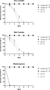
Fig 1. Susceptibility of Aedes aegypti larvae to blastospores, wet and dry conidia of Metarhizium brunneum ARSEF 4556.
Survival curves of Ae. aegypti (3rd-4th) instar larvae (n = 30 per treatment) inoculated with blastospores or conidia (wet and dry) of M. brunneum ARSEF 4556 applied at final dose of 107 spores ml-1. Blastospores significantly decreased survival compared to all other treatments (p < 0.001), but there were no statistical differences in the survival when comparing wet and dry conidia. The negative controls were either distilled water or 0.05% aqueous Tween. Error is represented as SE.
Fig 2
Fig 2. Comparison of Pr1 enzyme activity between blastospores, wet and dry conidia of Metarhizium brunneum ARSEF 4556.
Activity of Pr1 bound to the cell wall of conidia (dry and wet) and blastospores in culture media. Letters denote statistical differences. Error is represented as 95% ci.
Fig 3
Fig 3. Survival of Aedes aegypti larvae exposed to conidia or blastospores at 20°C and 27°C.
Survival curves of Ae. aegypti larvae (n = 30) exposed to conidia (wet & dry) and blastospores of M. brunneum at a concentration of 107 spores ml-1 incubated at 20°C and 27°C. Survival was similar for larvae inoculated with blastospores at 20°C or 27°C (P = 0.317) but significant differences were observed for larvae exposed to either dry (P = 0.022) or wet (P = 0.011) conidia when comparing these two temperatures. Controls consisted of distilled water or 0.05% aqueous Tween. Error is represented as SE.
Fig 4
Fig 4. Survival of Aedes aegypti larvae exposed to blastospores in the presence of different protease inhibitors.
Ae. aegypti larvae (n = 72) were exposed to M. brunneum blastospores with and without the addition of the protease inhibitors chicken egg white (CEW, a Pr1 specific inhibitor) and α2-macroglobulin (global protease inhibitor). The Kaplan-Meier method was used to plot survival curves of larvae; Log-rank test was used to assess difference in survival between treatments. Controls consisted of either distilled water or distilled water with protease inhibitors. Error is represented as SE.
Fig 5
Fig 5. Scanning electron microscopy of Aedes aegypti larvae infected with Metarhizium brunneum blastospores.
Larvae were inoculated with 1X10⁷ blastospores ml-1 and prepared for SEM 20 hrs post inoculation. (A): Head of Ae. aegypti larva showing blastospores (BS) attached to the surface of the cuticle. (B): Blastospores at different stages of germination attached to surface of abdomen. (C): Germinating and non-germinating blastospores surrounded by a mucilaginous matrix (M). (D). Cross section of infected larva showing blastospores of M. brunneum occluding the gut lumen (GL).
Fig 6
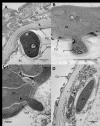
Fig 6. Transmission electron microscopy of Aedes aegypti larvae infected with Metarhizium brunneum blastospores 24 hr post-inoculation.
(A) Blastospore (BS) firmly adhering to surface of the cuticle (CU). The blastospore has dense cytoplasm, a relatively thin wall and a coating of mucilage (MU) which extends beyond the fungus. The mucilage consists of heterogeneous electron opaque material. (B) Penetration of the larval cuticle. Vacuoles (V) containing electron dense material evident in the penetration hyphae. Cuticle readily distorted by penetration hypha. (C) Blastospores in gut lumen penetrating the peritrophic membrane (PM). (D) One blastospore has penetrated the midgut epithelium and has entered the haemocoel (H).
Fig 7
Fig 7. Cross section of Aedes aegypti larvae 24 hrs post infection with Metarhizium brunneum blastospores.
(A) Blastospores of M. brunneum mostly confined to gut lumen. (B) Blastospores adjacent to the peritrophic membrane are swollen with some having penetrated the peritrophic membrane and midgut epithelium. Cells colonizing the haemocoel consisted of short filaments or hyphal bodies as well as yeast like cells. There was no evidence of branched filamentous hyphae. EP: Epithelium, PM: peritrophic membrane, MV: Microvilli, N: Nuclei, BS: Blastospores, GL: Gut lumen.
Fig 8
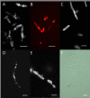
Fig 8. Fluorescence microscopy of Metarhizium brunneum blastospores.
(A) Calcofluor White staining of blastospores. Cell walls fluoresced weakly except at apices and septa. (B) Rhodamine 123 was used to visualise mitochondria in blastospores. (C) Filipin staining of ergosterol present in the plasma membrane. (D) Fluorescent staining of nuclei with DAPI. (E) FITC staining of proteins (F) Blastospores as seen in bright-field (F). Scale bar = 5 μm.
Fig 9
Fig 9. Expression of pathogenicity related genes in Metarhizium brunneum blastospores 20.3 hours post-infection.
Gene expression analysed by quantitative PCR included: proteases (Pr1A, Pr2), adhesins (Mad1, Mad2), an osmosensor (Mos1),regulators of G-protein signalling (Cag8) and nitrogen (nrr1). X axis shows: 1: infected living larvae, 2: infected dead larvae, 3: blastospores in the presence of Ae. aegypti larvae 4: blastospores in absence of the larvae, 5: Tenebrio molitor (terrestrial host) positive control. Boxes denote interquartile range, bisected horizontally by median values; whiskers extend to 1.5× interquartile range beyond boxes; outliers are marked as dots beyond whiskers. Expression is shown as the inverse of the number of amplification cycles to reach Critical Threshold values (CT-1).
Fig 10
Fig 10. Expression of Aedes aegypti antimicrobial peptides (AMP) and stress management genes during infection with Metarhizium brunneum blastospores.
Quantitative real time PCR used to analyse expression of stress management and AMP genes in Ae. aegypti larvae inoculated with M. brunneum blastospores at 1 = 0hr, 2 = 12hr and 3 = 20:30 hrs post-inoculation. AMP genes included; AeDA (Defensin A), AeDB1 (Defensin B), Ada-defD (Defensin D), AeCA2 (Cecropin A), and Ada-ccg (Cecropin G). Stress management genes include; HSP70 (Heatshock protein 70), HSP83 (Heatshock protein 83), GPX (Glutathione peroxidase), Cyp6Z6 (Cytochrome P450), and TPX10 (Thiol peroxidase 10). Boxes denote interquartile range, bisected horizontally by median values; whiskers extend to 1.5× interquartile range beyond boxes; outliers are marked as dots beyond whiskers. Expression is shown as the inverse of number of amplification cycles to reach Critical Threshold values (CT-1).

References

    1. WHO. Yellow fever fact sheet N°100. Weekly Epidemiological Record. 2010;85:33–6. - PubMed
    1. WHO. Global strategy for dengue prevention and control 2012–2020: World Health Organization; 2012.
    1. Tolle MA. Mosquito-borne diseases. Current Problems in Pediatric and Adolescent Health Care. 2009;39(4):97–140. 10.1016/j.cppeds.2009.01.001 - DOI - PubMed
    1. Marcondes CB, Ximenes MdFFd. Zika virus in Brazil and the danger of infestation by Aedes (Stegomyia) mosquitoes. Revista da Sociedade Brasileira de Medicina Tropical. 2015;(AHEAD):0-. - PubMed
    1. WHO. Epidemiological update on Zika virus infection—16 October 2015 2015 [cited 2015 14-12-2015]. Available from: http://www.paho.org/hq/index.php?option=com_docman&task=doc_view&Itemid=....

LinkOut - more resources