. 2016 Aug 25;128(8):1144-51.
doi: 10.1182/blood-2016-03-705228. Epub 2016 Jul 7.

Biochemical and structural analysis of the interaction between β-amyloid and fibrinogen

Affiliations

Biochemical and structural analysis of the interaction between β-amyloid and fibrinogen

Daria Zamolodchikov et al. Blood. .

Abstract

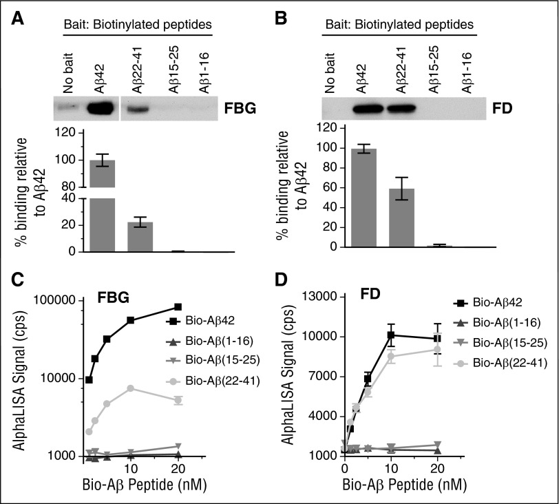
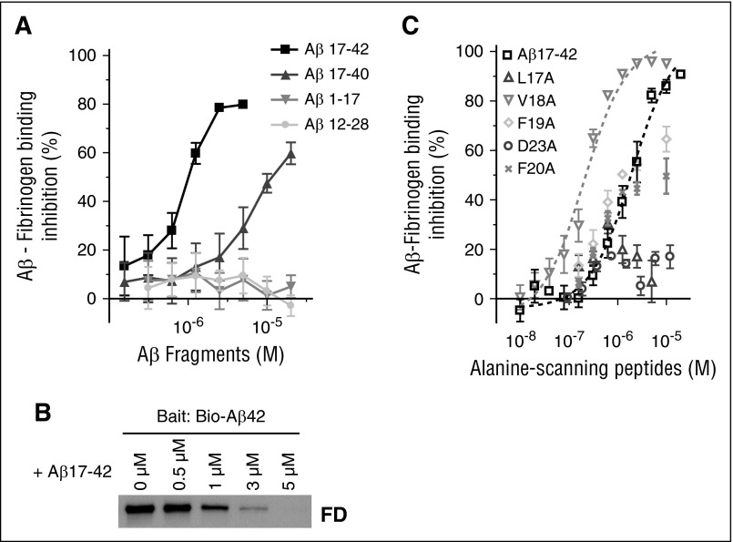
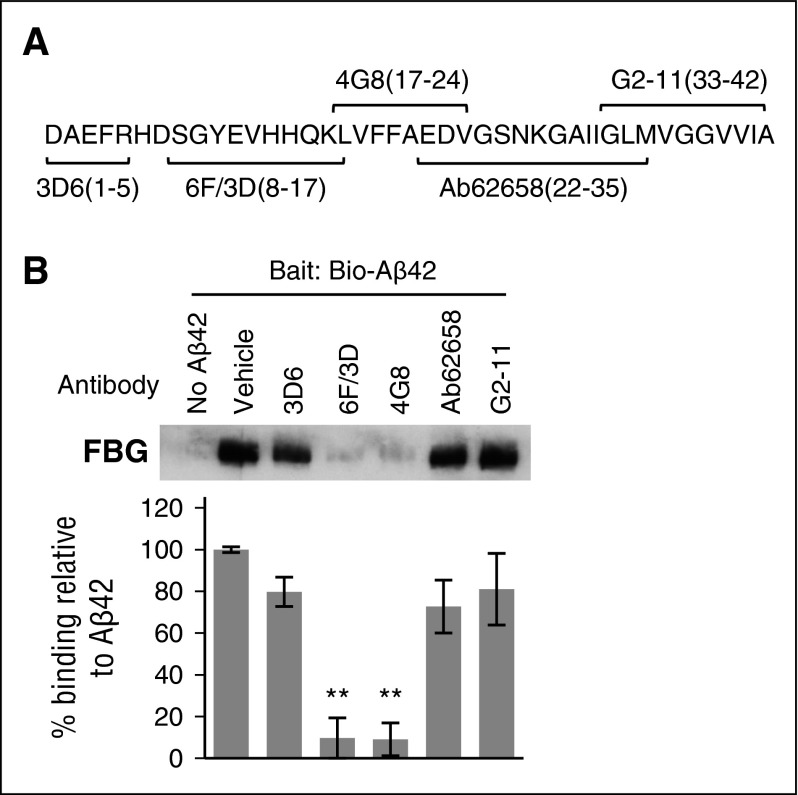
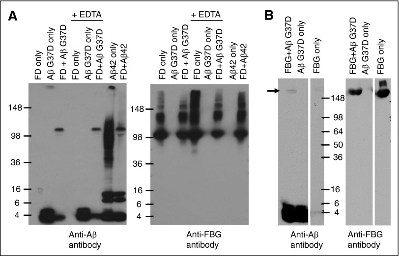
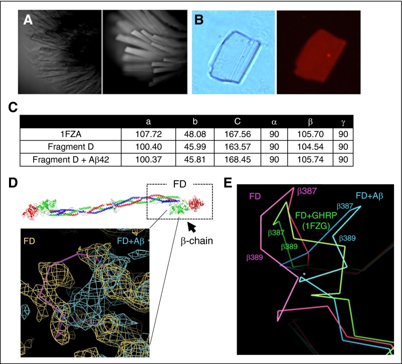
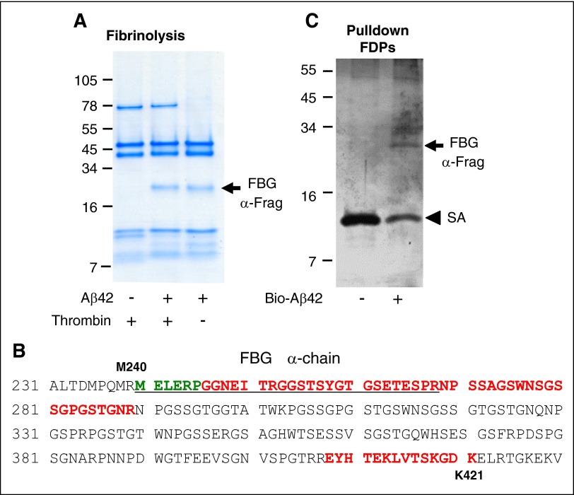
The majority of patients with Alzheimer disease (AD) suffer from impaired cerebral circulation. Accumulating evidence suggests that fibrinogen, the main protein component of blood clots, plays an important role in this circulatory dysfunction in AD. Fibrinogen interacts with β-amyloid (Aβ), forming plasmin-resistant abnormal blood clots, and increased fibrin deposition is found in the brains of AD patients and mouse models. In this study, we investigated the biochemical and structural details of the Aβ-fibrinogen interaction. We identified the central region of Aβ42 as the most critical region for the interaction, which can be inhibited by specific antibodies against the central region of Aβ and by naturally occurring p3 peptides, Aβ17-40 and Aβ17-42. X-ray crystallographic analysis revealed that Aβ42 binding to fragment D of fibrinogen induced a structural change in the C-terminal region of the fibrinogen β-chain (β384-393). Furthermore, we identified an additional Aβ-binding site within the αC region of fibrinogen. Aβ binding to this αC region blocked plasmin-mediated fibrin cleavage at this site, resulting in the generation of increased levels of a plasmin-resistant fibrin degradation fragment. Overall, our study elucidates the Aβ-fibrinogen interaction and clarifies the mechanism by which Aβ-fibrinogen binding delays fibrinolysis by plasmin. These results may facilitate the development of effective therapeutics against the Aβ-fibrinogen interaction to treat cerebrovascular abnormalities in AD.


Figures

Figure 1
Figure 1
Aβ22-41 binds to fibrinogen and fragment D. (A-B) Biotin-labeled Aβ42, Aβ1-16, Aβ15-25, and Aβ22-41 were incubated with fibrinogen (FBG) or fragment D (FD), and pulldown assays were carried out using streptavidin-coated magnetic beads. All samples were analyzed by western blot in unreduced condition using an anti-fibrinogen antibody. Only Aβ22-41 showed binding to both fibrinogen (A) and fragment D (B). When no Aβ peptides were added, the level of bound fibrinogen or fragment D was negligible. Images and graphs are representative of 4 experiments. (C-D) The binding between biotin-labeled Aβ42 or Aβ fragments with fibrinogen or fragment D was determined by AlphaLISA (n = 3). Controls and other lanes in panel A are from the same gel with some lanes omitted for clarity. Results presented in graphs are mean ± standard error of the mean (SEM).
Figure 2
Figure 2
Naturally occurring p3 peptides, Aβ17-40 and Aβ17-42, inhibit the Aβ-fibrinogen interaction. (A) Biotinylated Aβ42 was incubated with fibrinogen in the presence of various concentrations (0.05-20 µM) of 16 nonbiotinylated Aβ fragments listed in supplemental Figure 1. The inhibitory efficacy of the Aβ fragments on the Aβ42-fibrinogen interaction was analyzed using AlphaLISA. Of the 16 Aβ fragments tested, only Aβ17-40 (IC50 = 13.4 µM) and Aβ17-42 (IC50 = 1.03 µM) showed inhibitory efficacy (n = 3). (B) Western blot analysis with anti-fibrinogen antibody shows that Aβ17-42 blocks the ability of biotinylated Aβ42 to pull down fragment D (FD) in a dose-dependent manner. (C) Various concentrations (0.01-20 µM) of 5 alanine-scanning Aβ peptides (L17A, V18A, F19A, F20A, and D23A) were incubated with biotinylated Aβ42 and fibrinogen, and their ability to inhibit the Aβ42-fibrinogen interaction was analyzed using AlphaLISA (n = 3-6). Aβ L17A and D23A had almost no inhibitory activity (IC50 > 20 µM), whereas F19A (IC50 = 3.7 µM) and F20A (IC50 = 6.8 µM) showed a compatible inhibitory efficacy to original Aβ17-42. Interestingly, V18A (IC50 = 0.26 µM) had fivefold greater inhibitory efficacy than Aβ17-42. Results presented in graphs are mean ± SEM.
Figure 3
Figure 3
Specific antibodies against the central region of Aβ block the Aβ-fibrinogen interaction. (A) The epitopes for several antibodies against Aβ are illustrated in the schematic and include epitopes 1 to 5 (3D6; Elan), 8 to 17 (6F/3D; Dako), 17 to 24 (4G8; Covance), 22 to 35 (ab62658; Abcam), and 33 to 42 (G2-11; Abcam). (B) Antibodies at concentrations listed in “Methods” were incubated with fibrinogen and biotinylated Aβ42. Pulldown of biotinylated Aβ42 revealed that antibodies 6F/3D and 4G8 are able to interfere with the Aβ-fibrinogen interaction. Results presented in graphs are mean ± SEM, and statistical significance was determined using 1-way analysis of variance (ANOVA) and Bonferroni post hoc test (**P < .01; n = 3).
Figure 4
Figure 4
Long-term incubation of fibrinogen/fragment D with Aβ42 forms a SDS-stable complex. (A) Aβ42 G37D or Aβ42 was incubated with fragment D (FD) for 5 days at 37°C in the presence or absence of EDTA. Western blots were analyzed with antibody 6E10 against Aβ (left panel) and an antibody against fibrinogen (Dako; right panel). Aβ-fragment D SDS-stable complex was detected by 6E10. (B) Aβ42 G37D was incubated with fibrinogen (FBG) for 24 hours at 37°C. Western blots were analyzed with antibody 6E10 against Aβ (left panel) and an antibody against fibrinogen (right panel). Aβ-fibrinogen SDS-stable complex was also detected by 6E10 (arrow). Controls and other lanes in panel B are from the same gel with some lanes omitted for clarity.
Figure 5
Figure 5
Fragment D structure is altered in the presence of Aβ42. The fragment D crystals soaked with Aβ42 were analyzed by X-ray crystallography. (A) Brightfield (left) and UV fluorescence (right) images of fragment D crystals, indicating crystals are proteinaceous. (B) Left, Brightfield image of a fragment D crystal that had been subjected to soaking in TAMRA-Aβ42 followed by extensive washing. Right, Persistent red fluorescence after washing indicated that TAMRA-Aβ42 was binding within the crystal. (C) Unit cell dimensions of published (1FZA), nonsoaked, and Aβ42-soaked fragment D crystals. (D) Diagram of human fibrinogen with fragment D (FD) marked with a box. The location of altered structure in Aβ42-soaked fragment D crystals is indicated by the solid pink line. Superimposed 2Fo-Fc maps from nonsoaked (yellow; Rwork/Rfree = 0.24/0.33) and Aβ42-soaked (teal; Rwork/Rfree = 0.28/0.39) fragment D crystals with coordinates of nonsoaked crystals. Human fibrinogen schematic was generated from PDB file 3GHG. (E) Protein backbone diagram showing the shift of the β384-393 loop from nonsoaked fragment D (pink) to b-hole peptide (GHRP)-bound (1FZG; green) fragment D and Aβ42-soaked fragment D (blue). GHRP, Gly-His-Arg-Pro-amide peptide.
Figure 6
Figure 6
Aβ interacts with the α-chain of fibrinogen, producing a plasmin-resistant fibrin fragment during fibrinolysis. (A) Fibrin was digested with plasmin in the presence or absence of Aβ42. A plasmin-resistant fragment was observed only in the presence of Aβ42 (arrow). The same experiment was done without thrombin. In the absence of thrombin, plasmin degradation-resistant PRFF was also observed in the presence of Aβ42. Images are representative of ≥3 experiments. (B) Mass spectrometry analysis of the fragments in panels A and C showed they were derived from the α-chain of fibrinogen. Green residues were identified by N-terminal sequencing of band in panel A, red residues were identified by mass spectrometry analysis of band in panel A, and underlined residues were identified by mass spectrometry analysis of band in panel C. (C) Fibrinogen was partially digested with plasmin, incubated with biotinylated Aβ42, and Aβ42 was pulled down with streptavidin (SA)-coated beads. A fibrinogen fragment that bound to Aβ was observed (top arrow).

References

    1. Cortes-Canteli M, Paul J, Norris EH, et al. Fibrinogen and beta-amyloid association alters thrombosis and fibrinolysis: a possible contributing factor to Alzheimer’s disease. Neuron. 2010;66(5):695–709. - PMC - PubMed
    1. Ahn HJ, Glickman JF, Poon KL, et al. A novel Aβ-fibrinogen interaction inhibitor rescues altered thrombosis and cognitive decline in Alzheimer’s disease mice. J Exp Med. 2014;211(6):1049–1062. - PMC - PubMed
    1. Narayan PJ, Kim SL, Lill C, et al. Assessing fibrinogen extravasation into Alzheimer’s disease brain using high-content screening of brain tissue microarrays. J Neurosci Methods. 2015;247:41–49. - PubMed
    1. Weisel JW, Litvinov RI. Mechanisms of fibrin polymerization and clinical implications. Blood. 2013;121(10):1712–1719. - PMC - PubMed
    1. Davalos D, Akassoglou K. Fibrinogen as a key regulator of inflammation in disease. Semin Immunopathol. 2012;34(1):43–62. - PubMed

Publication types

Associated data