

Antennas of organ morphogenesis: the roles of cilia in vertebrate kidney development
- PMID: 27389733
- PMCID: PMC5053263
- DOI: 10.1002/dvg.22957
Antennas of organ morphogenesis: the roles of cilia in vertebrate kidney development
Abstract
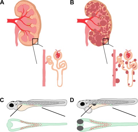
Cilia arose early during eukaryotic evolution, and their structural components are highly conserved from the simplest protists to complex metazoan species. In recent years, the role of cilia in the ontogeny of vertebrate organs has received increasing attention due to a staggering correlation between human disease and dysfunctional cilia. In particular, the presence of cilia in both the developing and mature kidney has become a deep area of research due to ciliopathies common to the kidney, such as polycystic kidney disease (PKD). Interestingly, mutations in genes encoding proteins that localize to the cilia cause similar cystic phenotypes in kidneys of various vertebrates, suggesting an essential role for cilia in kidney organogenesis and homeostasis as well. Importantly, the genes so far identified in kidney disease have conserved functions across species, whose kidneys include both primary and motile cilia. Here, we aim to provide a comprehensive description of cilia and their role in kidney development, as well as highlight the usefulness of the zebrafish embryonic kidney as a model to further understand the function of cilia in kidney health.
Keywords: ciliopathy; development; kidney; mesonephros; metanephros; motile cilia; multiciliated cell; primary cilia; pronephros; zebrafish.
© 2016 The Authors. genesis Published by Wiley Periodicals, Inc.
Figures
Similar articles
-
Cilia-driven fluid flow in the zebrafish pronephros, brain and Kupffer's vesicle is required for normal organogenesis.Development. 2005 Apr;132(8):1907-21. doi: 10.1242/dev.01772. Development. 2005. PMID: 15790966
-
Kidney organogenesis in the zebrafish: insights into vertebrate nephrogenesis and regeneration.Wiley Interdiscip Rev Dev Biol. 2013 Sep-Oct;2(5):559-85. doi: 10.1002/wdev.92. Epub 2012 Oct 16. Wiley Interdiscip Rev Dev Biol. 2013. PMID: 24014448 Free PMC article. Review.
-
The role of cilia during organogenesis in zebrafish.Open Biol. 2023 Dec;13(12):230228. doi: 10.1098/rsob.230228. Epub 2023 Dec 13. Open Biol. 2023. PMID: 38086423 Free PMC article. Review.
-
Growing kidney in the frog.Nephron Exp Nephrol. 2006;103(3):e81-5. doi: 10.1159/000092192. Epub 2006 Mar 22. Nephron Exp Nephrol. 2006. PMID: 16554664 Review.
-
Visualizing multiciliated cells in the zebrafish.Methods Cell Biol. 2023;175:129-161. doi: 10.1016/bs.mcb.2022.12.001. Epub 2023 Jan 6. Methods Cell Biol. 2023. PMID: 36967138
Cited by
-
NEK9 regulates primary cilia formation by acting as a selective autophagy adaptor for MYH9/myosin IIA.Nat Commun. 2021 Jun 2;12(1):3292. doi: 10.1038/s41467-021-23599-7. Nat Commun. 2021. PMID: 34078910 Free PMC article.
-
Modeling Podocyte Ontogeny and Podocytopathies with the Zebrafish.J Dev Biol. 2023 Feb 20;11(1):9. doi: 10.3390/jdb11010009. J Dev Biol. 2023. PMID: 36810461 Free PMC article. Review.
-
gldc Is Essential for Renal Progenitor Patterning during Kidney Development.Biomedicines. 2022 Dec 12;10(12):3220. doi: 10.3390/biomedicines10123220. Biomedicines. 2022. PMID: 36551976 Free PMC article.
-
Principles of Zebrafish Nephron Segment Development.J Dev Biol. 2023 Mar 18;11(1):14. doi: 10.3390/jdb11010014. J Dev Biol. 2023. PMID: 36976103 Free PMC article. Review.
-
The tbx2a/b transcription factors direct pronephros segmentation and corpuscle of Stannius formation in zebrafish.Dev Biol. 2017 Jan 1;421(1):52-66. doi: 10.1016/j.ydbio.2016.10.019. Epub 2016 Nov 11. Dev Biol. 2017. PMID: 27840199 Free PMC article.
References
-
- Bisgrove, B. W. , Snarr, B. S. , Emrazian, A. , & Yost, H. J. (2005). Polaris and Polycystin‐2 in dorsal forerunner cells and Kupffer's vesicle are required for specification of the zebrafish left‐right axis. Developmental Biology, 287, 274–288. - PubMed
-
- Bisgrove, B. W. , & Yost, H. J. (2006). The roles of cilia in developmental disorders and disease. Development, 133, 4131–4143. - PubMed
-
- Boletta, A. , Qian, F. , Onuchic, L. F. , Bhunia, A. K. , Phakdeekitcharoen, B. , Hanaoka, … Germino, G. G. (2000). Polycystin‐1, the gene product of PKD1, induces resistance to apoptosis and spontaneous tubulogenesis in MDCK cells. Molecular Cell, 6, 1267–1273. - PubMed
Publication types
MeSH terms
Grants and funding
LinkOut - more resources
Full Text Sources
Other Literature Sources
