. 2016 Jul 8:7:12061.
doi: 10.1038/ncomms12061.

Wars2 is a determinant of angiogenesis

Affiliations

Wars2 is a determinant of angiogenesis

Mao Wang et al. Nat Commun. .

Abstract

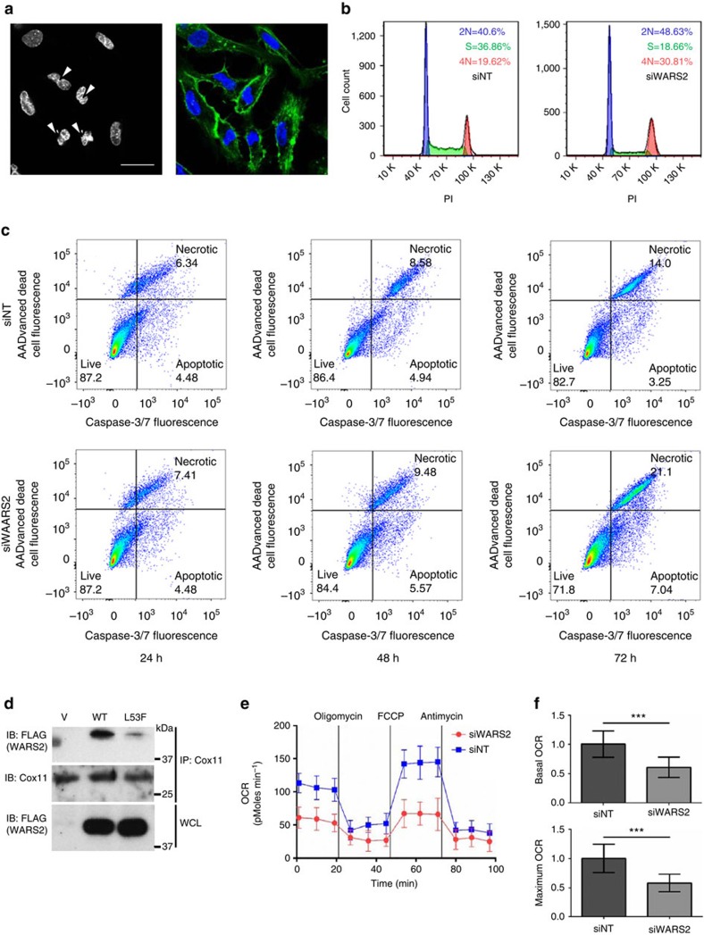
Coronary flow (CF) measured ex vivo is largely determined by capillary density that reflects angiogenic vessel formation in the heart in vivo. Here we exploit this relationship and show that CF in the rat is influenced by a locus on rat chromosome 2 that is also associated with cardiac capillary density. Mitochondrial tryptophanyl-tRNA synthetase (Wars2), encoding an L53F protein variant within the ATP-binding motif, is prioritized as the candidate at the locus by integrating genomic data sets. WARS2(L53F) has low enzyme activity and inhibition of WARS2 in endothelial cells reduces angiogenesis. In the zebrafish, inhibition of wars2 results in trunk vessel deficiencies, disordered endocardial-myocardial contact and impaired heart function. Inhibition of Wars2 in the rat causes cardiac angiogenesis defects and diminished cardiac capillary density. Our data demonstrate a pro-angiogenic function for Wars2 both within and outside the heart that may have translational relevance given the association of WARS2 with common human diseases.


Figures

Figure 1
Figure 1. Mapping of coronary flow to the rat 2q34 locus and identification of Wars2 as the candidate gene at the locus.
(a) Coronary flow (CF) under intrinsic heart rate conditions in the Brown Norway (BN) and Spontaneously Hypertensive rat (SHR) (left panel, n=12) and under cardiac pacing conditions (right panel, n=20). (b) Genome-wide Evolutionary Stochastic Search (ESS) mapping of CF at three distinct experimental time points in an F2 cross (n=172) between the SHR and BN strains (x axis, rat autosomes; posterior probabilities for the peak SNP shown). (c) Replication of the CF QTL by congenic rescue in an SHR strain (SHR.BN2q34) encoding the BN genotype at the CF locus (P=3.2 × 10⁻⁶; n=6, SHR and 8, congenic; two-way analysis of variance (ANOVA) with Tukey's multiple comparisons test). (d) Capillary density (shown in arbitrary optical units) in the heart in SHR.BN2q34 strain (n=8) and the parental SHR (n=6) strain. (e) Capillary density (shown in arbitrary optical units) in F2 rats by BN/SHR genotype of the peak-associated SNP (n=10–16 per genotype). One-way ANOVA with Tukey's multiple comparisons test. (f) Refinement of the 2q34 locus using flanking markers to the QTL (Chromosome 2: 189.3 Mb and 194.5 Mb) and all protein damaging variation at the locus in SHR, Salt Sensitive (SS) or Fawn Hooded Hypertensive (FHH) rats (black squares; grey squares, no variation). (g) Candidate gene prioritization using BN, SHR, SS and FHH whole-genome sequences and PolyPhen2 prediction of protein damaging variants effect (y-axis) from (f). Grey dashed lines indicate physical position of genes. (h) Sequence alignments of Wars2(L53F) protein variation in the ‘HXGH motif' in the SHR, the BN rat and other species (zebrafish, ZFish). (i) Western blot of wild-type WARS2 and WARS2(L53F) protein. Arrows indicate WARS2- specific immunoreactive band (upper arrow, high molecular weight isoform; lower arrow, low molecular weight isoform); bottom panel, GAPDH immunoblot as loading control. (j) In vitro assay of canonical enzymatic activities (arbitrary optical units) of WARS2 and WARS2(L53F) (red line, mean). t-test: *P<0.05; ***P<0.001.
Figure 2
Figure 2. WARS2 regulates endothelial cell morphology and angiogenic potential.
(a) Bright field micrographs of endothelial cells (ECs) following transfection with either control siRNA (siNT) or siRNA against WARS2 (siWARS2). Scale bar=200 μm. (b) Confocal microscopy of ECs transfected with siNT or siWARS2 (red, mitochondria; green, actin; blue, nucleus; scale bar =30 μm). (c) EC number in EC cultures transfected with siNT or siWARS2 (n=3 per condition, t-test). (d) Super-resolution microscopy of ECs transfected with siNT or siWARS2 and stained for actin (left), mitochondria (middle) and composite images with nuclear stain (right). Scale bar =25 μm. (e–j) Effects of WARS2 in an in vitro model of EC angiogenesis. (eg) WARS2 loss of function; (h–j) WARS2 gain-of-function. Total tubes (e, h), total tube length (in pixel) (f, i) and total branching points (g, j). n=8, t-test. **, P<0.01; ***, P<0.001.
Figure 3
Figure 3. Effects of inhibition of wars2 in the zebrafish and rat.
(a) Zebrafish embryos injected with morpholinos (MO; 0.5 ng, 1 ng or 2 ng) against wars2 or non-targeting MO (Control) and imaged at 72 h post fertilization (72 hpf). Scale bar=1 mm. (b, c) Percentage of fish with cardiac oedema (b) and normal, decreased or absent blood flow (c) at 72hpf. n=55, 62, 54, and 42 fish for doses of 0, 0.5, 1, and 2 ng wars2 MO, respectively; Chi-square test. (df) Inhibition effect of wars2 on heart rate (d), cardiac stroke volume (e) and cardiac output (f). n=15, 13, 16, and 15 fish for doses of 0, 0.25, 0.5, and 1 ng wars2 MO, respectively; Dunnett's multiple comparison test after one-way analysis of variance (ANOVA). (g) 3D reconstruction of 2-photon Z-stack images of 24–26 hpf (top) and 48–50 hpf (bottom) Tg(flk:EGFP) transgenic zebrafish embryos. Green fluorescent trunk vessels are shown from controls and embryos with 0.5 or 1 ng of the wars2 morpholino. Defects of intersegmental vessels (ISV, arrowheads) become obvious at 24 h.p.f.; disruptions of dorsal longitudinal anastomotic vessels (DLAV, arrows) occur at 48 hpf Fish anterior end is located to the left in all images. DA: dorsal aorta; PCV: posterior cardinal vein. Scale bar=100 μm. (h) Optical slice through 3D-reconstructed images of hearts of zebrafish embryos 5 days post fertilization using in vivo 2-photon microscopy. Tg(myl7:GFP;flk:dsRed) zebrafish, showing green fluorescence in myocardium and red fluorescence in endocardium, with separation (arrows) of cell layers after wars2 knockdown (a: atrium, o: outflow tract, v: ventricle.). Scale bar, 30 μm. (im), Effects of Wars2 loss-of-function on capillary density and coronary flow in rat heart. (i) Histological section of rat hearts stained for CD31 showing very large sub-epicardial veins in F1(Wars2−/L53F) rats but not F1(Wars2+/L53F) rats. Scale bar=500μm. (j) Sub-epicardial venule areas (log scale, arbitrary units) in F1 rats (n=5, red line, mean). (k) Relative capillary density in F1 rats (n=5). (l) Relative capillary area in the heart F1 rats (n=5). (m) Ex vivo quantification of coronary flow under paced conditions in wild-type BN(Wars2+/+), n=9; BN(Wars2−/+), n=7; F1(Wars2+/L53F), n=7 and F1(Wars2−/L53F), n=11. j–i, t-test; m, one-way ANOVA with Tukey's multiple comparisons test. **P<0.01; ***P<0.001.
Figure 4
Figure 4. Effects of WARS2 on endothelial cell viability and oxygen consumption.
(a) SiRNA-mediated inhibition of WARS2 in endothelial cells (ECs) results in multiple cells with two or more incompletely separated nuclei. Left panel, nuclei (arrow heads); right panel, nuclei (blue) and actin (green). Scale bar=50 μm. (b) FACS analysis of the cell cycle in proliferating ECs treated with non-targeting siRNA (siNT) or siRNA against WARS2 (siWARS2) shows a reduction in ECs in the S phase and an increased number in the G2/M phase with siW2. The experiment was repeated (n=3) with similar results (c) FACS analysis of caspase-3/7 activation and cell death over a 72-h time course. ECs were transfected with non-targeting siRNA (siNT) or siRNA against WARS2 (siWARS2). The experiment was repeated with similar results (and see Supplementary Fig. 14). (d) Immunoblot (IB) of COX11 and Flag in Cox11 immunoprecipitates (IP: COX11) and of whole cell lysates (WCL) from cells expressing either vector alone (V), Flag-tagged wild-type WARS2 (WT) or Flag-tagged mutant WARS2(L53F) (L53F). (e) Oxygen consumption rates (OCRs) in ECs transfected with non-targeting siRNA (siNT, blue boxes) or siRNA against WARS2 (siWARS2, red circles). A representative experiment is shown; the experiment repeated 5 times with similar results. (f) Quantification of basal (upper panel) and maximal (lower panel) OCRs. n=3, t-test. **, P<0.01. ***, P<0.001.

References

    1. Simons M. Angiogenesis: where do we stand now? Circulation 111, 1556–1566 (2005). - PubMed
    1. Folkman J. Angiogenesis: an organizing principle for drug discovery? Nat. Rev. Drug Discov. 6, 273–286 (2007). - PubMed
    1. Carmeliet P. & Jain R. K. Molecular mechanisms and clinical applications of angiogenesis. Nature 473, 298–307 (2011). - PMC - PubMed
    1. Sano M. et al.. p53-induced inhibition of Hif-1 causes cardiac dysfunction during pressure overload. Nature 446, 444–448 (2007). - PubMed
    1. Oka T., Akazawa H., Naito A. T. & Komuro I. Angiogenesis and cardiac hypertrophy: maintenance of cardiac function and causative roles in heart failure. Circ. Res. 114, 565–571 (2014). - PubMed

Publication types

MeSH terms

Substances