

Value of History, Physical Examination, and Radiographic Findings in the Diagnosis of Symptomatic Meniscal Tear Among Middle-Aged Subjects With Knee Pain
- PMID: 27390312
- PMCID: PMC5219865
- DOI: 10.1002/acr.22975
Value of History, Physical Examination, and Radiographic Findings in the Diagnosis of Symptomatic Meniscal Tear Among Middle-Aged Subjects With Knee Pain
Abstract
Objective: To evaluate the utility of clinical history, radiographic findings, and physical examination findings in the diagnosis of symptomatic meniscal tear (SMT) in patients over age 45 years, in whom concomitant osteoarthritis is prevalent.
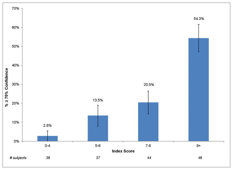
Methods: In a cross-sectional study of patients from 2 orthopedic surgeons' clinics, we assessed clinical history, physical examination findings, and radiographic findings in patients age >45 years with knee pain. The orthopedic surgeons rated their confidence that subjects' symptoms were due to meniscal tear; we defined the diagnosis of SMT as at least 70% confidence. We used logistic regression to identify factors independently associated with diagnosis of SMT, and we used the regression results to construct an index of the likelihood of SMT.
Results: In 174 participants, 6 findings were associated independently with the expert clinician having ≥70% confidence that symptoms were due to meniscal tear: localized pain, ability to fully bend the knee, pain duration <1 year, lack of varus alignment, lack of pes planus, and absence of joint space narrowing on radiographs. The index identified a low-risk group with 3% likelihood of SMT.
Conclusion: While clinicians traditionally rely upon mechanical symptoms in this diagnostic setting, our findings did not support the conclusion that mechanical symptoms were associated with the expert's confidence that symptoms were due to meniscal tear. An index that includes history of localized pain, full flexion, duration <1 year, pes planus, varus alignment, and joint space narrowing can be used to stratify patients according to their risk of SMT, and it identifies a subgroup with very low risk.
© 2016, American College of Rheumatology.
Conflict of interest statement
Figures

References
-
- Noble J, Turner PG. The function, pathology, and surgery of the meniscus. Clin Orthop Relat Res. 1986:62–68. - PubMed
-
- Englund M, Roos EM, Lohmander LS. Impact of type of meniscal tear on radiographic and symptomatic knee osteoarthritis: a sixteen-year followup of meniscectomy with matched controls. Arthritis Rheum. 2003;48:2178–2187. - PubMed
-
- Bhattacharyya T, Gale D, Dewire P, Totterman S, Gale ME, McLaughlin S, et al. The clinical importance of meniscal tears demonstrated by magnetic resonance imaging in osteoarthritis of the knee. J Bone Joint Surg Am. 2003;85-A:4–9. - PubMed
-
- Boden SD, Davis DO, Dina TS, Stoller DW, Brown SD, Vailas JC, et al. A prospective and blinded investigation of magnetic resonance imaging of the knee. Abnormal findings in asymptomatic subjects. Clin Orthop Relat Res. 1992:177–185. - PubMed
Publication types
MeSH terms
Grants and funding
LinkOut - more resources
Full Text Sources
Other Literature Sources
Medical
