Review
. 2016 Jul 8;119(2):330-40.
doi: 10.1161/CIRCRESAHA.116.307971.

Noninvasive Molecular Imaging of Disease Activity in Atherosclerosis

Affiliations
Review

Noninvasive Molecular Imaging of Disease Activity in Atherosclerosis

Marc R Dweck et al. Circ Res. .

Abstract

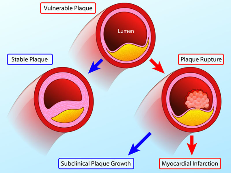
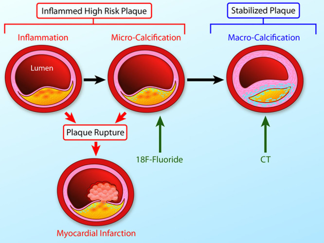
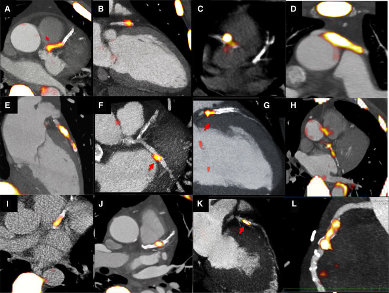
Major focus has been placed on the identification of vulnerable plaques as a means of improving the prediction of myocardial infarction. However, this strategy has recently been questioned on the basis that the majority of these individual coronary lesions do not in fact go on to cause clinical events. Attention is, therefore, shifting to alternative imaging modalities that might provide a more complete pan-coronary assessment of the atherosclerotic disease process. These include markers of disease activity with the potential to discriminate between patients with stable burnt-out disease that is no longer metabolically active and those with active atheroma, faster disease progression, and increased risk of infarction. This review will examine how novel molecular imaging approaches can provide such assessments, focusing on inflammation and microcalcification activity, the importance of these processes to coronary atherosclerosis, and the advantages and challenges posed by these techniques.

Keywords: 18F-Fluorodeoxyglucose; atherosclerosis; disease progression; inflammation; myocardial infarction.


Figures

Figure 1.
Figure 1.
Natural history of the vulnerable plaque. Vulnerable atherosclerotic plaques are thought to account for the majority of myocardial infarctions and are characterized by macrophage inflammation, a thin fibrous cap, positive remodeling, microcalcification, and angiogenesis. Histological and imaging studies conducted post myocardial infarction (MI) have consistently associated these plaques with rupture and myocardial infarction. However, in prospective observational studies, only a minority of these plaques go on to cause adverse clinical events (red arrows). This is because many vulnerable plaques will in fact heal and stabilize via multiple processes, including calcification. Although a proportion will go on to rupture, the majority of such events remain subclinical resulting in plaque growth rather than MI. As a consequence, the number of vulnerable plaques seems to greatly outnumber the clinical events that ensue (Illustration credit: Ben Smith).
Figure 2.
Figure 2.
Molecular imaging of vascular inflammation activity. A, 18F-fluorodeoxyglucose (18F-FDG) positron emission tomography (PET) image fused with contrast computed tomographic (CT) angiogram of the thoracic aorta, demonstrating regions of increased activity in the ascending aorta (blue), aortic arch (green), and descending aorta (red). B, 18F-FDG PET/magnetic resonance (MR) image of the carotid arteries demonstrating increased activity in the left carotid artery and the excellent soft tissue contrast provided by MR. C, 18-FDG PET fused with a CT coronary angiogram demonstrating increased uptake in the left ventricle but also in a remote plaque in the mid right coronary artery. Image in panel C reproduced from Cheng et al with permission of the publisher. Copyright © 2012, the Society of Nuclear Medicine and Molecular Imaging, Inc. D, 68Ga-DOTATATE PET/CT image with increased activity localizing to a plaque in the mid left anterior descending artery. E, T2* Map from patients with an abdominal aortic aneurysm that had been administered ultra small particles of iron oxide (USPIO). A hot spot is observed in the anterior wall of the aneurysm (arrow). F, In a second patient, focal areas of increased USPIO uptake can be observed (the increased signal adjacent to the lumen is considered normal because of high signal in the blood pool). Images in panels E and F reproduced from Richards et al with permission of the publisher. Copyright © 2011, Wolters Kluwer Health, Inc.
Figure 3.
Figure 3.
The link between inflammation, microcalcification, and macrocalcification. A large necrotic core, a thin fibrous cap, and an intense inflammation are key precipitants of acute plaque rupture and myocardial infarction. Intimal calcification is thought to occur as a healing response to this intense necrotic inflammation. However, the early stages of microcalcification (detected by 18F-fluoride positron emission tomography) are conversely associated with an increased risk of rupture. In part, this is because of residual plaque inflammation and in part because microcalcification itself increases mechanical stress in the fibrous cap further increasing propensity to rupture. With progressive calcification, plaque inflammation becomes pacified and the necrotic core walled off from the blood pool. The latter stages of macrocalcification (detected by computed tomographic) are, therefore, associated with plaque stability and a lower risk of that plaque rupturing (Illustration credit: Ben Smith).
Figure 4.
Figure 4.
18F-Fluoride preferentially binds microcalcification beyond the resolution of computed tomography (CT). Images are taken ex vivo of a carotid endarterectomy specimen excised from a patient who had a recent stroke. A, Histological section of the excised plaque stained for calcium with Alizarin red. B, Filled black arrow shows an area of dense macroscopic calcification that is visible on micro-CT. By comparison, the empty black arrow head demonstrates areas of microcalcification that are beyond the resolution of the micro-CT but by comparison demonstrate avid binding with 18F-fluoride on both autoradiography (C) and micro–positron emission tomography imaging (D). E, A second carotid endarterectomy sample from a patient post stroke demonstrates a large macro calcific deposit on micro-CT. F, Autoradiography shows that although 18F-fluoride is able to bind to the surface of the plaque, it is unable to penetrate in to the center. As a consequence of this effect, 18-fluoride binds preferentially to regions of microcalcification compared with macroscopic deposits. Reprinted from Irkle et al with permission. Copyright © 2015, Macmillan Publishers Limited.
Figure 5.
Figure 5.
18F-fluoride PET imaging in the coronary arteries. Multiple examples of 18F-fluoride localizing to individual coronary plaques on fused positron emission tomography/computed tomography images of the heart. This can be observed in the left anterior descending artery (A, B, E, G, I, J, and K), circumflex (F and H) and right coronary arteries (C and L), and in a saphenous vein graft (D).

References

    1. Naghavi M, Libby P, Falk E, et al. From vulnerable plaque to vulnerable patient: a call for new definitions and risk assessment strategies: Part I. Circulation. 2003;108:1664–1672. doi: 10.1161/01.CIR.0000087480.94275.97. - PubMed
    1. Ambrose JA, Tannenbaum MA, Alexopoulos D, Hjemdahl-Monsen CE, Leavy J, Weiss M, Borrico S, Gorlin R, Fuster V. Angiographic progression of coronary artery disease and the development of myocardial infarction. J Am Coll Cardiol. 1988;12:56–62. - PubMed
    1. Falk E, Shah PK, Fuster V. Coronary plaque disruption. Circulation. 1995;92:657–671. - PubMed
    1. Stone GW, Maehara A, Lansky AJ, de Bruyne B, Cristea E, Mintz GS, Mehran R, McPherson J, Farhat N, Marso SP, Parise H, Templin B, White R, Zhang Z, Serruys PW PROSPECT Investigators. A prospective natural-history study of coronary atherosclerosis. N Engl J Med. 2011;364:226–235. doi: 10.1056/NEJMoa1002358. - PubMed
    1. Calvert PA, Obaid DR, O’Sullivan M, Shapiro LM, McNab D, Densem CG, Schofield PM, Braganza D, Clarke SC, Ray KK, West NE, Bennett MR. Association between IVUS findings and adverse outcomes in patients with coronary artery disease: the VIVA (VH-IVUS in Vulnerable Atherosclerosis) Study. JACC Cardiovasc Imaging. 2011;4:894–901. doi: 10.1016/j.jcmg.2011.05.005. - PubMed

Publication types

MeSH terms