

Clinical Trial
doi: 10.3324/haematol.2016.146092.
Epub 2016 Jul 6.
Baseline bone involvement in multiple myeloma - a prospective comparison of conventional X-ray, low-dose computed tomography, and 18flourodeoxyglucose positron emission tomography in previously untreated patients
Affiliations
- PMID: 27390357
- PMCID: PMC5046664
- DOI: 10.3324/haematol.2016.146092
Item in Clipboard
Clinical Trial
Baseline bone involvement in multiple myeloma - a prospective comparison of conventional X-ray, low-dose computed tomography, and 18flourodeoxyglucose positron emission tomography in previously untreated patients
Haematologica.
2016 Oct.
No abstract available
Keywords: CT; Myeloma; PET; X-ray; bone marker.
Figures
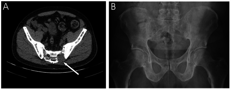
Low-dose CT versus conventional X-ray. Low-dose CT (A) and conventional X-ray (B) of a 67-year-old man with newly diagnosed multiple myeloma. X-ray performed 7 days after low-dose CT-scanning. The CT-scan shows a significant osteolytic lesion, not visualised at X-ray, of the pelvic area.
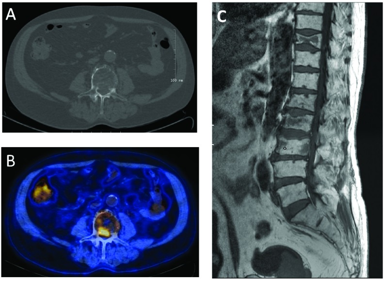
Imminent medullary compression revealed by low-dose CT. The low-dose CT-scan reveals tumor growth in the vertebral canal (A). This lesion was PET-positive (B). MR-scan confirmed the intra-spinal growth (C). The patient was asymptomatic.
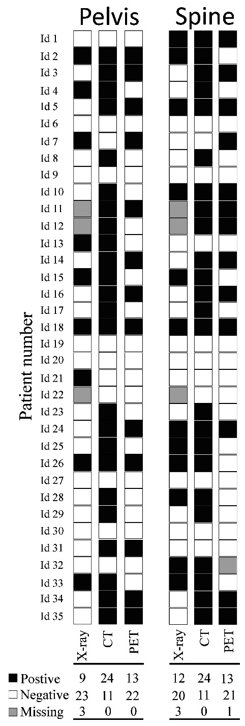
Results of X-ray, low-dose CT and PET. The individual results of the different imaging methods performed at baseline for the 35 previously untreated multiple myeloma patients. Below the columns are the numbers of positive, negative and missing results of each imaging modality shown. Low-dose CT identifies most patients with osteolysis. Black squares illustrate osteolysis at low-dose CT (CT), conventional X-rays (X-ray), and focal activity at 18FDG-PET (PET), while white squares illustrate imaging without osteolysis of focal activity. Grey squares represent missing data.
References
-
- Rajkumar SV, Dimopoulos MA, Palumbo A, et al. International Myeloma Working Group updated criteria for the diagnosis of multiple myeloma. Lancet Oncol. 2014;15(12):e538–e548. - PubMed
-
- Horger M, Claussen CD, Bross-Bach U, et al. Whole-body low-dose multidetector row-CT in the diagnosis of multiple myeloma: an alternative to conventional radiography. Eur J Radiol. 2005;54(2):289–297. - PubMed
-
- Regelink JC, Minnema MC, Terpos E, et al. Comparison of modern and conventional imaging techniques in establishing multiple myeloma-related bone disease: a systematic review. Br J Haematol. 2013;162(1):50–61. - PubMed
-
- Gleeson TG, Moriarty J, Shortt CP, et al. Accuracy of whole-body low-dose multidetector CT (WBLDCT) versus skeletal survey in the detection of myelomatous lesions, and correlation of disease distribution with whole-body MRI (WBMRI). Skeletal Radiol. 2009;38(3):225–236. - PubMed
Publication types
MeSH terms
Substances
LinkOut - more resources
Full Text Sources
Other Literature Sources
Medical
