Review
. 2016:4:38.
doi: 10.1007/s40134-016-0167-7. Epub 2016 May 20.

Advanced Analysis Techniques for Intra-cardiac Flow Evaluation from 4D Flow MRI

Affiliations
Review

Advanced Analysis Techniques for Intra-cardiac Flow Evaluation from 4D Flow MRI

Rob J van der Geest et al. Curr Radiol Rep. 2016.

Abstract

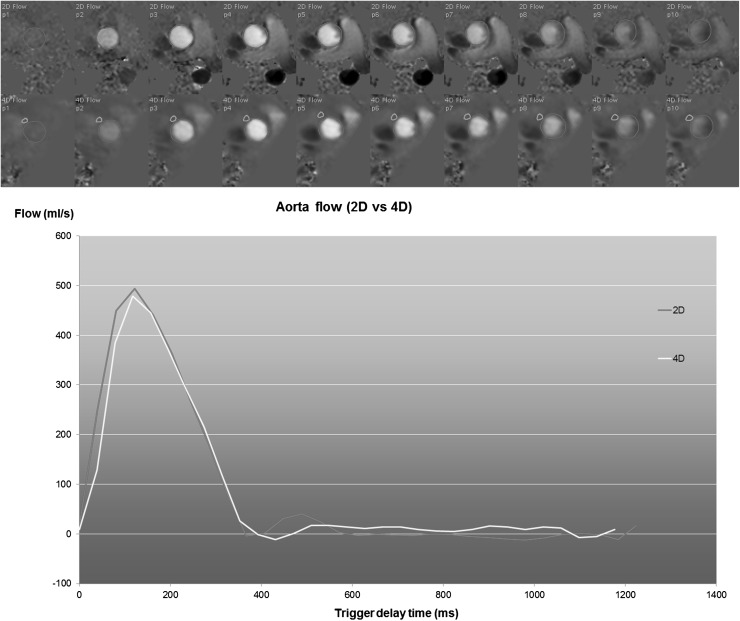
Purpose of the review: Time-resolved 3D velocity-encoded MR imaging with velocity encoding in three directions (4D Flow) has emerged as a novel MR acquisition technique providing detailed information on flow in the cardiovascular system. In contrast to other clinically available imaging techniques such as echo-Doppler, 4D Flow MRI provides the 3D Flow velocity field within a volumetric region of interest over the cardiac cycle. This work reviews the most recent advances in the development and application of dedicated image analysis techniques for the assessment of intra-cardiac flow features from 4D Flow MRI.

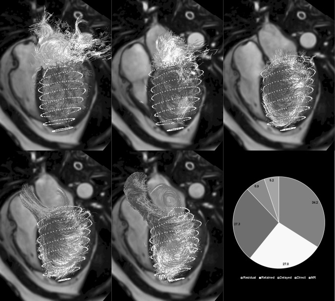
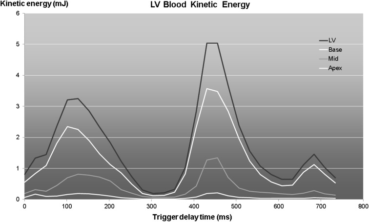
Recent findings: Novel image analysis techniques have been developed for extraction of relevant intra-cardiac flow features from 4D Flow MRI, which have been successfully applied in various patient cohorts and volunteer studies. Disturbed flow patterns have been linked with valvular abnormalities and ventricular dysfunction. Recent technical advances have resulted in reduced scan times and improvements in image quality, increasing the potential clinical applicability of 4D Flow MRI.

Summary: 4D Flow MRI provides unique capabilities for 3D visualization and quantification of intra-cardiac blood flow. Contemporary knowledge on 4D Flow MRI shows promise for further exploration of the potential use of the technique in research and clinical applications.

Keywords: 4D Flow; CMR; Flow components; Image processing; Kinetic energy; Path lines; Streamlines; Vortex.


Figures

Fig. 1
Fig. 1
Long-axis view obtained by conventional balanced FFE with color overlay derived from 4D Flow MRI. The coloring applied encodes for the magnitude of velocity in the direction of the indicated arrow, simulating Color Doppler flow imaging. The left panel shows the moment of peak early LV filling. The right panel, showing a systolic phase, clearly reveals the regurgitant jet distal to the mitral valve in the left atrium
Fig. 2
Fig. 2
Example of 2D velocity vector display in a patient with mitral valve regurgitation. Systolic four-chamber image showing a central high-velocity jet in the left atrium resulting in a clockwise recirculating flow in the left atrium
Fig. 3
Fig. 3
Left ventricular outflow tract view with 2D streamlines as overlay. Left moment of late diastolic filling. The presence of a 3D vortex ring can be seen as two counter-rotating recirculating flow regions distal to the mitral valve leaflets. Middle end-diastolic moment showing preserved rotational flow. Right early systolic moment showing redirection of flow toward the left ventricular outflow tract
Fig. 4
Fig. 4
Three-dimensional visualization of intra-cardiac flow used to show mitral inflow velocity at the moment of peak filling. Left vector display. Right streamline display with seeds defined in spherical region with a radius of 15 mm centered at the mitral valve annulus. LV endocardial surface is displayed with red dots and RV endocardial surface with yellow dots. The LV outflow tract cine view image is shown in the background as anatomical reference (Color figure online)
Fig. 5
Fig. 5
Example of aortic flow curves of a healthy subject derived from conventional 2D phase-contrast MRI (2D Flow) and from a reformatted 4D Flow acquisition. The 4D Flow acquisition was reformatted into a through-plane encoded view, identical to the corresponding 2D Flow acquisition. The top two rows depict the systolic through-plane velocity-encoded images (the first 10 frames out of 30) for 2D Flow and 4D Flow MRI, respectively. Contours are defined around the aortic lumen (shown in red). In the 4D Flow-derived images, an additional contour is defined in a region of tissue adjacent to the aorta for velocity offset correction. A good agreement is observed between the two flow curves. The stroke volume derived from 2D Flow is 105 ml and from 4D Flow is 103 ml (Color figure online)
Fig. 6
Fig. 6
Result of flow component analysis using particle tracing of a patient with mitral valve regurgitation. Displayed path lines are color coded according to flow component classification. Top row from left to right start of early filling, peak early filling, end diastole; Bottom row early systole, late systole, pie depicting percentage of LV for each of the five defined flow components. Regurgitant jet results in recirculating flow pattern in the left atrium
Fig. 7
Fig. 7
Temporal evolution of kinetic energy of blood in the LV for a healthy subject computed for multiple levels of the LV. The red curve shows the total LV kinetic energy

References

    1. Westenberg JJ, Roes SD, Ajmone Marsan N, Binnendijk NM, Doornbos J, Bax JJ, Reiber JH, de Roos A, van der Geest RJ. Mitral valve and tricuspid valve blood flow: accurate quantification with 3D velocity-encoded MR imaging with retrospective valve tracking. Radiology. 2008;249(3):792–800. doi: 10.1148/radiol.2492080146. - DOI - PubMed
    1. Wigström L, Sjöqvist L, Wranne B. Temporally resolved 3D phase-contrast imaging. Magn Reson Med. 1996;36:800–803. doi: 10.1002/mrm.1910360521. - DOI - PubMed
    1. Eriksson J, Carlhall CJ, Dyverfeldt P, Engvall J, Bolger AF, Ebbers T. Semiautomatic quantification of 4D left ventricular blood flow. J Cardiovasc Magn Reson. 2010;12:19. doi: 10.1186/1532-429X-12-9. - DOI - PMC - PubMed
    1. Markl M, Kilner PJ, Ebbers T. Comprehensive 4D velocity mapping of the heart and great vessels by cardiovascular magnetic resonance. J Cardiovasc Magn Reson. 2011;7:13. - PMC - PubMed
    1. • Dyverfeldt P, Bissell M, Barker AJ, Bolger AF, Carlhäll CJ, Ebbers T, Francios CJ, Frydrychowicz A, Geiger J, Giese D, Hope MD, Kilner PJ, Kozerke S, Myerson S, Neubauer S, Wieben O, Markl M. 4D flow cardiovascular magnetic resonance consensus statement. J Cardiovasc Magn Reson 2015;17:22. Consensus statement paper providing guidelines for 4D Flow MRI acquisition and analysis methods for evaluation of the heart and greater vessels. - PMC - PubMed