. 2015 Dec 23;16(1):96.
doi: 10.1186/s12868-015-0229-4.

Characterization of a novel zebrafish (Danio rerio) gene, wdr81, associated with cerebellar ataxia, mental retardation and dysequilibrium syndrome (CAMRQ)

Affiliations

Characterization of a novel zebrafish (Danio rerio) gene, wdr81, associated with cerebellar ataxia, mental retardation and dysequilibrium syndrome (CAMRQ)

Fusun Doldur-Balli et al. BMC Neurosci. .

Abstract

Background: WDR81 (WD repeat-containing protein 81) is associated with cerebellar ataxia, mental retardation and disequilibrium syndrome (CAMRQ2, [MIM 610185]). Human and mouse studies suggest that it might be a gene of importance during neurodevelopment. This study aimed at fully characterizing the structure of the wdr81 transcript, detecting the possible transcript variants and revealing its expression profile in zebrafish, a powerful model organism for studying development and disease.

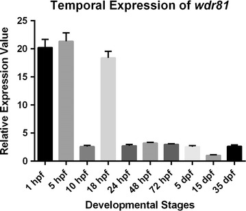
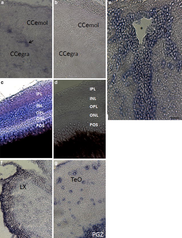
Results: As expected in human and mouse orthologous proteins, zebrafish wdr81 is predicted to possess a BEACH (Beige and Chediak-Higashi) domain, a major facilitator superfamily domain and WD40-repeats, which indicates a conserved function in these species. We observed that zebrafish wdr81 encodes one open reading frame while the transcript has one 5' untranslated region (UTR) and the prediction of the 3' UTR was mainly confirmed along with a detected insertion site in the embryo and adult brain. This insertion site was also found in testis, heart, liver, eye, tail and muscle, however, there was no amplicon in kidney, intestine and gills, which might be the result of possible alternative polyadenylation processes among tissues. The 5 and 18 hpf were critical timepoints of development regarding wdr81 expression. Furthermore, the signal of the RNA probe was stronger in the eye and brain at 18 and 48 hpf, then decreased at 72 hpf. Finally, expression of wdr81 was detected in the adult brain and eye tissues, including but not restricted to photoreceptors of the retina, presumptive Purkinje cells and some neurogenic brains regions.

Conclusions: Taken together these data emphasize the importance of this gene during neurodevelopment and a possible role for neuronal proliferation. Our data provide a basis for further studies to fully understand the function of wdr81.

Keywords: In situ hybridization; RACE; Zebrafish; qRT-PCR; wdr81.


Figures

Fig. 1
Fig. 1
Genomic structure of the zebrafish wdr81 and location of primers. The genomic structure of zebrafish wdr81 was derived from the predicted sequence information released by the Ensembl database and the open reading frame was amplified based on this information. Exons are shown as boxes and introns are shown as lines. Location of RNA oligos, ligated to the transcript by employing the RACE kit are shown in gray boxes. Arrows in the top box illustrate the approximate binding sites of the primers used in the RACE experiments. Arrows in the lower box indicate the approximate binding sites of primers used to amplify the open reading frame and of qRT-PCR primers
Fig. 2
Fig. 2
Agarose gel electrophoretic separation of overlapping amplicons of the zebrafish wdr81 open reading frame. Amplification of the open reading frame from 24 hpf embryo cDNA and adult brain cDNA resulted in one amplicon per reaction indicating that there is one open reading frame of zebrafish wdr81. Since the experiment was designed based on the predicted sequence, this result also confirms the prediction. a Amplification of 24 hpf embryo cDNA. b Amplification of adult brain cDNA. Lanes 1, 3, 5, 7, 9, 11, 13, 15, 17 and 19 were loaded with the PCR products of primer pairs 1–10, respectively. Lanes 2, 4, 6, 8, 10, 12, 14, 16, 18 and 20 were loaded with the PCR products of −RT controls amplified with primer pairs 1–10, respectively. Lanes indicated as M are loaded with pUC mix DNA Marker (SM0301, Thermo Scientific)
Fig. 3
Fig. 3
Relative temporal expression of wdr81 as determined by qRT-PCR. (1) 1 hpf embryo, (2) 5 hpf embryo, (3) 10 hpf embryo, (4) 18 hpf embryo, (5) 24 hpf embryo, (6) 48 hpf embryo, (7) 72 hpf larva, (8) 5 dpf larva, (9) 15 dpf larva, (10) 35 dpf juvenile zebrafish. Error bars represent +SE
Fig. 4
Fig. 4
Whole mount in situ hybridization revealed differential expression of wdr81 transcript during embryonic development. Our results from the WMISH method are in parallel with the qRT-PCR data. The signal is high during the first 3 developmental timepoints (6–18 hpf), it is decreased and maintained during the rest of the development periods (24–72 hpf). OV optic vesicle, Mb midbrain, Le lens, H hindbrain, Di diencephalon, MLF medial longitudinal fascicle
Fig. 5
Fig. 5
Transverse sections through the head regions of whole mount in situ hybridization specimens at 3 embryonic timepoints. The expression of wdr81 was observed in a regionally-specific manner by 48 hpf (ac), whereas it was ubiquitously expressed at 18 hpf (d), and decreased by 72 hpf (e, f). Arrows indicate the optic nerve, asterisk the region of the nucleus of the medial longitudinal fascicle, and arrowhead the retina. Po preoptic area, Di diencephalon, T. midbrain tegmentum, Le lens, OV optic vesicle, Yolk yolk sac. Scale bar equals 100 μm
Fig. 6
Fig. 6
Relative spatial expression graphic of wdr81 as determined by qRT-PCR. (1) Brain, (2) testis, (3) heart, (4) kidney, (5) liver, (6) intestine, (7) eye, (8) gills, (9) tail, (10) muscle. Error bars indicate +SE
Fig. 7
Fig. 7
wdr81 expression in the adult brain and eye tissues. wdr81 expression was detected in the cerebellum (a), retina (c), tectal ventricle (e), brain stem (f), and optic tectum (g). Results with a sense probe in both cerebellum (b) and retina (d), which demonstrate no staining, indicates the specificity of the signal obtained with an antisense probe and both cerebellum (b) and retina (d) are shown. CCe mol Cerebellar molecular layer, Cce gra cerebellar granular layer, POS photoreceptor outer segments, ONL outer nuclear layer, OPL outer plexiform layer, INL inner nuclear layer, IPL inner plexiform layer, LX lobus vagus, TeO optic tectum, PGZ periventricular gray zone of the optic tectum. The arrow indicates the Purkinje cell layer and the asterisk the tectal ventricle. Scale bar equals 200 μm
Fig. 8
Fig. 8
Agarose gel electrophoretic separation of the 5′RACE and overlapping 3′RACE products of zebrafish wdr81 transcript. a Expected size of the 5′RACE amplicon based on the experimental design was obtained and one amplicon was observed as a PCR product indicating that there is one 5′ UTR structure of zebrafish wdr81. The band of ~692 bp was excised, cloned and sequenced. Lane 1 5′RACE product of 24 hpf embryo wdr81 cDNA; lane 2 5′RACE product of brain wdr81 cDNA; lane 3 negative control without template, M pUC mix marker 8 (SM0301, Thermo Scientific). Expected size of the overlapping 3′RACE amplicons based on the experiment design were obtained from the PCR experiments with primer pairs 1 (data not shown) and 3 from the Table 3. The resulting amplicon obtained with primer pair 2 was longer than the expected amplicon size indicating presence of an insertion site. One amplicon per reaction was obtained as PCR products concluding that there is one transcript of zebrafish wdr81. b Amplification with primer pair 3 from Table 3. c Amplification with primer pair 2 from Table 3. In both gels, b and c, lane 1 24 hpf embryo RACE ready cDNA, lane 2 brain RACE ready cDNA, lane 3 no template negative control. M MassRuler Mix DNA Marker (SM0403, Thermo Scientific)
Fig. 9
Fig. 9
A 266 bp long insertion was determined in the 3′ UTR of the zebrafish wdr81 transcript. Results of 3′ UTR characterization experiments indicated presence of an insertion site in 24 hpf embryo and adult brain templates. When the insertion site was further analyzed, cloned and sequenced, the sequence of the insertion site from 6 single colonies was revealed as being 266 bp in length. Twenty-four hpf embryo sample is the insert of plasmids 1–1, 1–5, and 1–8 and brain sample is the insert of plasmids 2–3, 2–4, and 2–6
Fig. 10
Fig. 10
Detection of the insertion in the tested developmental and tissue samples except kidney, intestine, gills. a Agarose gel electrophoretic separation of the amplicons as products of a PCR experiment in which the presence of the detected insertion site in wdr81 3′ UTR was tested among templates from several developmental stages (top gel picture). Presence and integrity of the cDNA samples were tested with beta-actin amplification (lower gel picture). b Agarose gel electrophoretic separation of the amplicons as products of a PCR experiment in which the presence of the detected insertion site in wdr81 3′ UTR was tested among templates from several adult tissues (top gel picture). Presence and integrity of the cDNA samples were tested with beta-actin amplification (lower gel picture). In all gels, lanes were labelled with the cDNA source of each reaction loaded. Lane indicated as M was loaded with pUC mix DNA Marker (SM0301, Thermo Scientific). “No t.” indicates the no template negative control sample in which water was used instead of a template

References

    1. Gulsuner S, Tekinay AB, Doerschner K, Boyaci H, Bilguvar K, Unal H, Onat OE, Atalar E, Basak N, Topaloglu H, Kansu T, Tan M, Tan U, Gunel M, Ozcelik T. Homozygosity mapping and targeted genomic sequencing reveal the gene responsible for cerebellar hypoplasia and quadrupedal locomotion in a consanguineous kindred. Genome Res. 2011;21(12):1995–2003. doi: 10.1101/gr.126110.111. - DOI - PMC - PubMed
    1. Turkmen S, Demirhan O, Hoffmann K, Diers A, Zimmer C, Sperling K, Mundlos S. Cerebellar hypoplasia and quadrupedal locomotion in humans as a recessive trait mapping to chromosome 17p. J Med Genet. 2006;43(October 2008):461–464. - PMC - PubMed
    1. Tan U. New syndrome with quadrupedal gait, primitive speech, and severe mental retardation as a live model for human evolution. Int J Neurosci. 2006;116(3):361–369. doi: 10.1080/00207450500455330. - DOI - PubMed
    1. Donnard E, Asprino PF, Correa BR, Bettoni F, Koyama FC, Navarro FCP, Perez RO, Mariadason J, Sieber OM, Strausberg RL, Simpson AJG, Jardim DLF, Reis LFL, Parmigiani RB, Galante PAF, Camargo AA. Mutational analysis of genes coding for cell surface proteins in colorectal cancer cell lines reveal novel altered pathways, druggable mutations and mutated epitopes for targeted therapy. Oncotarget. 2014;5(19):9199–9213. doi: 10.18632/oncotarget.2374. - DOI - PMC - PubMed
    1. Traka M, Millen KJ, Collins D, Elbaz B, Kidd GJ, Gomez CM, Popko B. WDR81 is necessary for purkinje and photoreceptor cell survival. J Neurosci. 2013;33(16):6834–6844. doi: 10.1523/JNEUROSCI.2394-12.2013. - DOI - PMC - PubMed

Publication types

Substances

Supplementary concepts