. 2016 Jul 8;11(7):e0158692.
doi: 10.1371/journal.pone.0158692. eCollection 2016.

Identification of Novel and Recurrent Disease-Causing Mutations in Retinal Dystrophies Using Whole Exome Sequencing (WES): Benefits and Limitations

Affiliations

Identification of Novel and Recurrent Disease-Causing Mutations in Retinal Dystrophies Using Whole Exome Sequencing (WES): Benefits and Limitations

Amit Tiwari et al. PLoS One. .

Abstract

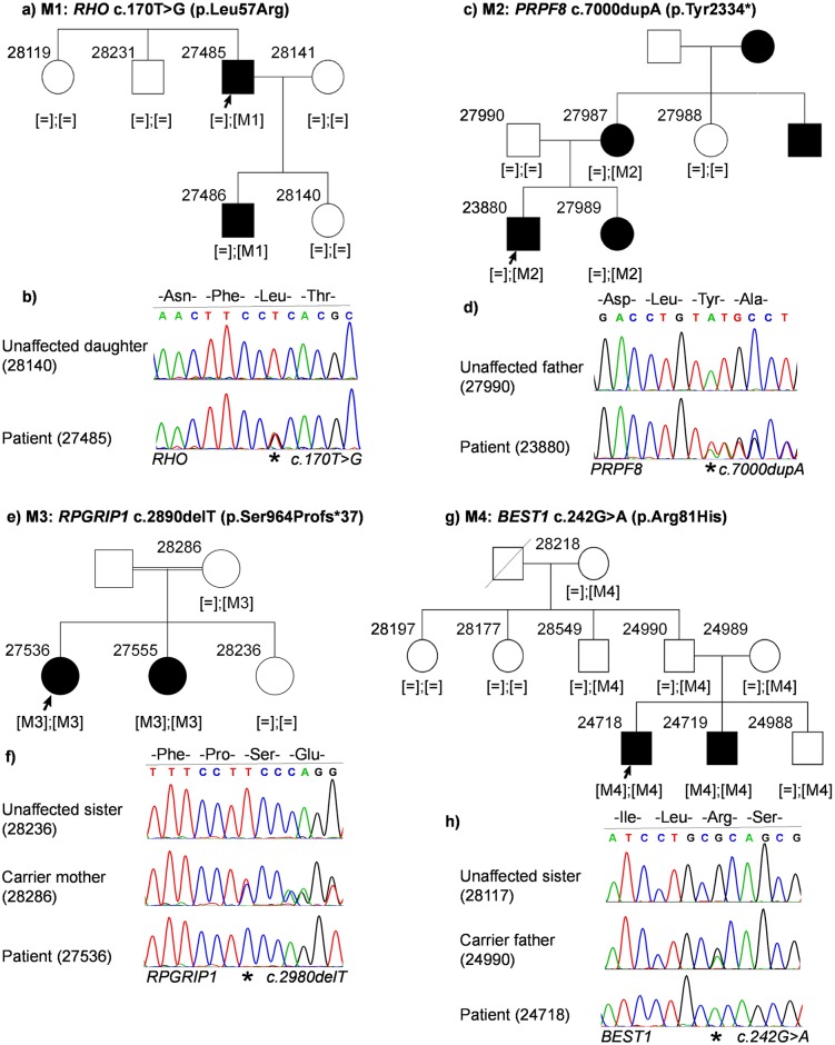
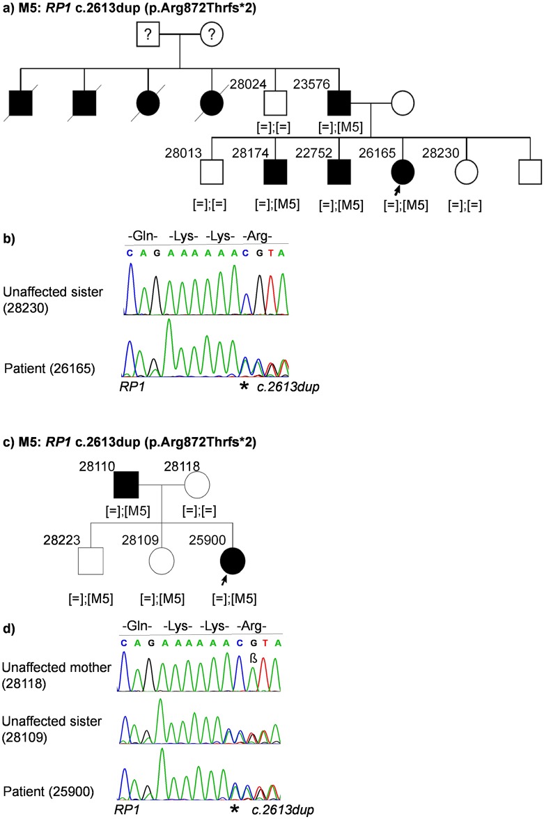
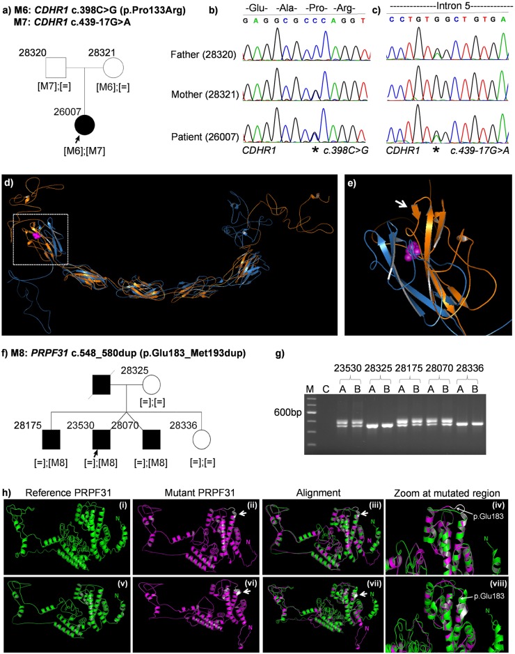
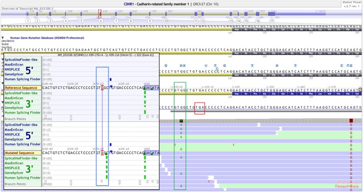
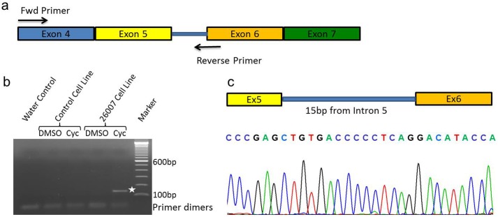
Inherited retinal dystrophies (IRDs) are Mendelian diseases with tremendous genetic and phenotypic heterogeneity. Identification of the underlying genetic basis of these dystrophies is therefore challenging. In this study we employed whole exome sequencing (WES) in 11 families with IRDs and identified disease-causing variants in 8 of them. Sequence analysis of about 250 IRD-associated genes revealed 3 previously reported disease-associated variants in RHO, BEST1 and RP1. We further identified 5 novel pathogenic variants in RPGRIP1 (p.Ser964Profs*37), PRPF8 (p.Tyr2334Leufs*51), CDHR1 (p.Pro133Arg and c.439-17G>A) and PRPF31 (p.Glu183_Met193dup). In addition to confirming the power of WES in genetic diagnosis of IRDs, we document challenges in data analysis and show cases where the underlying genetic causes of IRDs were missed by WES and required additional techniques. For example, the mutation c.439-17G>A in CDHR1 would be rated unlikely applying the standard WES analysis. Only transcript analysis in patient fibroblasts confirmed the pathogenic nature of this variant that affected splicing of CDHR1 by activating a cryptic splice-acceptor site. In another example, a 33-base pair duplication in PRPF31 missed by WES could be identified only via targeted analysis by Sanger sequencing. We discuss the advantages and challenges of using WES to identify mutations in heterogeneous diseases like IRDs.


Conflict of interest statement

Competing Interests: The authors have declared that no competing interests exist.

Figures

Fig 1
Fig 1. Patient pedigrees and sequence chromatography of identified disease-associated variants.
Affected individuals are indicated with filled symbols and unaffected individuals are indicated with open symbols. Index patients are indicated with an arrow and were subjected to WES. Variants are denoted as M1, M2, M3 and M4 and zygosity of variants are indicated in third brackets below every analyzed family member. Sequence chromatography images of patients and representative family members are shown below the reference sequence. Mutated position on the chromatograph is depicted with an asterix. (a) Family pedigree of patient 27485. (b) Sequence chromatography of identified heterozygous RHO variant in patient (bottom) and unaffected daughter (top). (c) Family pedigree of patient 23880. (d) Sequence chromatography of identified heterozygous PRPF8 variant in the patient (bottom) and unaffected father (top). (e) Family pedigree of patient 27536. (f) Sequence chromatography of identified homozygous RPGRIP1 variant in the patient (bottom), heterozygous unaffected mother (middle) and unaffected sister (top). (g) Family pedigree of patient 24718. (h) Sequence chromatography of identified homozygous BEST1 variant in patient (bottom), unaffected heterozygous father (middle) and unaffected sister (top).
Fig 2
Fig 2. Patient pedigrees and sequence chromatography of identified disease-associated variants.
Variants are denoted as M5. (a) Family pedigree of patient 26165. (b) Sequence chromatography of identified heterozygous RP1 variant in patient (bottom) and unaffected sister (top). (c) Family pedigree of patient 25900. (d) Sequence chromatography of identified heterozygous RP1 variant in patient (bottom), unaffected sister carrying the variant (middle) and unaffected mother (top). β: A known polymorphism at position c.2618 in RP1 gene with a frequency of 26.98% in Europeans (Source: Exome Aggregation Consortium).
Fig 3
Fig 3. Patient pedigrees and sequence chromatography of identified disease-associated variants.
Variants are denoted as M6, M7 and M8. (a) Family pedigree of patient 26007. (b) Sequence chromatography of heterozygous CDHR1 variant c.398C>G in patient (bottom), carrier mother (middle) and father (top). (c) Sequence chromatography of heterozygous CDHR1 variant c.439-17G>A in patient (bottom), mother (middle) and carrier father (top). (d) Predicted structure of CDHR1 reference protein (in blue) aligned to mutant CDHR1 (p.Pro133Arg) (in orange). The mutation is shown by magenta spheres and is localized within the first cadherin domain (white rectangle). (e) A zoomed image of the first cadherin domain of CDHR1 shows an additional beta-sheet (white arrow) close to the mutation (f) Family pedigree of patient 23530. (g) Agarose gel image of PRPF31 exon 7 PCR shows a larger band only in affected members indicating a duplication. C = Water control in PCR. (h) Comparison of predicted models of the PRPF31 reference protein sequence (i & v, in green), mutant PRPF31 (ii & vi, in magenta), alignment of reference and mutant PRPF31(iii & viii) and zoomed image of the alignment at the mutation site (iv & viii). An additional turn of the mutant in the coiled-coil domain is depicted in white. The first amino acid of the 11bp duplication is shown by a white arrow. “N” denotes the N-terminus of the protein.
Fig 4
Fig 4. CDHR1 intronic variant.
Snapshot of Alamut visual showing an intronic variant (c.439-17G>A) in patient 26007 (green rectangle). The bam alignment file clearly shows a heterozygous variant 17bp upstream from exon 6. Inset: Snapshot of splicing prediction algorithms (in silico) shows a strong cryptic splice acceptor gain at the site of the variant (blue rectangle). Values are comparable to that of canonical splice acceptor site. Red rectangle shows a stop codon in-frame to the cryptic splice activator site.
Fig 5
Fig 5. Analysis of alternative splicing in vitro.
(a) Primer design to capture putative intron retention due to c.439-17G>A variant in CDHR1. (b) Agarose gel electrophoresis of RT-PCR showing a 140bp PCR product only in the patient cell-line (white asterix). No products were seen in control cell line or water control. (c) Bottom: Sequence chromatograph shows a clear retention of 15bp from intron 5 in the RT-PCR product, generated due to the cryptic splice-site activation. Top: Schematic representing the exon-intron boundaries as observed in the RT-PCR product.
Fig 6
Fig 6. Pedigrees of families where underlying genetic mutations were not identified.
(a) Family pedigree of patient 22538 diagnosed with CORD. (b) Family pedigree of patient 26309 diagnosed with RP. (c) Family pedigree of patient 23609 diagnosed with CORD.

References

    1. Berger W, Kloeckener-Gruissem B, Neidhardt J. The molecular basis of human retinal and vitreoretinal diseases. Progress in retinal and eye research. 2010;29(5):335–75. 10.1016/j.preteyeres.2010.03.004 . - DOI - PubMed
    1. Ellingford JM, Barton S, Bhaskar S, O'Sullivan J, Williams SG, Lamb JA, et al. Molecular findings from 537 individuals with inherited retinal disease. Journal of medical genetics. 2016. 10.1136/jmedgenet-2016-103837 . - DOI - PMC - PubMed
    1. Corton M, Nishiguchi KM, Avila-Fernandez A, Nikopoulos K, Riveiro-Alvarez R, Tatu SD, et al. Exome sequencing of index patients with retinal dystrophies as a tool for molecular diagnosis. PloS one. 2013;8(6):e65574 10.1371/journal.pone.0065574 - DOI - PMC - PubMed
    1. Huang L, Xiao X, Li S, Jia X, Wang P, Sun W, et al. Molecular genetics of cone-rod dystrophy in Chinese patients: New data from 61 probands and mutation overview of 163 probands. Experimental eye research. 2016;146:252–8. 10.1016/j.exer.2016.03.015 . - DOI - PubMed
    1. Li H, Durbin R. Fast and accurate short read alignment with Burrows-Wheeler transform. Bioinformatics. 2009;25(14):1754–60. 10.1093/bioinformatics/btp324 - DOI - PMC - PubMed

LinkOut - more resources