. 2016 Jul 8;11(7):e0159029.
doi: 10.1371/journal.pone.0159029. eCollection 2016.

Deep Whole-Genome Sequencing to Detect Mixed Infection of Mycobacterium tuberculosis

Affiliations

Deep Whole-Genome Sequencing to Detect Mixed Infection of Mycobacterium tuberculosis

Mingyu Gan et al. PLoS One. .

Abstract

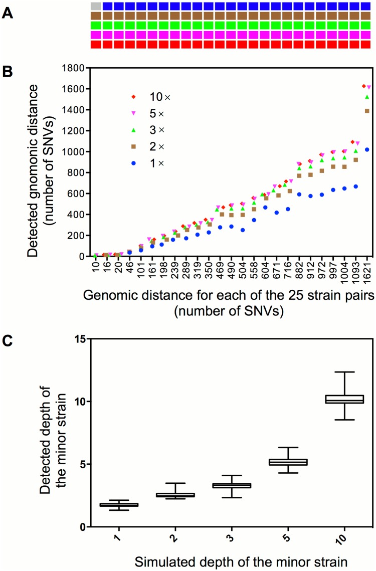
Mixed infection by multiple Mycobacterium tuberculosis (MTB) strains is associated with poor treatment outcome of tuberculosis (TB). Traditional genotyping methods have been used to detect mixed infections of MTB, however, their sensitivity and resolution are limited. Deep whole-genome sequencing (WGS) has been proved highly sensitive and discriminative for studying population heterogeneity of MTB. Here, we developed a phylogenetic-based method to detect MTB mixed infections using WGS data. We collected published WGS data of 782 global MTB strains from public database. We called homogeneous and heterogeneous single nucleotide variations (SNVs) of individual strains by mapping short reads to the ancestral MTB reference genome. We constructed a phylogenomic database based on 68,639 homogeneous SNVs of 652 MTB strains. Mixed infections were determined if multiple evolutionary paths were identified by mapping the SNVs of individual samples to the phylogenomic database. By simulation, our method could specifically detect mixed infections when the sequencing depth of minor strains was as low as 1× coverage, and when the genomic distance of two mixed strains was as small as 16 SNVs. By applying our methods to all 782 samples, we detected 47 mixed infections and 45 of them were caused by locally endemic strains. The results indicate that our method is highly sensitive and discriminative for identifying mixed infections from deep WGS data of MTB isolates.


Conflict of interest statement

Competing Interests: The authors have declared that no competing interests exist.

Figures

Fig 1
Fig 1. Principle of the phylogenomic-database based detection of mixed infection.
(A) WGS reads of a mixed infection by strain A and B were mapped to the ANC0 genome to call SNVs that include common SNVs (red color) shared by both strains and strain specific SNVs (green and blue color). (B) The evolutionary paths of the two strains were determined by mapping SNVs to the reference phylogenomic database. These two paths share a common segment (mapped by common SNVs) from ANC0 to ANC1 and diverged into two separated segments (mapped by strain specific SNVs) after ANC1.
Fig 2
Fig 2. The phylogenomic database of global MTB.
(A) Maximum-likelihood (ML) phylogeny of 652 global MTB strains based on the concatenated alignment of 68,639 genomic SNVs. The colors represent seven MTBC lineages. (B) Schematic diagram illustrating the components of the phylogenomic database. The nodes of the ML phylogeny (represented by a sub-branch outlined in red in panel A) were numbered to record the branching order. A branch was determined by its mother and descendant nodes. The SNVs and evolutionary route of the each branch were recorded.
Fig 3
Fig 3. Detection of mixed infections from artificial WGS reads.
(A) The cells represent 125 synthetic samples of mixed infection. Samples in the same column represent five different levels of mixed infections (depth from top to bottom is 1×, 2×, 3×, 5× or 10× for the minor strain) that are synthesized from the same pair of strains whose genomic distances are in correspondence with the X-axis of panel B. The gray cell indicates no detection of mixed infection and the cells in other five colors indicate detection of mixed infections in different mixed levels. (B) The detected genomic distance (defines as number of SNVs) between the mixed strains in each synthetic sample. (C) The estimated depth of the minor strain under the five simulated depths.

References

    1. (WHO) WHO. Global tuberculosis report 2015. World Health Organization, Geneva, Switzerland 2015; Report WHO/HTM/TB/2015.22.
    1. Theisen A, Reichel C, Rusch-Gerdes S, Haas WH, Rockstroh JK, Spengler U, et al. Mixed-strain infection with a drug-sensitive and multidrug-resistant strain of Mycobacterium tuberculosis. Lancet. 1995;345(8963):1512 . - PubMed
    1. Chaves F, Dronda F, Alonso-Sanz M, Noriega AR. Evidence of exogenous reinfection and mixed infection with more than one strain of Mycobacterium tuberculosis among Spanish HIV-infected inmates. AIDS. 1999;13(5):615–20. Epub 1999/04/15. . - PubMed
    1. Warren RM, Victor TC, Streicher EM, Richardson M, Beyers N, Gey van Pittius N, et al. Patients with active tuberculosis often have different strains in the same sputum specimen. Am J Respir Crit Care Med. 2004;169(5):610–4. 10.1164/Rccm.200305-714oc. WOS:000189249300015. - DOI - PubMed
    1. van Rie A, Victor TC, Richardson M, Johnson R, van der Spuy GD, Murray EJ, et al. Reinfection and mixed infection cause changing Mycobacterium tuberculosis drug-resistance patterns. Am J Respir Crit Care Med. 2005;172(5):636–42. 10.1164/rccm.200503-449OC - DOI - PMC - PubMed