. 2016 Nov 1;23(11):1765-1777.
doi: 10.1038/cdd.2016.65. Epub 2016 Jul 8.

Impaired DNA double-strand break repair contributes to the age-associated rise of genomic instability in humans

Affiliations

Impaired DNA double-strand break repair contributes to the age-associated rise of genomic instability in humans

Z Li et al. Cell Death Differ. .

Abstract

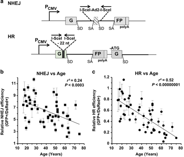
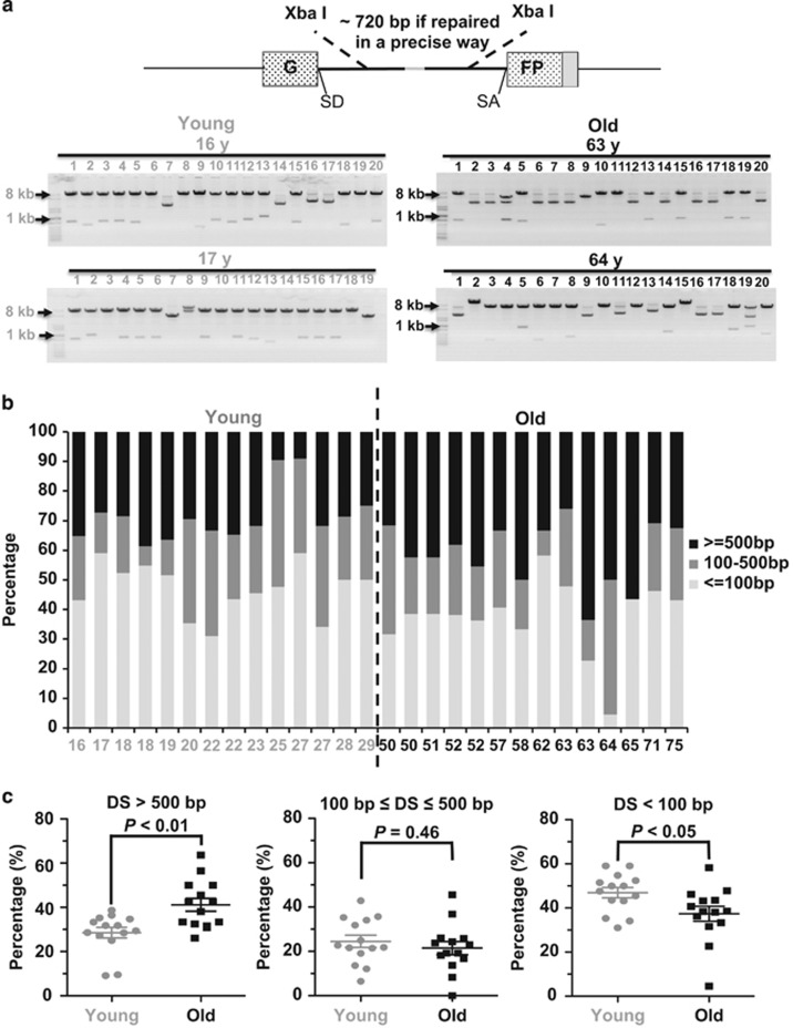
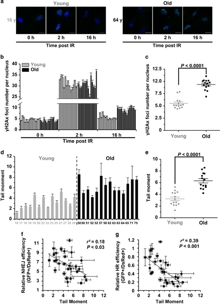
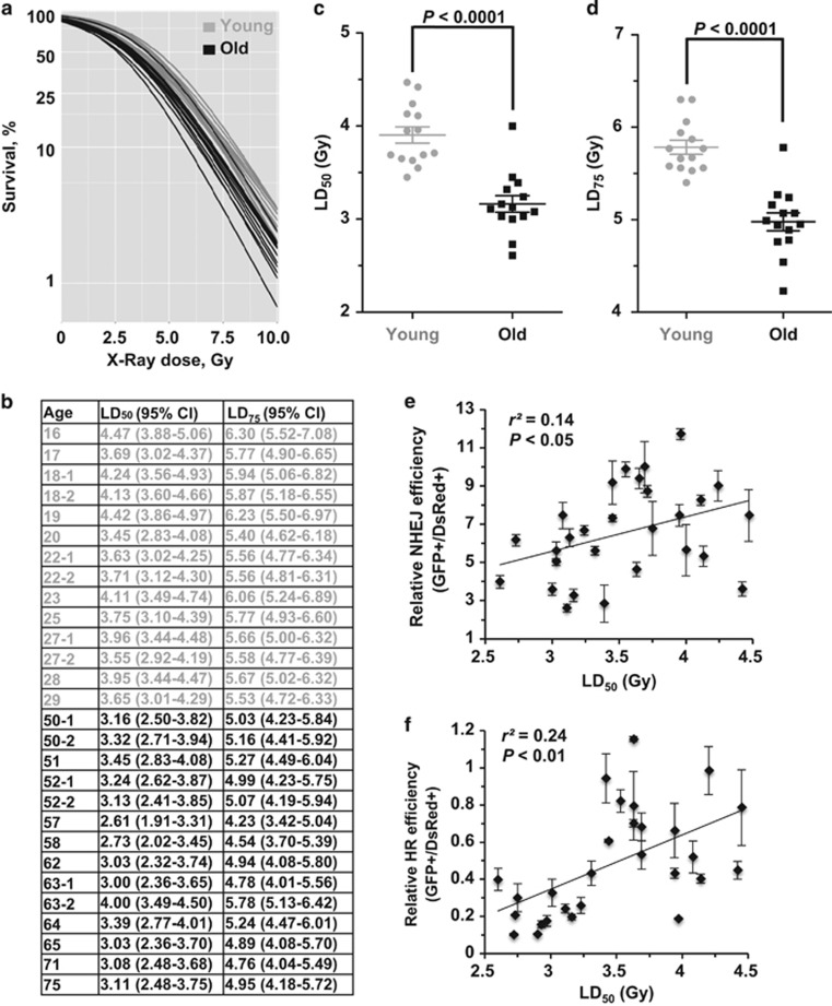
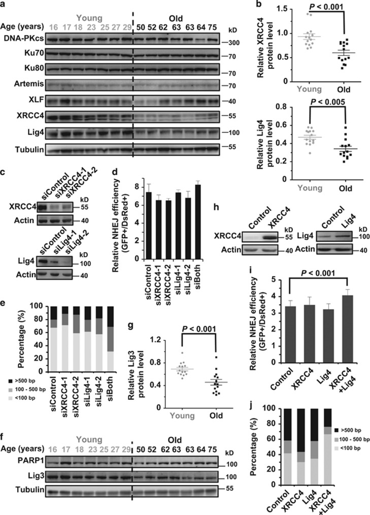
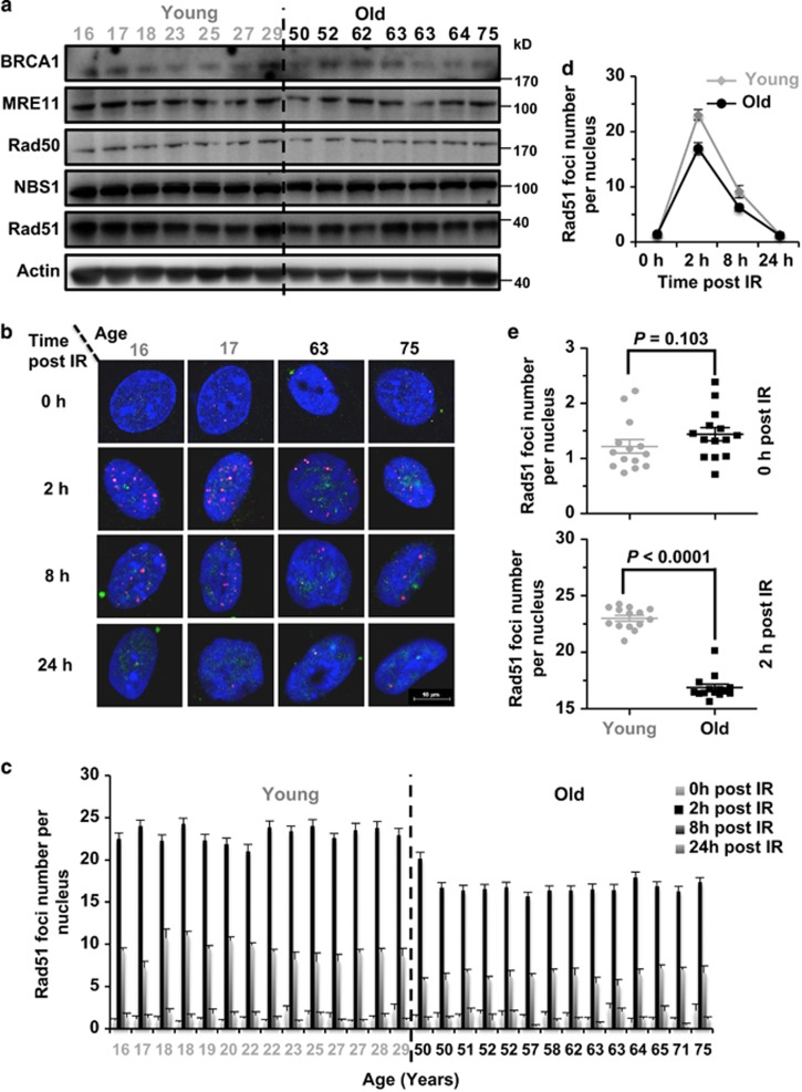
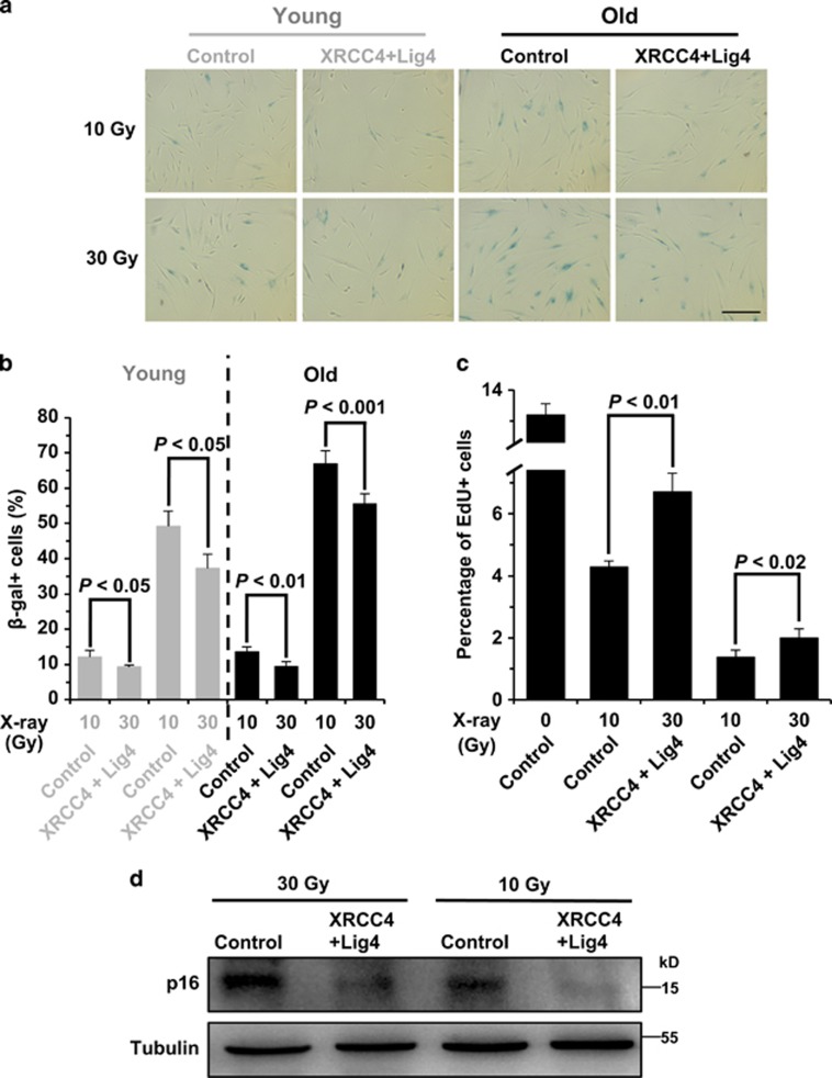
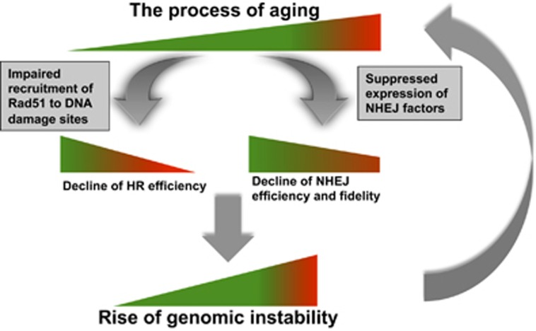
Failing to repair DNA double-strand breaks by either nonhomologous end joining (NHEJ) or homologous recombination (HR) poses a threat to genome integrity, and may have roles in the onset of aging and age-related diseases. Recent work indicates an age-related decrease of NHEJ efficiency in mouse models, but whether NHEJ and HR change with age in humans and the underlying mechanisms of such a change remain uncharacterized. Here, using 50 eyelid fibroblast cell lines isolated from healthy donors at the age of 16-75 years, we demonstrate that the efficiency and fidelity of NHEJ, and the efficiency of HR decline with age, leading to increased IR sensitivity in cells isolated from old donors. Mechanistic analysis suggests that decreased expression of XRCC4, Lig4 and Lig3 drives the observed, age-associated decline of NHEJ efficiency and fidelity. Restoration of XRCC4 and Lig4 significantly promotes the fidelity and efficiency of NHEJ in aged fibroblasts. In contrast, essential HR-related factors, such as Rad51, do not change in expression level with age, but Rad51 exhibits a slow kinetics of recruitment to DNA damage sites in aged fibroblasts. Further rescue experiments indicate that restoration of XRCC4 and Lig4 may suppress the onset of stress-induced premature cellular senescence, suggesting that improving NHEJ efficiency and fidelity by targeting the NHEJ pathway holds great potential to delay aging and mitigate aging-related pathologies.


Figures

Figure 1
Figure 1
The efficiency of DNA DSB repair by both NHEJ and HR declines significantly with age. (a) Reporter substrates for analysis of NHEJ and HR efficiency. Both reporter cassettes were constructed on the basis of EGFP. The NHEJ construct is composed of one copy of EGFP with a Pem1 intron and an Ad2 exon, which inactivates EGFP expression at the transcriptional level. DNA DSBs can be induced by the I-SceI restriction enzyme, which cuts the two I-SceI recognition sites in an opposite orientation flanking the Ad2 exon. Successful NHEJ restores the EGFP expression, which may be scored on FACS. The HR construct contains two copies of an inactivated EGFP-Pem1. In the first copy, the first exon of GFP was engineered with a 22-nt deletion and two inverted I-SceI recognition sites. The second copy lacks the ATG start codon and the second exon of GFP. After a DNA DSB is induced on the HR reporter, only gene conversion, which predominates HR pathway, restores EGFP activity. (b) A significantly negative correlation between NHEJ efficiency and age. Linearized NHEJ construct by I-SceI digestion at an amount of 0.2 μg together with 0.005 μg pDsRed2-N1 for normalizing transfection efficiency was electroporated into 3 × 10⁵ cells at exponentially growth stage. Cells were collected for the analysis on FACS at 72 h post transfection. Error bars, S.D. (c) A significantly negative correlation between HR efficiency and age. Similar to the analysis of NHEJ efficiency, 0.3 μg HR construct linearized by I-SceI and 0.005 μg pDsRed2-N1 was co-transfected to the rapidly growing cells. And the following analysis is identical to that of NHEJ. All the experiments were repeated 3–12 times. Error bars, S.D. SA, splicing acceptor; SD, splicing donor
Figure 2
Figure 2
NHEJ becomes more error-prone during aging. (a) A depiction of NHEJ fidelity analysis. At 72 h post transfection of 0.6 μg of linearized NHEJ construct into 106 cells, cells were collected for plasmid rescue. The rescued NHEJ plasmids were isolated and digested with XbaI restriction enzyme. Two XbaI recognition sites sit downstream and upstream of break sites, so the size of DNA bands after XbaI digestion would reflect the relative fidelity of NHEJ. Twenty rescued plasmids were isolated and digested with XbaI. Two representative gel pictures from two cell lines from each group are shown. (b) The frequency of acquiring different sizes of deletions in the 28 cell lines. For each cell line, at least 20 clones with deletions were sequenced at the junction areas. (c) The old group of cells are more likely to have large deletions but less prone to have small deletions than the young group of cells. The Mann–Whitney U (MWU) test indicates that the differences are significant. DS, deletion sizes
Figure 3
Figure 3
Age-related decline of DNA DSB repair efficiency strongly correlates with genomic instability. (a) Representative pictures of γH2Ax foci at different time points post IR (X-ray, 8 Gy) in one young and old cell line. Scale bars, 10 μm. (b) Quantification of γH2Ax foci numbers at the three time points in the 28 cell lines. At least 50 cells were counted for each time points. Error bars, S.E.M. (c) Statistical analysis (MWU test) reveals that the old group of cells have a significantly higher number of γH2Ax foci at 16 h post IR than the young group of cells. (d) Comparison of genomic instability measured by neutral comet assay between the two groups. The 28 cell lines were analyzed at PD 16–19 using a kit from Trevigen. For each cell line, the tail moments of at least 50 cells were quantified using the Cometscore software (Sumerduck, VA, USA). Error bars, S.E.M. (e) MWU test reveals that the old group of cells have significantly higher tail moments than the young group. (f) There is a significant negative correlation between NHEJ efficiency and tail moment. Error bars of tail moment, S.E.M. Error bars of NHEJ efficiency, S.D. (g) There is a significant negative correlation between HR efficiency and tail moment. Error bars of tail moment, S.E.M. Error bars of HR efficiency, S.D.
Figure 4
Figure 4
Age-related decline of DNA DSB repair strongly correlates with radiosensitivity. (a) Young and old cells were irradiated with increasing doses of X-ray. Survival was calculated as the relative plating efficiencies of irradiated versus control, unirradiated cells. Logistic regression was performed separately on the 28 samples to estimate the cell's sensitivity to IR. The experiment was performed in triplicate for each sample. (b) Sensitivity of eyelid fibroblasts to X-ray (Gy). The lethal dose that kills 50 and 75% of the population of a test sample's cells were calculated by the logistic regression. (c,d) The MWU test indicates that the young group of cells has a statistically significant higher LD50 and LD75 than that of the old cells. (e) NHEJ efficiency positively correlates with LD50. (f) HR efficiency positively correlates with LD50
Figure 5
Figure 5
Age-related reduction of XRCC4, Lig4 and Lig3 expression causes the decline of NHEJ capacity. (a) Western blot analysis of c-NHEJ factors in the two groups of cells. Cells were collected for the whole-cell lysate extraction and western blot analysis at 48 h post splitting. (b) Statistical comparison of XRCC4 and Lig4 expression between the two groups. Western blot results were further analyzed with ImageJ software (Bethesda, MD, USA) for quantification. (c) XRCC4 and Lig4 were successfully depleted in the 17-year-old cell line using siRNA transfections. Cells were transfected with siRNA twice with two days interval. On day 3 post the second siRNA transfection, cells were collected for protein extraction and western blot analysis. (d) No significant effect on NHEJ efficiency was observed with the two proteins depleted. On day 2 post the second siRNA transfection, 3 × 10⁵ cells were transfected with 0.2 μg I-SceI linearized NHEJ reporter and 0.005 μg pDsRed2-N1. At 72 h post transfection, cells were collected for the FACS analysis. Error bars, S.D. (e) Depleting both proteins using siRNA causes a reduction of NHEJ fidelity. Similar to the analysis in (d), cells depleted with the indicated proteins using siRNA were further transfected with 0.2 μg I-SceI linearized NHEJ reporter, followed by DNA extraction and plasmid rescue. The repaired NHEJ constructs were further sequenced at the junction area. At least 20 clones with deletions were sequenced and quantified. (f) Western blot analysis of alt-NHEJ factors in the two groups of cells. (g) Statistical comparison of Lig3 expression between the two groups. (h) Western blot analysis of XRCC4 and Lig4 overexpression in the 63-year-old cell line. Cells at the amount of 1 × 10⁶ were transfected with 5 μg XRCC4 or Lig4 expression vector. At 24 h post transfection, cells were collected for protein extraction and western blot analysis. (i) Simultaneous overexpression of XRCC4 and Lig4 significantly elevates NHEJ efficiency. Together with 5 μg the control vector, 2.5 μg XRCC4+2.5 μg control vector, 2.5 μg Lig4+2.5 μg control vector or 2.5 μg XRCC4+2.5 μg Lig4 vector, cells were transfected with 5 μg I-SceI vector and 0.015 μg pDsRed2-N1 vector. At 72 h cells were collected for analysis of NHEJ efficiency. All experiments were repeated at least six times. Error bars, S.D. (j) Overexpression of both XRCC4 and Lig4 rescues the decline of NHEJ fidelity in the 63-year-old cell line. Similar to the experiments performed in (i), at 72 h post co-transfection, cells were collected for DNA extraction and plasmid rescue. For each group, at least 20 clones with deletions were sequenced and further analyzed
Figure 6
Figure 6
The recruitment of Rad51 to DNA damage sites is impaired in the old group of cells. (a) Western blot analysis of major HR factors in the 28 cell lines. Cells were pre-seeded and grown for 48 h before being collected for the protein extraction and the western blot analysis. (b) Representative pictures of Rad51 foci at 0, 8 and 24 h post IR in two young and two old cell lines. At 48 h post splitting, cells were treated with X-ray at a dosage of 8 Gy. At different time points post the treatment, cells were fixed and co-stained with Geminin and Rad51 antibodies. Pictures were taken on a confocal microscope. Scale bars, 10 μm. (c) Quantification of Rad51 foci post IR in the 28 cell lines. At least 50 Geminin-positive cells were counted on the fluorescence microscope. Error bars, S.E.M. (d) The kinetics of Rad51 recruitment to DNA damage sites. Each point represents the average number of Rad51 foci per nucleus for each of the 14 cell lines at different time points post IR. Error bars, S.D. (e) Statistical comparison of Rad51 foci number at 0 h post IR between the two groups of cells. MWU test was performed to compare the difference. (f) Statistical comparison of Rad51 foci number at 2 h post IR between the two groups of cells. MWU test was performed to compare the difference
Figure 7
Figure 7
Overexpression of both XRCC4 and Lig4 suppresses the onset of SIPS. (a) Representative pictures of β-gal staining on 10 or 30 Gy X-ray-treated 17-year-old and 63-year-old fibroblasts. Cells were transfected with control vector or XRCC4+Lig4 expression vectors, and irradiated at indicated dosages, then kept in the incubator for 10 days before being stained. Scale bars, 100 μm. (b) Comparison of β-gal+ cell numbers between the control and XRCC4+Lig4 overexpressing cells. The experiments were repeated 3–6 times and a t-test was performed to compare the difference. Error bars, S.D. (c) Analysis of cell division rates on 10 or 30 Gy X-ray-treated 63-year-old fibroblasts using the EdU incorporation assay. (d) Western blot analysis of p16 expression in irradiated cells with control and XRCC4+Lig4 vectors transfected. On day 10 post IR, cells were collected for protein extraction and western blot analysis
Figure 8
Figure 8
Model for aging, change of DNA DSB repair and the rise of genomic instability. During the process of aging, the accumulation of mutations gradually impairs the recruitment of essential HR factors to DNA damage sites, and the expression of NHEJ factors, causing a decline o DNA DSB repair capacity. The age-related decline of HR is much shaper than that of NHEJ, possibly acting as a tumor suppressing mechanism. However, it also forces cells to choose the more error-prone NHEJ repair pathway. Decrease of both pathways eventually introduces more DNA mutations to genomes, leading to age-related to genomic instability, accelerating the process of aging

References

    1. Lopez-Otin C, Blasco MA, Partridge L, Serrano M, Kroemer G. The hallmarks of aging. Cell 2013; 153: 1194–1217. - PMC - PubMed
    1. Tigges J, Krutmann J, Fritsche E, Haendeler J, Schaal H, Fischer JW et al. The hallmarks of fibroblast ageing. Mech Ageing Dev 2014; 138: 26–44. - PubMed
    1. Dolle ME, Giese H, Hopkins CL, Martus HJ, Hausdorff JM, Vijg J. Rapid accumulation of genome rearrangements in liver but not in brain of old mice. Nat Genet 1997; 17: 431–434. - PubMed
    1. Gorbunova V, Seluanov A, Mao Z, Hine C. Changes in DNA repair during aging. Nucleic Acids Res 2007; 35: 7466–7474. - PMC - PubMed
    1. Rube CE, Fricke A, Widmann TA, Furst T, Madry H, Pfreundschuh M et al. Accumulation of DNA damage in hematopoietic stem and progenitor cells during human aging. PLoS ONE 2011; 6: e17487. - PMC - PubMed

Publication types

MeSH terms