. 2016 Sep;46(9):2137-48.
doi: 10.1002/eji.201546171. Epub 2016 Aug 2.

Expression of heavy chain-only antibodies can support B-cell development in light chain knockout chickens

Affiliations

Expression of heavy chain-only antibodies can support B-cell development in light chain knockout chickens

Benjamin Schusser et al. Eur J Immunol. 2016 Sep.

Abstract

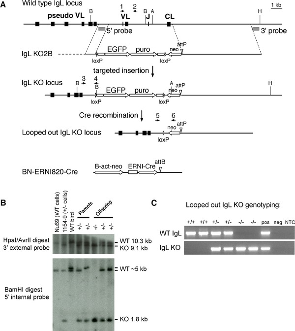
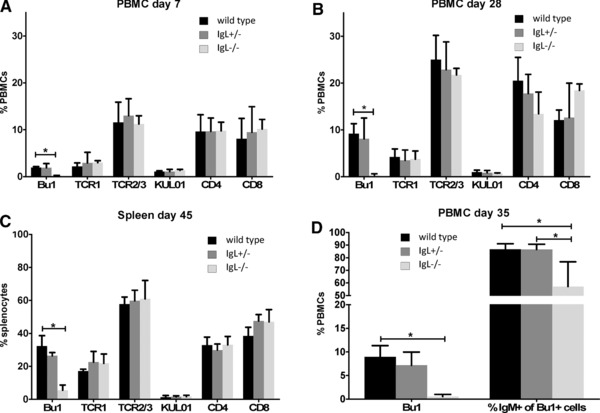
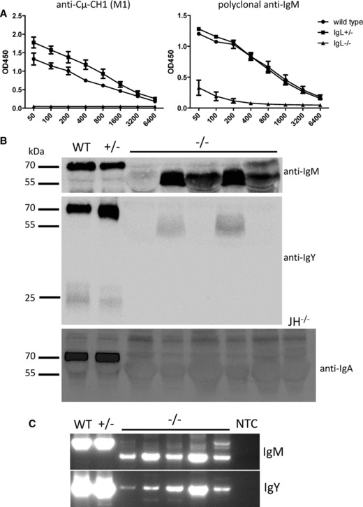
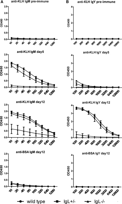
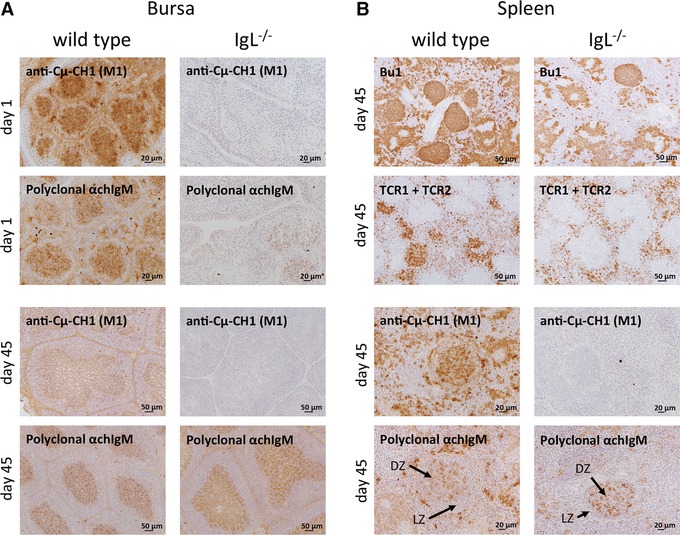
Since the discovery of antibody-producing B cells in chickens six decades ago, chickens have been a model for B-cell development in gut-associated lymphoid tissue species. Here we describe targeting of the immunoglobulin light chain locus by homologous recombination in chicken primordial germ cells (PGCs) and generation of VJCL knockout chickens. In contrast to immunoglobulin heavy chain knockout chickens, which completely lack mature B cells, homozygous light chain knockout (IgL(-/-) ) chickens have a small population of B lineage cells that develop in the bursa and migrate to the periphery. This population of B cells expresses the immunoglobulin heavy chain molecule on the cell surface. Soluble heavy-chain-only IgM and IgY proteins of reduced molecular weight were detectable in plasma in 4-week-old IgL(-/-) chickens, and antigen-specific IgM and IgY heavy chain proteins were produced in response to immunization. Circulating heavy-chain-only IgM showed a deletion of the CH1 domain of the constant region enabling the immunoglobulin heavy chain to be secreted in the absence of the light chain. Our data suggest that the heavy chain by itself is enough to support all the important steps in B-cell development in a gut-associated lymphoid tissue species.

Keywords: Antibodies; B-cell development; Immunoglobulins; Knockout chickens.


Figures

Figure 1
Figure 1
Targeting the IgL locus in chicken PGCs, removal of the selectable marker cassette, and generation of IgL−/− chickens. (A) The chicken IgL locus (top line) is targeted by vector IgL KO2B (second line) with a 5′ homology region upstream of the functional V (VL) and a 3′ homology region downstream of the constant region (CL). The upstream pseudogene array (pseudo VL) is left intact in the targeted allele (third line). The positions of the 5′ and 3′ probes and restriction sites used in the Southern blot in (C) are shown. A, AvrII; B, BamHI; H, HpaI. The structure of the looped out allele obtained after Cre recombination is shown (fourth line), with the remaining promoterless neo and attP site. The ERNI‐Cre construct stably integrated into the IgL KO PGCs is shown at bottom. Scale bar, 1 kb. (B) Southern blot analysis of the original, nonlooped out IgL KO PGCs and germline progeny derived from them. The parental cell line Nu69, IgL KO clone 1154–9, and samples from wild‐type and transgenic animals are shown. Analysis of IgL+/− male and female (parents) and three of their offspring are shown, with the genotypes as indicated. The light chain locus is polymorphic in our birds, and the wild‐type 5′ BamHI fragment was observed to be either 5 or 5.2 kb. (C) Genotyping looped out IgL KO birds. PCR for the wild‐type and knockout alleles was used to determine genotypes of progeny from crosses of IgL+/− males and females. Two representative progeny of each genotype are shown, as indicated. Pos and neg refer to positive and negative genomic controls for the PCR reactions. NTC, no template control.
Figure 2
Figure 2
Bu1+ cells are detectable in blood and spleen of IgL−/− chickens. (A, B) PBMCs of (A) 7‐day, (B) 28‐day‐old chickens, and (C) spleen cells of 45‐day‐old chickens were isolated and stained for B cells using Bu1‐Alexa 647, or stained for gamma/delta T cells (TCR1), alpha/beta T cells (TCR2/3), monocytes/macrophages (KUL01), and CD4+ and CD8+ cells, and detected using goat‐anti‐mouse IgG‐Alexa 647. (D) PBMCs of 35‐day‐old birds were double stained with polyclonal goat‐anti‐chIgM/donkey anti‐goat Alexa 647 and Bu1/goat‐anti‐mouse IgG‐FITC. Stained cells were analyzed by flow cytometry. The left set of bars shows the percentages of Bu1+ cells for the three genotypes, and the right set of bars shows the percentage of Bu1+ cells that are also IgM+. Data are shown as mean and SEM of at least four animals per genotype at each time point, following the same animals over time. Significance was calculated by Kruskal–Wallis test followed by Bonferroni correction. *p ≤ 0.05.
Figure 3
Figure 3
The IgM and IgY heavy chain CH1 domain is deleted in IgL−/‐ chickens. (A) ELISA plates were coated with anti‐chicken‐Cμ‐CH1 (M1) which binds the CH1 domain. Plasma of 28‐day‐old birds was incubated on the plates and captured IgM was detected with polyclonal goat‐anti‐chicken‐IgM‐POD. For each graph, data are shown as mean and SD of at least four birds per genotype. The IgL−/− ELISA from Supporting Information Fig. 5B, using a polyclonal anti‐IgM as capture antibody, is reproduced here for comparison. (B) Plasma samples of one wild‐type (WT), one IgL+/−, and five IgL−/− birds were tested by Western blot for IgM, IgY, and IgA. Serum from a heavy chain knockout (JH−/−) bird is included as a control for background. (C) RNA was isolated from PBMCs of the same 28‐day‐old chickens used in (B) and one‐Step‐RT‐PCR was performed spanning the region from the heavy chain V to the CH2 domain of IgM or IgY. NTC, no template control. (B, C) PCR data shown are from single experiments representative of eight performed.
Figure 4
Figure 4
IgL−/− chickens produce antigen specific immunoglobulin. (A, B) 35‐day‐old chickens were immunized with keyhole limpet hemocyanin (KLH) and boosted 7 days after the initial immunization. Levels of antigen‐specific (A) IgM and (B) IgY were analyzed 5 days and 12 days after immunization. Data are shown as mean and SEM of at least four birds per genotype, following the same animals over time.
Figure 5
Figure 5
Formation of B‐cell follicles in the bursa of IgL−/− chickens. Sections of paraffin‐embedded bursa tissue from 1‐day and 28‐day‐old wild‐type, IgL+/− and IgL−/− chickens were stained with hematoxylin and eosin (H&E). Sections from day 28 were also stained with a chicken B‐cell marker (anti‐chicken Bu1a and Bu1b) and the basement membrane forming the cortico‐medullary junction was stained using a cross‐reactive anti‐human‐desmin antibody. Cortex (C) and medulla (M) are indicated. Antibodies were detected using the Vector ABC Kit followed by the Vector DAB Kit. One representative picture per group and staining is shown. Three bursas per genotype with a minimum of ten sections per organ were analyzed.
Figure 6
Figure 6
A single chain immunoglobulin heavy chain supports B‐cell development in IgL−/− chickens. (A, B) Fresh frozen sections of (A) bursa and (B) spleen from 1‐day and 45‐day‐old wild‐type and IgL−/− chickens were stained for B cells (anti‐chicken‐Bu1 (AV20)), T cells (TCR1 + TCR2), anti‐Cμ‐CH1 domain (M1) and IgM (polyclonal anti‐chIgM). Antibodies were detected using goat‐anti‐mouse‐HRP followed by the Vector DAB Kit. One representative picture per group and staining is shown. Three spleens and bursas per genotype with a minimum of ten sections per organ were analyzed.

References

    1. Schusser, B. , Collarini, E. J. , Yi, H. , Izquierdo, S. M. , Fesler, J. , Pedersen, D. , Klasing, K. C. et al., Immunoglobulin knockout chickens via efficient homologous recombination in primordial germ cells. Proc. Natl. Acad. Sci. U. S. A. 2013. 110: 20170–20175. - PMC - PubMed
    1. Kitamura, D. , Roes, J. , Kuhn, R. and Rajewsky, K. , A B cell‐deficient mouse by targeted disruption of the membrane exon of the immunoglobulin mu chain gene. Nature 1991. 350: 423–426. - PubMed
    1. Feige, M. J. , Groscurth, S. , Marcinowski, M. , Shimizu, Y. , Kessler, H. , Hendershot, L. M. and Buchner, J. , An unfolded CH1 domain controls the assembly and secretion of IgG antibodies. Mol. Cell 2009. 34: 569–579. - PMC - PubMed
    1. Haas, I. G. and Wabl, M. , Immunoglobulin heavy chain binding protein. Nature 1983. 306: 387–389. - PubMed
    1. Melchers, F. , Karasuyama, H. , Haasner, D. , Bauer, S. , Kudo, A. , Sakaguchi, N. , Jameson. et al., The surrogate light chain in B‐cell development. Immunol. Today 1993. 14: 60–68. - PubMed

Publication types

MeSH terms