. 2016 Jul 6;53(4):1485-97.
doi: 10.3233/JAD-150882.

The Anti-Prion Antibody 15B3 Detects Toxic Amyloid-β Oligomers

Affiliations
Free PMC article

The Anti-Prion Antibody 15B3 Detects Toxic Amyloid-β Oligomers

Matteo Stravalaci et al. J Alzheimers Dis. .
Free PMC article

Abstract

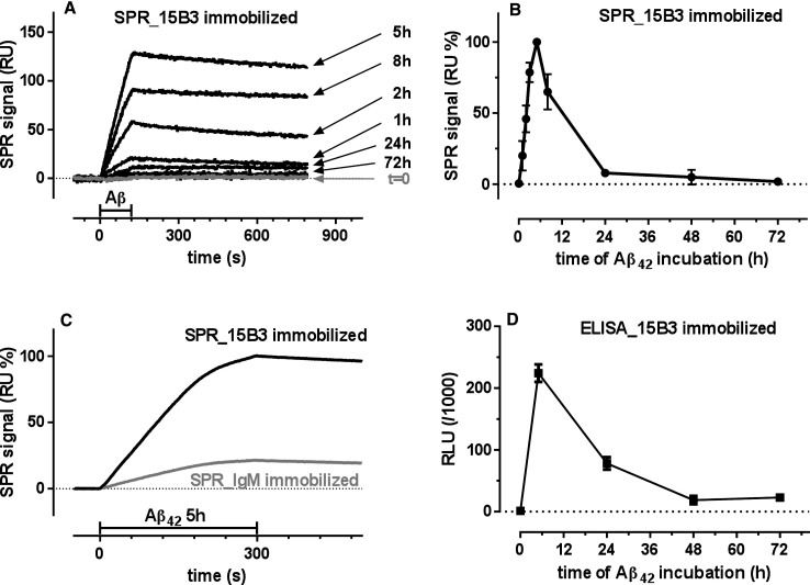
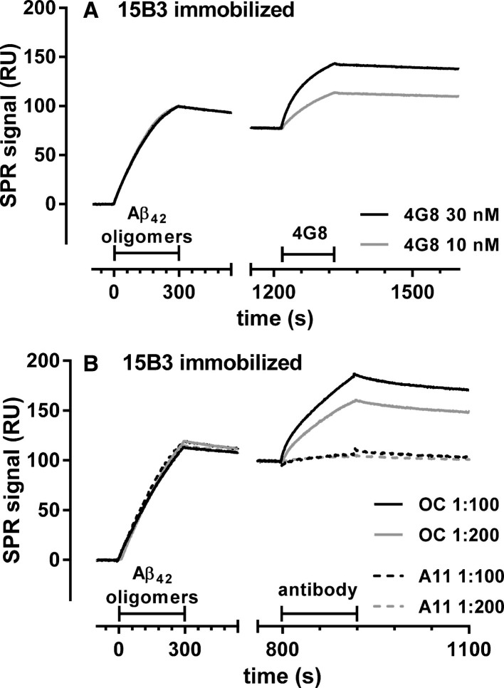
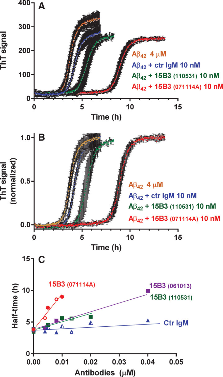
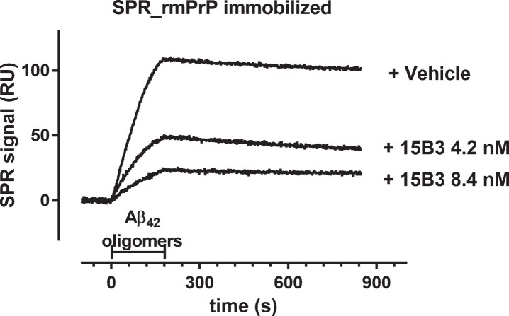
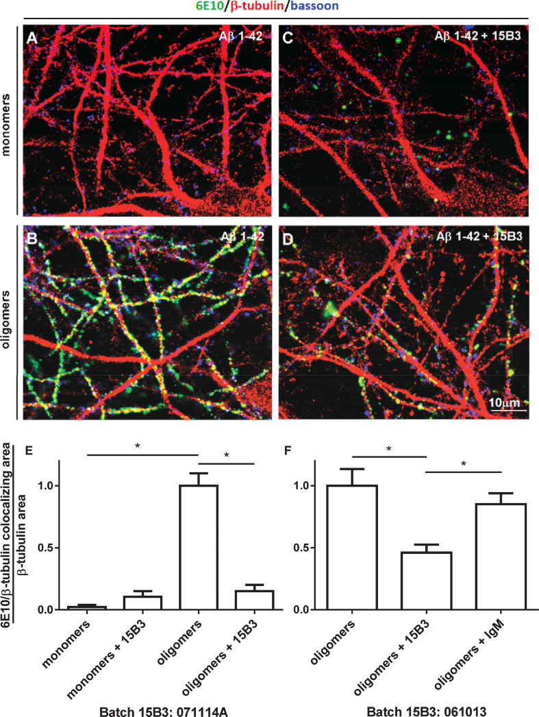
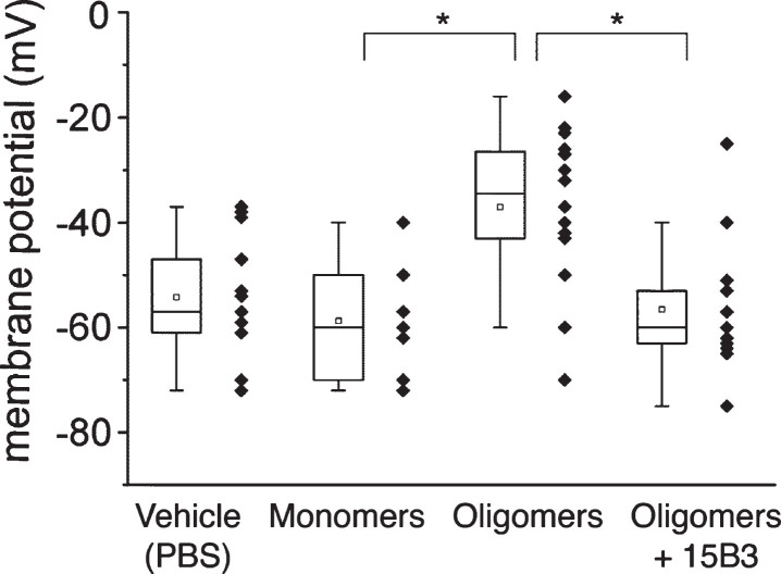
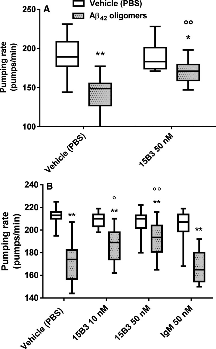
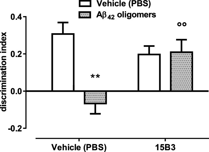
15B3 is a monoclonal IgM antibody that selectively detects pathological aggregates of the prion protein (PrP). We report the unexpected finding that 15B3 also recognizes oligomeric but not monomeric forms of amyloid-β (Aβ)42, an aggregating peptide implicated in the pathogenesis of Alzheimer's disease (AD). The 15B3 antibody: i) inhibits the binding of synthetic Aβ42 oligomers to recombinant PrP and neuronal membranes; ii) prevents oligomer-induced membrane depolarization; iii) antagonizes the inhibitory effects of oligomers on the physiological pharyngeal contractions of the nematode Caenorhabditis elegans; and iv) counteracts the memory deficits induced by intracerebroventricular injection of Aβ42 oligomers in mice. Thus this antibody binds to pathologically relevant forms of Aβ, and offers a potential research, diagnostic, and therapeutic tool for AD.

Keywords: 15B3 antibody; Alzheimer’s disease; amyloid beta-protein (1– 42); oligomers; prion protein; prions.


Figures

Fig.1
Fig.1
Surface plasmon resonance (SPR) and ELISA studies. Synthetic Aβ42 (100 μM) was incubated at 25°C, and samples were taken at different times (from 0 to 72 h), diluted to 1 μM in 10 mM PBS, pH 7.4, and flowed (SPR) or incubated (ELISA) on immobilized 15B3. A) Sensorgrams (time course of the SPR signal expressed in resonance units, RU) obtained from a representative experiment in which Aβ42 solutions were flowed for 2 min (bar), followed by 11 min of dissociation. B) Effect of the length of incubation of Aβ42 on its binding to 15B3 immobilized on the SPR sensor chip. Each value is the mean ± SD of three different experiments. C) Sensorgrams obtained flowing the 5-h Aβ42 solution over parallel sensor surfaces on which 15B3 or control IgM had been previously immobilized (immobilization levels respectively 7040 and 7050 RU); this experiment was replicated twice with identical results. D) Effect of the length of incubation of Aβ42 on its binding to 15B3 immobilized on ELISA plates. Mean ± SD of three replicates.
Fig.2
Fig.2
SPR studies showing binding of 4G8 and OC, but not A11, to 15B3-captured Aβ42 oligomers—Synthetic Aβ42 (100 μM) was incubated at 25°C, sampled after 5 h, diluted to 1 μM in 10 mM PBS, pH 7.4, and injected over immobilized 15B3 (batch # 071114A) for 5 min, followed by injection of two different concentrations of 4G8 (A), OC or A11 (B) antibodies for 2 min (bars). The binding of 4G8 to captured oligomers (A) confirms the data shown in Fig. 1D. The experiment shown in B was replicated three times with very similar results.
Fig.3
Fig.3
Effect of 15B3 on Aβ42 fibrillogenesis, evaluated by ThT fluorescence—Synthetic Aβ42 (4 μM) was incubated with ThT (20 μM) with or without 15B3 or control IgM, and ThT fluorescence was monitored every 2.5 min. Three batches of 15B3 were used for these studies, as indicated. A) Representative raw fluorescent values. B) Normalization of the data in A on the corresponding maximal values to illustrate better the shift in the half-time of transition, i.e., the time corresponding to half the maximum ThT signal. C) Half-time of transition of Aβ42 in the presence of different concentrations of the antibodies. Antibodies are identified by the colors; open or solid symbols indicate results of independent experiments.
Fig.4
Fig.4
Effect of 15B3 on the binding of Aβ42 oligomers to recombinant mouse PrP (rmPrP) immobilized on the sensor chip—Synthetic Aβ42 (100 μM) was incubated at 25°C, sampled after 5 h, diluted to 1 μM in 10 mM PBS, pH 7.4, and incubated for another 30 min with or without 15B3 (4.2 and 8.4 nM, lot# 061013). Aliquots were then injected for 3 min (bar) over immobilized rmPrP. The figure shows the sensorgrams (time course of the SPR signal expressed in resonance units, RU) from a representative experiment. This study was replicated twice with very similar results.
Fig.5
Fig.5
Effect of 15B3 on the binding of Aβ42 oligomers to rat hippocampal neurons—A–D) Representative images obtained exposing 12–15 DIV hippocampal neurons for 1 h to solutions containing (A) Aβ42 monomers or (B) Aβ42 oligomers. The final concentration of Aβ42 was 1 μM in both cases. C, D) Neurons exposed to 1 μM Aβ42 monomers or oligomers pre-incubated for 30 min with 15B3 (batch # 071114A, 10 nM). Neurons were washed, fixed with 4% paraformaldehyde and stained using the following antibodies: mouse anti-Aβ, 6E10 (green), rabbit anti-β tubulin (red) and guinea pig anti-Bassoon (blue). E) Corresponding quantification of 6E10 binding to cultured neurons expressed as colocalizing area between 6E10 and β-tubulin, relative to total β-tubulin. Mean ± SEM of 20 fields from two independent experiments, *p < 0.05 Dunn’s test after Kruskal-Wallis One Way Analysis of Variance on Ranks (this statistical analysis was used because the normality test failed). F) Quantification of a third experiment with 15B3 batch # 061013 and control IgM, both 10 nM. Mean ± SEM of 10 fields, *p < 0.05 Holm-Sidak test after One Way Analysis of Variance (used because the normality test passed).
Fig.6
Fig.6
Effect of 15B3 on membrane potential depolarization caused by Aβ42 oligomers—HEK293T cells were perfused with solutions containing 10 μM Aβ42 monomers, 10 μM Aβ42 oligomers, or 10 μM Aβ42 oligomers pre-incubated for 30 min with 10 nM 15B3, batch # 071114A. Control cells were treated with the vehicle. Data are expressed as median and interquartile range, as well as single values (12, 16, 14, and 11 cells for the four groups). *p < 0.05; one-way ANOVA, Tukey’s post hoc test.
Fig.7
Fig.7
Effect of 15B3 on the ability of Aβ42 oligomers to reduce the pharyngeal motility of C. elegans—Synthetic Aβ42 (100 μM) was incubated at 25°C for 5 h, diluted to 10 μM and incubated with 10 nM or 50 nM 15B3 antibody or 50 nM control IgM, or the vehicle (PBS). The solutions were incubated for another 30 min before being given to the worms. Nematodes were fed for 2 h with these solutions, then plated on Nematode Growth Medium plates seeded with OP50 E. coli. The pharyngeal pumping rate was scored 2 h after plating. Data are expressed as minimum to maximum box and whisker plots (10–20 worms/group from one or two independent experiments). Panels A and B show the results with 15B3 batches # 071114A and # 110531, respectively. **p < 0.01 effect of Aβ42 oligomers versus corresponding vehicle; °p < 0.05, °°p < 0.01 effect of 15B3 versus corresponding vehicle, Bonferroni’s test after two-way ANOVA.
Fig.8
Fig.8
42 oligomer-induced memory impairment in mice is prevented by pretreatment with 15B3—Bars show the discrimination index, calculated as follows: (time on novel object – time on familiar object)/(total time on objects). Data are expressed as the mean ± standard error (SE) (n = 5–7). Two-way ANOVA showed a significant interaction between Aβ42 and 15B3 (p < 0.01). **p = 0.01 effect of Aβ42 oligomers versus corresponding vehicle; °°p < 0.01 effect of 15B3 versus corresponding vehicle, post hoc Bonferroni’s test. Batch # 071114A of 15B3 was used for these experiments.

References

    1. Cleary JP, Walsh DM, Hofmeister JJ, Shankar GM, Kuskowski MA, Selkoe DJ, Ashe KH (2005) Natural oligomers of the amyloid-beta protein specifically disrupt cognitive function. Nat Neurosci 8, 79–84. - PubMed
    1. Lambert MP, Barlow AK, Chromy BA, Edwards C, Freed R, Liosatos M, Morgan TE, Rozovsky I, Trommer B, Viola KL, Wals P, Zhang C, Finch CE, Krafft GA, Klein WL (1998) Diffusible, nonfibrillar ligands derived from Abeta1-42 are potent central nervous system neurotoxins. Proc Natl Acad Sci U S A 95, 6448–6453. - PMC - PubMed
    1. Walsh DM (2002) Naturally secreted oligomers of amyloid [beta] protein potently inhibit hippocampal long-term potentiation in vivo. Nature 416, 535–539. - PubMed
    1. Shankar GM, Bloodgood BL, Townsend M, Walsh DM, Selkoe DJ, Sabatini BL (2007) Natural oligomers of the Alzheimer amyloid-beta protein induce reversible synapse loss by modulating an NMDA-type glutamate receptor-dependent signaling pathway. J Neurosci 27, 2866–2875. - PMC - PubMed
    1. Balducci C, Beeg M, Stravalaci M, Bastone A, Sclip A, Biasini E, Tapella L, Colombo L, Manzoni C, Borsello T, Chiesa R, Gobbi M, Salmona M, Forloni G (2010) Synthetic amyloid-beta oligomers impair long-term memory independently of cellular prion protein. Proc Natl Acad Sci U S A 107, 2295–2300. - PMC - PubMed

Publication types

MeSH terms