Case Reports
. 2016 Oct;133(2):129-138.
doi: 10.1007/s10633-016-9552-z. Epub 2016 Jul 8.

Diagnostic methods in ocular argyrosis: case report

Affiliations
Case Reports

Diagnostic methods in ocular argyrosis: case report

Monika Sarnat-Kucharczyk et al. Doc Ophthalmol. 2016 Oct.

Abstract

Purpose: The aim of this report is to present a case of a patient, metal foundry worker, who had been exposed to industrial silver salts for over 20 years. It is well established that chronic exposure to silver compounds can cause accumulation of silver deposits in various tissues. This condition is referred to as argyrosis or argyria, whereas changes related to eye tissues are defined as ocular argyrosis.

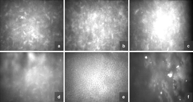
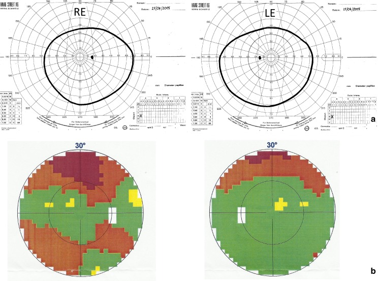
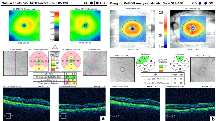
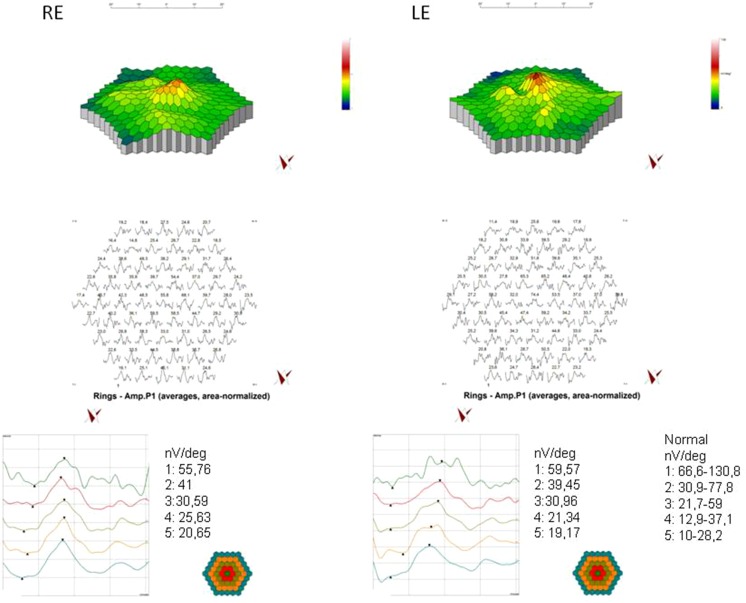
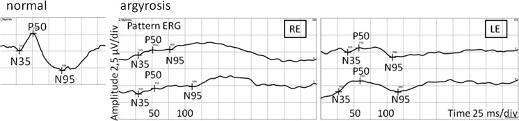
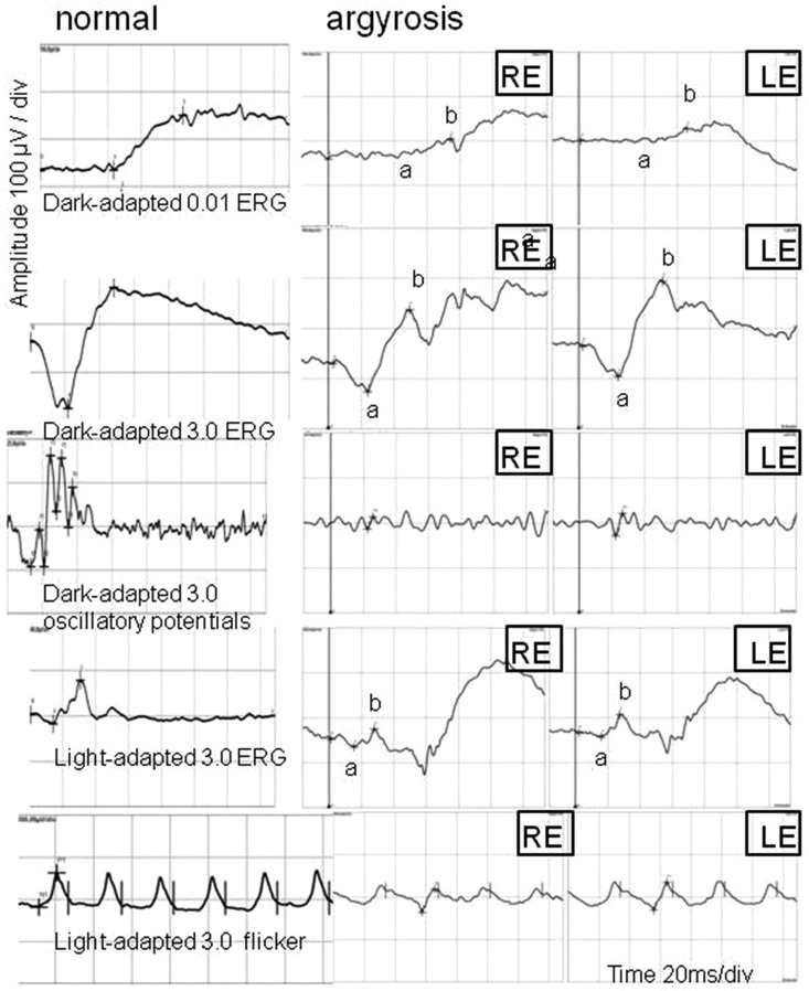
Methods: A complete eye examination, corneal confocal microscopy, kinetic and static visual field test, posterior segment optical coherent tomography, pattern visual evoked potentials (PVEP), flash visual evoked potentials, multifocal electroretinogram, pattern electroretinogram (PERG), full-field electroretinography (FERG) and electrooculogram were all performed.

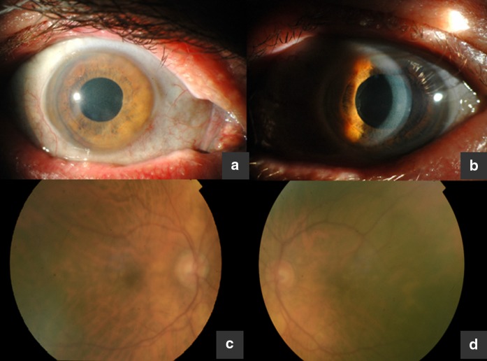
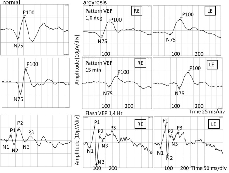
Results: Eye examination revealed decreased visual acuity, corneal deposits and drusenoid changes within the macula. Although electrophysiology tests did not show changes in the function of retinal pigment epithelium, they revealed abnormal function of photoreceptors in the central and peripheral retina. PERG abnormalities and delayed latency of P100 wave in PVEP confirmed impaired function of the inner layers of the retina in the macular region.

Conclusions: Corneal confocal microscopy and electrophysiological tests may help confirm the diagnosis of ocular argyrosis.

Keywords: Argyria; Argyrosis; Ocular argyrosis; Silver nitrate.


Conflict of interest statement

All authors certify that they have no affiliations with or involvement in any organization or entity with any financial interest (such as honoraria; educational grants; participation in speakers’ bureaus; membership, employment, consultancies, stock ownership, or other equity interests; and expert testimony or patent-licensing arrangements), or non-financial interest (such as personal or professional relationships, affiliations, knowledge or beliefs) in the subject matter or materials discussed in this manuscript. Ethical approval All procedures performed in studies involving human participants were in accordance with the ethical standards of the institutional and/or national research committee and with the 1964 Declaration of Helsinki and its later amendments or comparable ethical standards. The study was approved by the Ethics Committee of Medical University of Silesia, Katowice, Poland. Informed consent Informed consent was obtained from all individual participants included in the study. Statement of human rights and statement on the welfare of animals The study protocol was adhered to the tenets of the Declaration of Helsinki. 10.1007/s10633-016-9552-z

Figures

Fig. 1
Fig. 1
a Conjunctival discoloration, grayish-black pigmentation in the area of the medial canthus. b Silver deposits scattered in corneal layers, pseudoexfoliative syndrome. c Right eye fundus. d Left eye fundus
Fig. 2
Fig. 2
Corneal confocal microscopy. a, b Highly reflective punctiform deposits in the stroma, hyper-reflective keratocytes, changes in the density and shape of the keratocytes. c, d Confluent, dense deposits e Normal basal layer of corneal epithelium f Obscured view of corneal endothelium
Fig. 3
Fig. 3
a Kinetic visual fields of the right and left eyes. b Static visual fields of the right and left eyes
Fig. 4
Fig. 4
OCT of the right and left eyes. a Small retinal pigment detachment secondary to drusenoid changes in the macula. b Ganglion cells complex
Fig. 5
Fig. 5
Pattern and flash VEP. RE right eye; LE left eye. Pattern VEP: P100 amplitude: 1.0° RE = 12.2 µV, LE = 12.4 µV (95 % reference interval: 7.0–9.2 µV); 15 min RE = 8.47 µV, LE = 12.3 µV (95 % reference interval: 6.2–9.4 µV); P100 latency: 1.0° RE = 113 ms, LE = 121 ms (95 % reference interval: 103.2–106.8 ms); 15 min RE = 150 ms, LE = 151 ms (95 % reference interval: 102–117 ms); flash VEP P2 latency RE = LE = 115 ms (95 % reference interval: 109.6–126.2 ms)
Fig. 6
Fig. 6
mfERG of the right and left eyes. RE right eye; LE left eye; P1 amplitude in rings
Fig. 7
Fig. 7
PERG of the right and left eyes. RE right eye; LE left eye; P50 amplitude: RE < 1 µV, LE = 1.58 µV (95 % reference interval: 1.4–2.6 µV); N95 amplitude RE < 1 µV, LE = 1.64 µV (95 % reference interval: 2.2–4.6 µV); P50 implicit time RE = 52 ms, LE = 59 ms (95 % reference interval: 49–54 ms); N95 implicit time RE = 111 ms; LE = 110 ms (95 % reference interval: 93–102 ms)
Fig. 8
Fig. 8
Full-field ERG of the right and left eyes. RE right eye; LE left eye; RE/LE: dark-adapted 0.01 ERG b-wave amplitude 46/27 µV (95 % reference interval: 95–305 µV); implicit time 75/67 ms (95 % reference interval: 67–91 ms); dark-adapted 3.0 ERG a-wave amplitude 56/60 µV (95 % reference interval: 155–356 µV); b-wave amplitude 163/190 µV (95 % reference interval: 290–654 µV); a-wave implicit time 24 ms (95 % reference interval: 14–22 ms); b-wave implicit time 50 ms (95 % reference interval: 33–46 ms); OP2 (oscillatory potential) amplitude 7/12 µV (95 % reference interval: 21–46 µV); OP2 implicit time 26 ms (95 % reference interval: 22–24 ms); light-adapted 3.0 ERG a-wave amplitude 17/9 µV (95 % reference interval: 26–62 µV); b-wave amplitude 40/50 µV (95 % reference interval: 103–250 µV); a-wave implicit time 16 ms (95 % reference interval: 13–16 ms); b-wave implicit time 28 ms (95 % reference interval: 29–33 ms); light-adapted 3.0 flicker b-wave amplitude 55/65 µV (95 % reference interval: 57–223 µV)

References

    1. Pala G, Fronterre A, Scafa F, et al. Ocular argyrosis in a silver craftsman. J Occup Health. 2008;50(6):521–524. doi: 10.1539/joh.N8001. - DOI - PubMed
    1. Wadhera A, Fung M. Systemic argyria associated with ingestion of colloidal silver. Dermatol Online J. 2005;11(1):12. - PubMed
    1. Drake PL, Hazelwood KJ. Exposure-related health effects of silver and silver compounds: a review. Ann Occup Hyg. 2005;49(7):575–585. doi: 10.1093/annhyg/mei019. - DOI - PubMed
    1. Stafeeva K, Erlanger M, Velez-Montoya R, Olson JL. Ocular argyrosis secondary to long-term ingestion of silver nitrate salts. Clin Ophthalmol. 2012;6:2033–2036. - PMC - PubMed
    1. Gallardo MJ, Randleman JB, Price KM, Johnson DA, Acosta S, Grossniklaus HE, Stulting RD. Ocular argyrosis after long-term self-application of eyelash tint. Am J Ophthalmol. 2006;141(1):198–200. doi: 10.1016/j.ajo.2005.07.054. - DOI - PubMed

Publication types

LinkOut - more resources