. 2016 Sep;132(3):361-75.
doi: 10.1007/s00401-016-1595-4. Epub 2016 Jul 8.

High-fat diet-induced brain region-specific phenotypic spectrum of CNS resident microglia

Affiliations

High-fat diet-induced brain region-specific phenotypic spectrum of CNS resident microglia

Caroline Baufeld et al. Acta Neuropathol. 2016 Sep.

Abstract

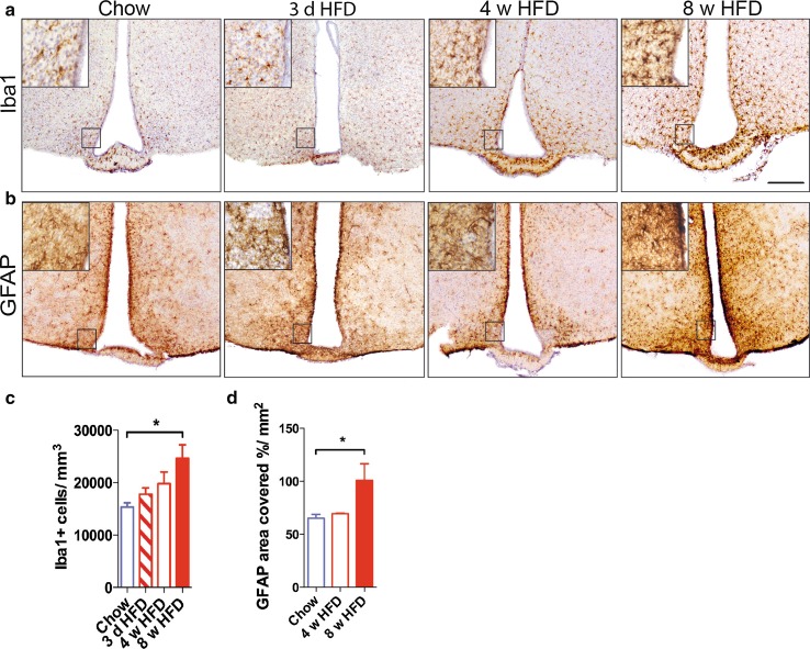
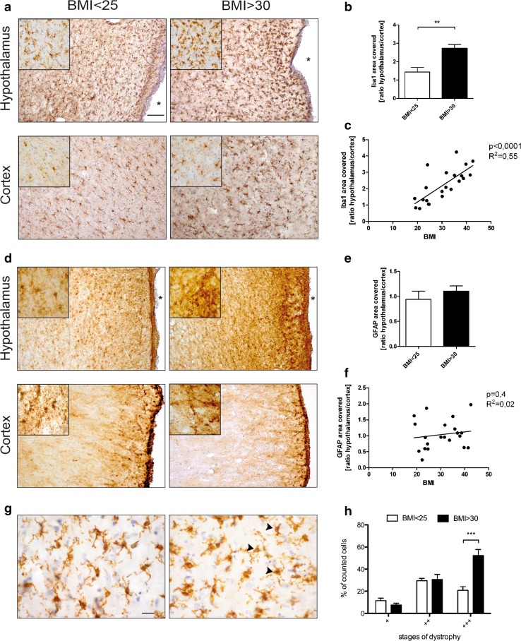
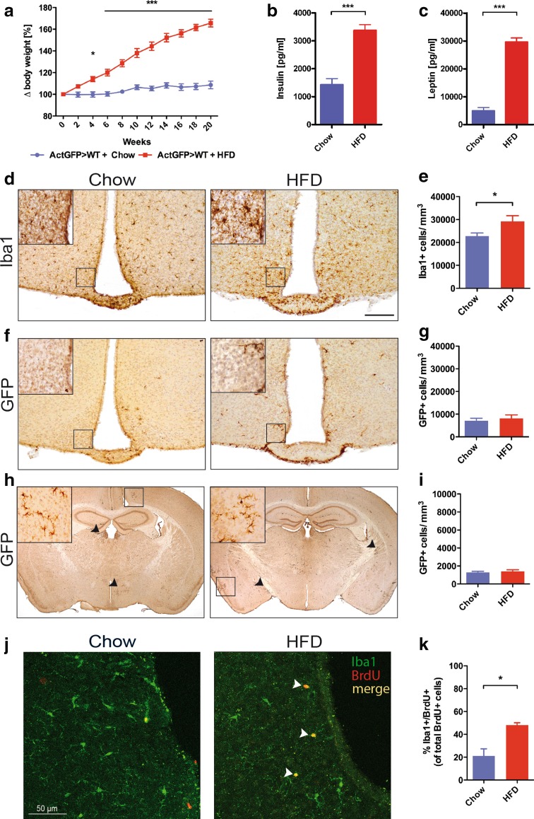
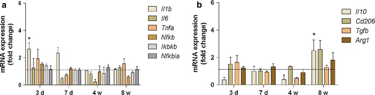
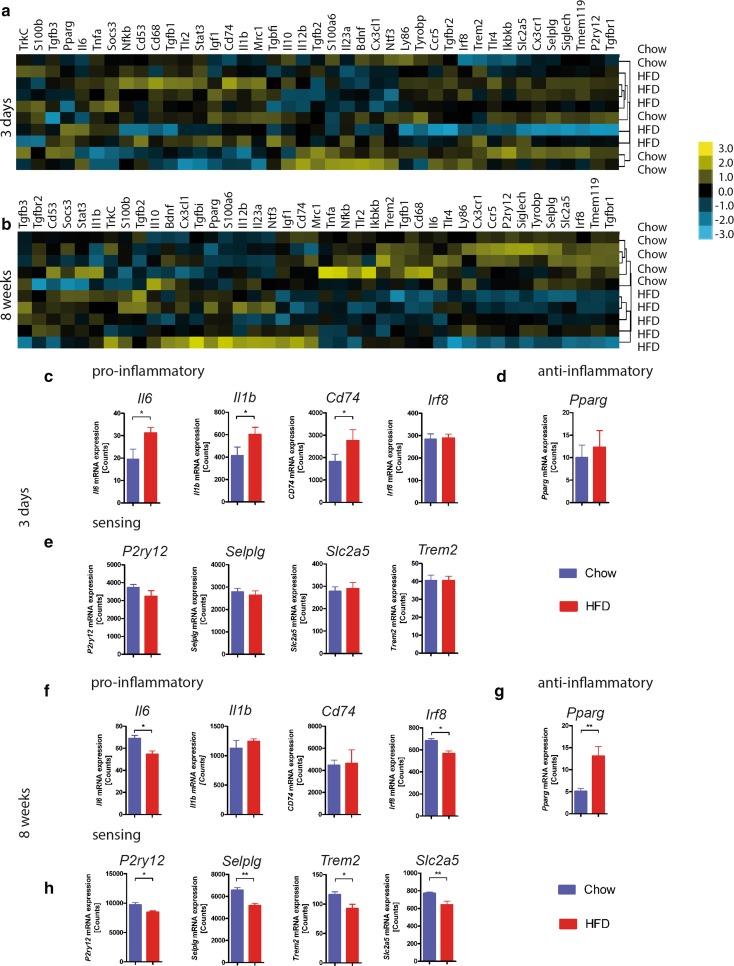
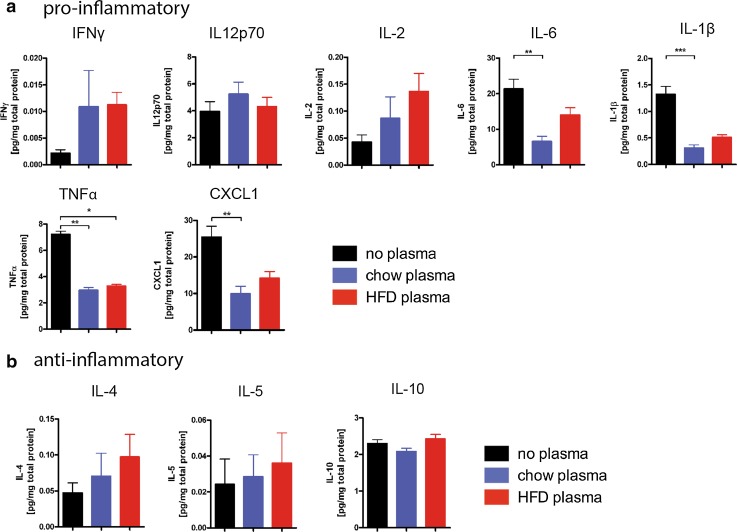
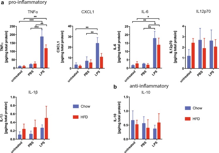
Diets high in fat (HFD) are known to cause an immune response in the periphery as well as the central nervous system. In peripheral adipose tissue, this immune response is primarily mediated by macrophages that are recruited to the tissue. Similarly, reactivity of microglia, the innate immune cells of the brain, has been shown to occur in the hypothalamus of mice fed a high-fat diet. To characterize the nature of the microglial response to diets high in fat in a temporal fashion, we studied the phenotypic spectrum of hypothalamic microglia of mice fed high-fat diet for 3 days and 8 weeks by assessing their tissue reaction and inflammatory signature. While we observed a significant increase in Iba1+ myeloid cells and a reaction of GFAP+ astrocytes in the hypothalamus after 8 weeks of HFD feeding, we found the hypothalamic myeloid cell reaction to be limited to endogenous microglia and not mediated by infiltrating myeloid cells. Moreover, obese humans were found to present with signs of hypothalamic gliosis and exacerbated microglia dystrophy, suggesting a targeted microglia response to diet in humans as well. Notably, the glial reaction occurring in the mouse hypothalamus was not accompanied by an increase in pro-inflammatory cytokines, but rather by an anti-inflammatory reaction. Gene expression analyses of isolated microglia not only confirmed this observation, but also revealed a downregulation of microglia genes important for sensing signals in the microenvironment. Finally, we demonstrate that long-term exposure of microglia to HFD in vivo does not impair the cell's ability to respond to additional stimuli, like lipopolysaccharide. Taken together, our findings support the notion that microglia react to diets high in fat in a region-specific manner in rodents as well as in humans; however, this response changes over time as it is not exclusively pro-inflammatory nor does exposure to HFD prime microglia in the hypothalamus.

Keywords: Glia; High-fat diet; Metabolism; Microglia.


Figures

Fig. 1
Fig. 1
Gliosis in the mouse hypothalamus in response to HFD. a Iba1 and b GFAP immunoreactivity in the hypothalamus of HFD- and chow-fed wild-type mice. c Quantification of Iba1-positive cells in the hypothalamus of HFD- vs. chow-fed wild-type mice. n = 5 (Chow), n = 5 (3 d HFD), n = 3 (4 w HFD), n = 7 (8 w HFD). d Quantification of area covered by GFAP-positive cells in the hypothalamus of HFD- vs. chow-fed wild-type mice. n = 5 (Chow), n = 3 (4 w HFD), n = 3 (8 w HFD). Statistical analyses: *P < 0.05 based on Kruskal–Wallis test with Dunn’s Multiple Comparison Post-Test. Scale bar 200 µm. Data represent mean ± SEM. HFD high-fat diet, d days, w weeks
Fig. 2
Fig. 2
Gliosis in the hypothalamus of human individuals with BMI > 30. a Iba1 immunoreactivity in the hypothalamus (upper panel) and cortex (lower panel) of individuals with BMI < 25 vs. BMI > 30. b Ratio of area covered by Iba1 immunoreactivity in the hypothalamus versus cortex of individuals with BMI < 25 vs. BMI > 30. Scale bar 100 µm. c Correlation of area covered by Iba1 immunoreactivity in the hypothalamus (normalized to the respective individual cortical Iba1 immunoreactivity) to BMI. d GFAP immunoreactivity in the hypothalamus (upper panel) and cortex (lower panel) of individuals with BMI < 25 vs. BMI > 30. Scale bar 100 µm. e Ratio of GFAP-covered area in the hypothalamus versus cortex of individuals with BMI < 25 vs. BMI > 30. f Correlation of area covered by GFAP immunoreactivity in the hypothalamus (normalized to the respective individual cortical GFAP immunoreactivity) to BMI. n = 9 (BMI < 25), 12 (BMI > 30). g Close-up of Iba1 + cells in the hypothalamus of individuals with BMI < 25 (left panel) vs. BMI > 30 (right panel). Arrowheads depict late stages of microglia dystrophy. Scale bar 20 µm. h Quantification of different stages of microglial dystrophy in the hypothalamus of individuals with BMI < 25 and BMI > 30. n = 9 (BMI < 25), 12 (BMI > 30). Statistical analyses: b **p < 0.01 based on unpaired student’s t test. h ***p < 0.001 based on two-way ANOVA with Bonferroni post hoc test; interaction: F (1, 54) = 9.162, p = 0.0038. Data represent mean ± SEM. Asterisk marks the third ventricle. BMI body mass index
Fig. 3
Fig. 3
HFD leads to proliferation of endogenous microglia in the hypothalamus. a Body weight development of Actin-GFP bone marrow chimeric mice fed either HFD or chow for 20 weeks. b Serum insulin and c serum leptin levels of Actin-GFP bone marrow chimeric mice fed either HFD or chow for 20 weeks. d Iba1 immunoreactivity of myeloid cells in the hypothalamus of Actin-GFP bone marrow chimeras fed HFD or chow for 20 weeks. e Quantification of Iba1-positive cells in the hypothalamus of Actin-GFP bone marrow chimeras fed HFD or chow for 20 weeks. f GFP immunoreactivity in the hypothalamus of Actin-GFP bone marrow chimeras fed HFD or chow for 20 weeks. g Quantification of GFP-positive cells in the hypothalamus of Actin-GFP bone marrow chimeras fed HFD or chow for 20 weeks. h GFP immunoreactivity in the whole brain of Actin-GFP bone marrow chimeras fed HFD or chow for 20 weeks. i Quantification of GFP-positive cells in the whole brain of Actin-GFP bone marrow chimeras fed HFD or chow for 20 weeks. Scale bar 200 µm. j Iba1 (green)/BrdU (red)-immunoreactivity in chow- and HFD-fed Actin-GFP bone marrow chimeras. k % of Iba1 +/BrdU + cells of all BrdU + cells in the hypothalamus of chow- and HFD-fed Actin-GFP bone marrow chimeras. n = 7 Chow, n = 8 HFD. Statistical analyses: a *p < 0.05 and ***p < 0.001 based on two-way ANOVA with Bonferroni post hoc test; interactions: F (1, 143) = 653.21, p < 0.0001; b, c, e, k *p < 0.05 and ***p < 0.001, respectively, based on unpaired student’s t test. Data represent mean ± SEM. HFD high-fat diet
Fig. 4
Fig. 4
Pro- and anti-inflammatory gene expression in the hypothalamus of HFD-fed mice. a Pro- and b anti-inflammatory cytokine gene expression in the mouse hypothalamus in response to HFD for 3 days, 7 days, 4 and 8 weeks. n = 6–7 per group. Statistical analyses: *P < 0.05 based on two-way ANOVA with Bonferroni post hoc test. Interactions: a F (3, 103) = 4.04, p = 0.0092; b F (3, 67) = 5.43, p = 0.0021. Data represent mean ± SEM. HFD high-fat diet, d days, w weeks
Fig. 5
Fig. 5
Quantitative gene expression analysis corroborates an anti-inflammatory signature of isolated microglia in response to prolonged HFD. Heat map showing non-biased clustering of normalized gene expression of microglia isolated from the hypothalamus of mice fed HFD or chow for a 3 days or b 8 weeks. mRNA counts of c pro-inflammatory genes (Il6, Il1b, Cd74 and Irf8), d Pparg and e microglia-specific sensing genes (P2ry12, Selplg, Slc2a5 and Trem2) in isolated hypothalamic microglia from mice fed HFD or chow for 3 days. mRNA counts of f pro-inflammatory genes (Il6, Il1b, Cd74 and Irf8), g Pparg and h microglia-specific sensing genes (P2ry12, Selplg, Slc2a5 and Trem2) in isolated hypothalamic microglia from mice fed HFD or chow for 8 weeks. n = 5 per group. Statistical analyses: *P > 0.05, **P > 0.01 based on unpaired student’s t test. Data represent mean ± SEM
Fig. 6
Fig. 6
Plasma of HFD-fed mice does not stimulate cytokine production in isolated adult microglia. Protein level of a pro-inflammatory factors (IFNγ, IL12p70, IL-2, IL-6, IL-1β, TNFα and CXCL1) and b anti-inflammatory factors (IL-4, IL-5, IL-10) in the supernatant of isolated microglia stimulated with 10 % plasma of mice fed HFD or chow for 16 weeks or no plasma as control. n = 3 independent experiments. Statistical analyses: *P < 0.05, **P < 0.01, ***P < 0.001 based on Kruskal–Wallis test with Dunn’s Multiple Comparison Post-Test. Data represent mean ± SEM. HFD high-fat diet
Fig. 7
Fig. 7
Microglia exposed to chronic HFD ex vivo react normally to LPS stimulation. Protein level of a pro-inflammatory factors (TNFα, CXCL1, IL-6, IL12p70 and IL-1 β) and b anti-inflammatory IL-10 in the supernatant of untreated, PBS- and LPS-stimulated isolated adult hypothalamic microglia of mice fed HFD or chow for 16 weeks. n = 3 independent experiments. Statistical analyses: *P < 0.05, **P < 0.01, ***P < 0.001 based on Two-Way ANOVA with Bonferroni’s Multiple Comparison Post-Test. Interactions: TNFα) F (2, 18) = 30.37, p < 0.0001; CXCL1) F (2, 18) = 9.69, p = 0.0014; IL-6) F (2, 18) = 16.57, p < 0.0001. Data represent mean ± SEM. LPS lipopolysaccharide, HFD high-fat diet

References

    1. Ajami B, Bennett JL, Krieger C, McNagny KM, Rossi FMV. Infiltrating monocytes trigger EAE progression, but do not contribute to the resident microglia pool. Nat Neurosci. 2011;14:1142–1149. doi: 10.1038/nn.2887. - DOI - PubMed
    1. Ajmone-Cat MA, Mancini M, De Simone R, Cilli P, Minghetti L. Microglial polarization and plasticity: evidence from organotypic hippocampal slice cultures. Glia. 2013;61:1698–1711. doi: 10.1002/glia.22550. - DOI - PubMed
    1. Ajmone-Cat MA, Nicolini A, Minghetti L. Prolonged exposure of microglia to lipopolysaccharide modifies the intracellular signaling pathways and selectively promotes prostaglandin E2 synthesis. J Neurochem. 2003;87:1193–1203. doi: 10.1046/j.1471-4159.2003.02087.x. - DOI - PubMed
    1. Baquedano E, Ruiz-Lopez AM, Sustarsic EG, Herpy J, List EO, Chowen JA, Frago LM, Kopchick JJ, Argente J. The absence of GH signaling affects the susceptibility to high-fat diet-induced hypothalamic inflammation in male mice. Endocrinology. 2014;155:4856–4867. doi: 10.1210/en.2014-1367. - DOI - PubMed
    1. Vom Berg J, Prokop S, Miller KR, Obst J, Kälin RE, Lopategui-Cabezas I, Wegner A, Mair F, Schipke CG, Peters O, Winter Y, Becher B, Heppner FL. Inhibition of IL-12/IL-23 signaling reduces Alzheimer’s disease-like pathology and cognitive decline. Nat Med. 2012;18:1812–1819. doi: 10.1038/nm.2965. - DOI - PubMed

Publication types