

Inflammation in Alzheimer's disease: Lessons learned from microglia-depletion models
- PMID: 27395435
- PMCID: PMC5218993
- DOI: 10.1016/j.bbi.2016.07.003
Inflammation in Alzheimer's disease: Lessons learned from microglia-depletion models
Abstract
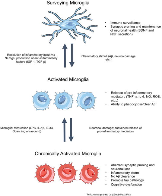
Microglia are the primary immune cell of the brain and function to protect the central nervous system (CNS) from injury and invading pathogens. In the homeostatic brain, microglia serve to support neuronal health through synaptic pruning, promoting normal brain connectivity and development, and through release of neurotrophic factors, providing support for CNS integrity. However, recent evidence indicates that the homeostatic functioning of these cells is lost in neurodegenerative disease, including Alzheimer's disease (AD), ultimately contributing to a chronic neuroinflammatory environment in the brain. Importantly, the development of compounds and genetic models to ablate the microglial compartment has emerged as effective tools to further our understanding of microglial function in AD. Use of these models has identified roles of microglia in several pathological facets of AD, including tau propagation, synaptic stripping, neuronal loss, and cognitive decline. Although culminating evidence utilizing these microglial ablation models reports an absence of CNS-endogenous and peripheral myeloid cell involvement in Aβ phagocytosis, recent data indicates that targeting microglia-evoked neuroinflammation in AD may be essential for potential therapeutics. Therefore, identifying altered signaling pathways in the microglia-devoid brain may assist with the development of effective inflammation-based therapies in AD.
Keywords: Alzheimer’s disease; Amyloid; Colony-stimulating factor 1 receptor; Inflammation; Microglia; Spines; Tau.
Copyright © 2016 Elsevier Inc. All rights reserved.
Conflict of interest statement
The authors E.E.S and K.N.G have no conflicting financial interests.
Figures
References
-
- Alzheimer A, Stelzmann RA, Schnitzlein HN, Murtagh FR. An English translation of Alzheimer’s 1907 paper, “Uber eine eigenartige Erkankung der Hirnrinde”. Clin Anat. 1995;8:429–431. - PubMed
-
- Bellinger FP, Madamba S, Siggins GR. Interleukin 1 beta inhibits synaptic strength and long-term potentiation in the rat CA1 hippocampus. Brain Res. 1993;628:227–234. - PubMed
-
- Ben-Menachem-Zidon O, Ben-Menahem Y, Ben-Hur T, Yirmiya R. Intrahippocampal transplantation of neural precursor cells with transgenic overexpression of IL-1 receptor antagonist rescues memory and neurogenesis impairments in an Alzheimer’s disease model. Neuropsychopharmacology. 2014;39:401–414. - PMC - PubMed
Publication types
MeSH terms
Substances
Grants and funding
LinkOut - more resources
Full Text Sources
Other Literature Sources
Medical
Research Materials
