Review
. 2016 Aug;14(8):479-93.
doi: 10.1038/nrmicro.2016.87. Epub 2016 Jul 11.

Influenza virus RNA polymerase: insights into the mechanisms of viral RNA synthesis

Affiliations
Review

Influenza virus RNA polymerase: insights into the mechanisms of viral RNA synthesis

Aartjan J W Te Velthuis et al. Nat Rev Microbiol. 2016 Aug.

Abstract

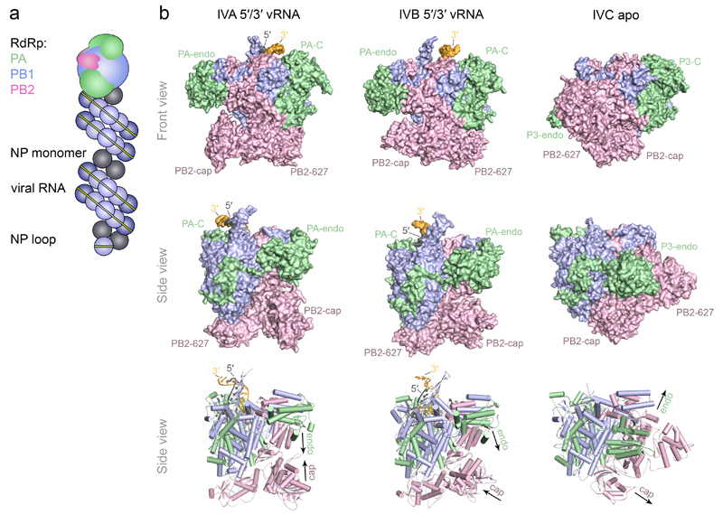
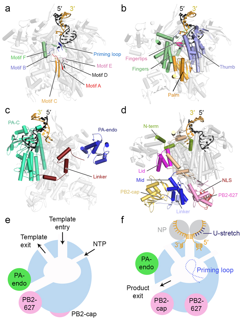
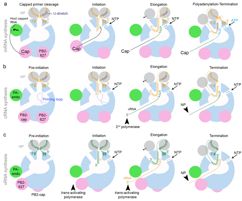
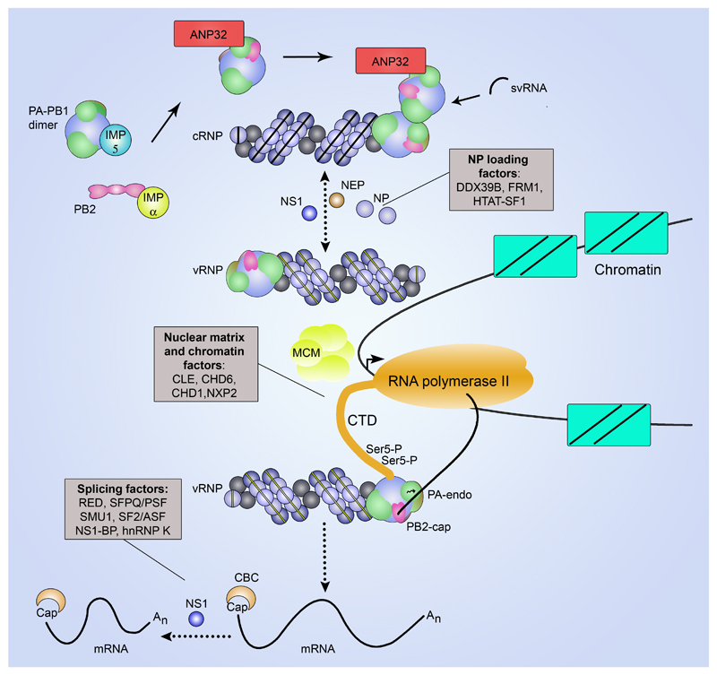
The genomes of influenza viruses consist of multiple segments of single-stranded negative-sense RNA. Each of these segments is bound by the heterotrimeric viral RNA-dependent RNA polymerase and multiple copies of nucleoprotein, which form viral ribonucleoprotein (vRNP) complexes. It is in the context of these vRNPs that the viral RNA polymerase carries out transcription of viral genes and replication of the viral RNA genome. In this Review, we discuss our current knowledge of the structure of the influenza virus RNA polymerase, and insights that have been gained into the molecular mechanisms of viral transcription and replication, and their regulation by viral and host factors. Furthermore, we discuss how advances in our understanding of the structure and function of polymerases could help in identifying new antiviral targets.


Conflict of interest statement

Competing interests statement

The authors declare no competing interests.

Figures

Figure 1
Figure 1. Model of the vRNP complex and structures of the influenza A, B and C virus RNA polymerases.
a Model of the vRNP complex. In the vRNP, the 5′ and 3′ termini of vRNA are bound by a heterotrimeric polymerase complex and the rest of the vRNA is coated by nucleoprotein. The complex is twisted into an anti-parallel double helix, the structure of which is maintained by contacts between NP monomers. The RNA forms a loop at the end opposite the polymerase-bound end, . b Front and side views of the influenza A (PDB: 4WSB), B (PDB: 4WSA) and C (PDB: 5D98) virus RNA polymerase structures shown in surface (top and middle row) or cartoon model (bottom row) representation. PB1, PB2 and PA/P3 subunits are coloured in blue, pink and light green, respectively. The PB2 cap-binding (PB2-cap), PB2 627-domain (PB2-627), PA/P3 endonuclease (PA-endo) and PA/P3 C-terminal (PA-C) domains are indicated. The 5′ and 3′ termini of vRNA in the influenza A and B virus polymerase structures are shown in dark grey and yellow, respectively. In the influenza A virus polymerase the cap-binding and endonuclease domains face each other, configured for cap-snatching (left, bottom row). In the influenza B virus polymerase structure the cap-binding domain is rotated to face the product exit channel consistent with insertion of the capped primer into the active site via the product exit channel (middle, bottom row). In the influenza C virus polymerase structure the cap-binding and endonuclease domains face opposite directions, incompatible with cap-snatching (right, bottom row).
Figure 2
Figure 2. Polymerase architecture and channels.
a-d Cartoon models of the influenza A virus polymerase (PDB: 4WSB), in different orientations, showing PB1 polymerase motifs A to F and the priming loop (a), the right handed arrangement of the PB1 fingers, palm and thumb subdomains and the fingertips (b), the PA (c) and PB2 (d) domain structures. The 5′ and 3′ termini of vRNA are coloured as in FIG. 1. e, f Models of the RNA polymerase in the ‘inactive’ (e) and ‘transcription pre-initiation’ states (f) showing the conformational re-arrangement of the flexible peripheral cap-binding, 627 and endonuclease (Endo) domains. The temple entry and exit, NTP entry and product exit channels, the priming loop near the active site as well as the binding of the 5′ and 3′ vRNA termini and the positions of nucleoproteins (NP) in the ‘transcription pre-initiation’ model are shown. The U-stretch near the 5′ end of the vRNA that acts as a poly(A) signal is indicated.
Figure 3
Figure 3. Models of viral transcription (mRNA synthesis) and replication (cRNA and vRNA synthesis).
a Model for mRNA synthesis. The polymerase is depicted as in FIG. 2f in the ‘transcription pre-initiation’ state. Host capped RNA is bound by the PB2 cap-binding domain and cleaved by the PA/P3 endonuclease domain. The cap-binding domain rotates to allow the insertion of the 3′ end of the capped RNA primer into the active site via the product exit channel. The 3′ end of the vRNA template also inserts into the active site and NTPs enter via the NTP entry channel. Transcription is initiated by the addition of a GTP to the 3′ end of the capped primer that is templated by the second residue in the vRNA template. During elongation, product and template are separated and they exit through their respective exit channels. As the template is pulled into the active site, NP detaches from the entering vRNA, translocates via the surface of the polymerase and joins the vRNA template as it emerges through the template exit channel. The 5′ cap is released from the PB2 cap-binding domain as the polymerase enters elongation. Termination is achieved through polyadenylation that occurs as the result of repeated copying of the U-stretch of the vRNA template. The 5′ end of the vRNA remains bound to its binding site and is stabilized by base-pairing with the 3′ end of the vRNA that re-binds at its binding site near the surface of the polymerase during elongation. b Model for cRNA synthesis. The polymerase is shown in the same conformation as for mRNA synthesis. The 3′ of the vRNA inserts into the active site and NTPs enter via the NTP entry channel. De novo initiation occurs at the first residue of the vRNA template (terminal initiation) and the initiating nucleotide is stabilized by the priming loop, , . During elongation, product and template are separated and they exit through their respective exit channels. NP translocates via the surface of the polymerase to join the vRNA template as it emerges through the template exit channel. The 5′ end of the cRNA product is bound by a second polymerase as it emerges from the product exit channel and the rest of the RNA associates with NP to assemble cRNPs. For termination, the 5′ end of the vRNA template is released from its binding site and is pulled through the polymerase active site while the 3′ end re-binds at the surface of the polymerase. c Model for vRNA synthesis. The polymerase is depicted as in FIG. 2e in the ‘inactive’ state consistent with the conformation of the polymerase bound to the 5′ end of cRNA. For replication initiation to take place the polymerase needs to interact with a trans-activating polymerase. The 3′ of the cRNA inserts into the active site and NTPs enter via the NTP entry channel. De novo initiation occurs at the fourth and fifth residue of the cRNA template (internal initiation) without the participation of the priming loop, , . The resulting pppApG dinucleotide re-locates to the 3′ end of the cRNA template (not shown) and is elongated by the polymerase. Elongation and termination proceed as described above for cRNA synthesis. The 5′ end of the vRNA product is bound by the trans-activating polymerase and the rest of the vRNA associates with NP to assemble vRNPs. Note that an alternative model, involving a trans-acting polymerase, has also been proposed for cRNA to vRNA replication (not shown), .
Figure 4
Figure 4. Regulation of transcription and replication.
Viral RNA synthesis is performed in association with the chromatin/nuclear matrix close to sites of host Pol II transcription. This association is mediated by nuclear matrix and chromatin factors. For transcription, vRNPs bind to the serine-5 phopshorylated CTD of the large subunit of Pol II to access nascent capped host RNA for cap-snatching, . Polyadenylation of viral mRNA is stimulated by the host splicing factor SFPQ/PSF; viral NS1 and host RED, SMU1, SF2/ASF, NS1-BP, hnRNP K participate in mRNA processing, , –. The host cap-binding complex (CBC) binds to the 5′ cap once it is released by PB2. vRNPs in close proximity to Pol II but not bound to the CTD of Pol II carry out cRNA synthesis. cRNA serves as template for vRNA synthesis. Free viral polymerase and NP are required for the assembly of cRNA and vRNA into cRNP and vRNP, respectively. DDX39B, HTAT-SF1 and FMR1 stimulate viral RNA replication by promoting NP interactions with the viral RNA polymerase or NP recruitment to nascent viral RNA during cRNP and vRNP assembly, , . Viral NS1 regulates viral RNA replication by interacting with NP– and viral NEP promotes viral RNA replication, possibly via the stimulation of svRNA synthesis–. svRNAs are implicated as viral co-factors in genome replication. The cellular DNA helicase MCM as well as proteins of the ANP32 family are implicated in genome replication, . ANP32A may possibly contribute by recruiting a trans-activating polymerase to the resident cRNP-bound polymerase in a host- and PB2 627-domain-specific manner to facilitate vRNA synthesis although the exact mechanism of its involvement remains unknown, , . Importin-α, which acts as import factor for PB2, is proposed to have a role in transcription and replication independent of its role as a nuclear import receptor, . Importin-5 (also known as RanBP5) acts as a co-factor for the nuclear import of the PB1-PA dimer, , .

References

    1. Molinari NA, et al. The annual impact of seasonal influenza in the US: measuring disease burden and costs. Vaccine. 2007;25:5086–96. - PubMed
    1. Muraki Y, Hongo S. The molecular virology and reverse genetics of influenza C virus. Jpn J Infect Dis. 2010;63:157–65. - PubMed
    1. Krammer F, Palese P. Advances in the development of influenza virus vaccines. Nat Rev Drug Discov. 2015;14:167–82. - PubMed
    1. Das K. Antivirals targeting influenza A virus. J Med Chem. 2012;55:6263–77. - PubMed
    1. Watanabe T, Kawaoka Y. Influenza virus-host interactomes as a basis for antiviral drug development. Curr Opin Virol. 2015;14:71–8. - PMC - PubMed

MeSH terms