Review
. 2016 Nov;95(11):449-464.
doi: 10.1016/j.ejcb.2016.06.007. Epub 2016 Jun 25.

Causes and consequences of nuclear envelope alterations in tumour progression

Affiliations
Review

Causes and consequences of nuclear envelope alterations in tumour progression

Emily S Bell et al. Eur J Cell Biol. 2016 Nov.

Abstract

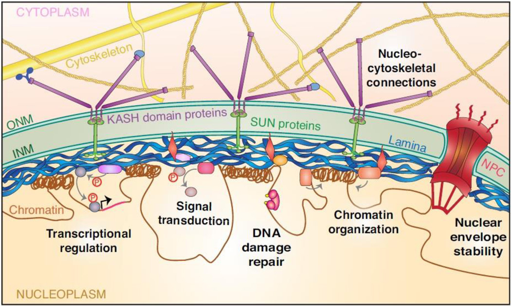
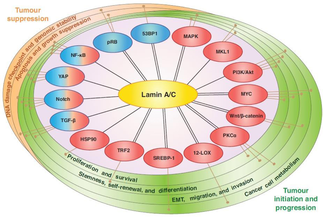
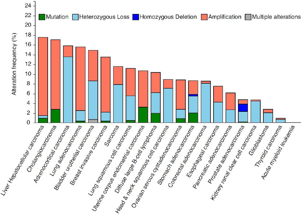
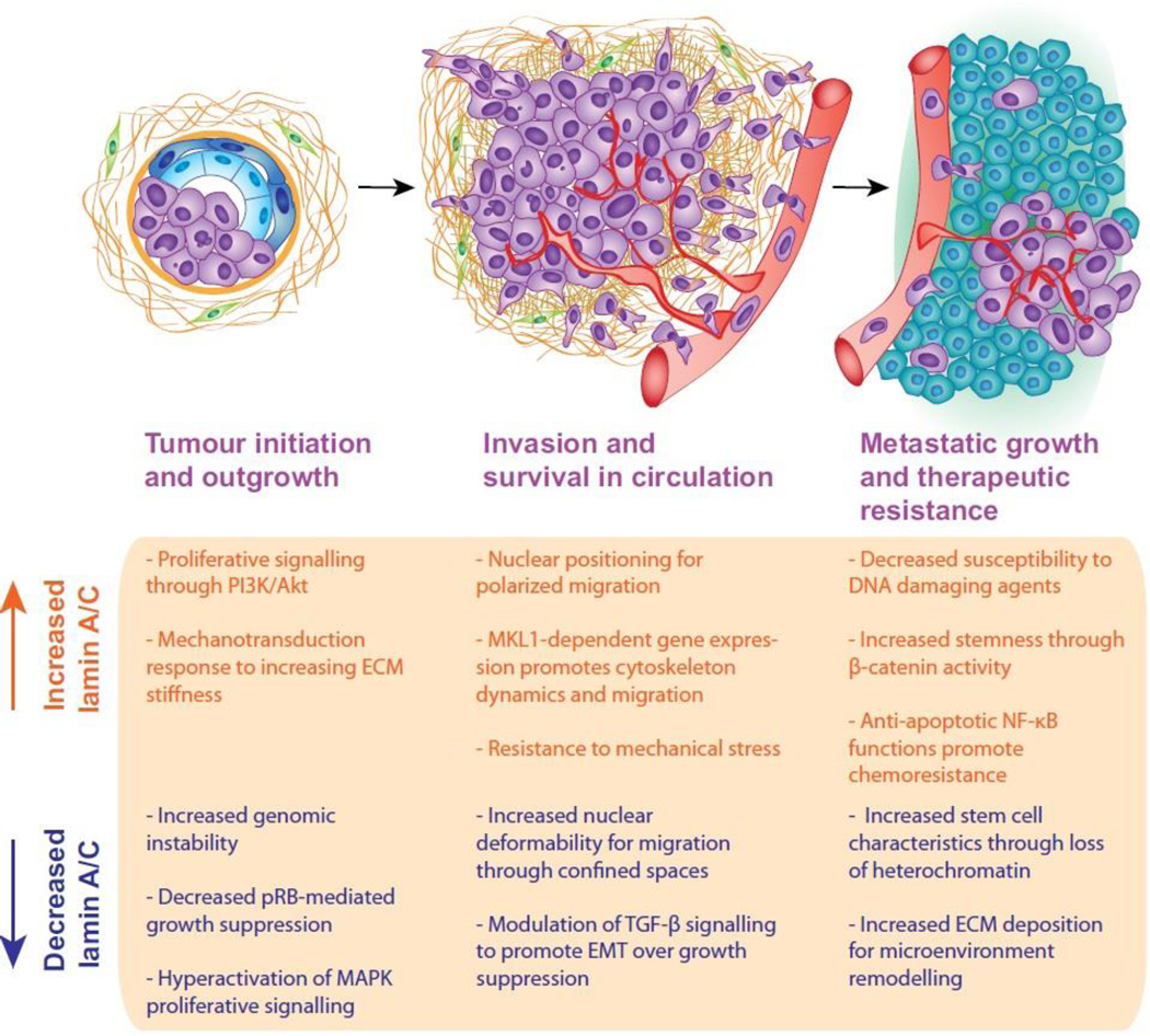
Morphological changes in the size and shape of the nucleus are highly prevalent in cancer, but the underlying molecular mechanisms and the functional relevance remain poorly understood. Nuclear envelope proteins, which can modulate nuclear shape and organization, have emerged as key components in a variety of signalling pathways long implicated in tumourigenesis and metastasis. The expression of nuclear envelope proteins is altered in many cancers, and changes in levels of nuclear envelope proteins lamins A and C are associated with poor prognosis in multiple human cancers. In this review we highlight the role of the nuclear envelope in different processes important for tumour initiation and cancer progression, with a focus on lamins A and C. Lamin A/C controls many cellular processes with key roles in cancer, including cell invasion, stemness, genomic stability, signal transduction, transcriptional regulation, and resistance to mechanical stress. In addition, we discuss potential mechanisms mediating the changes in lamin levels observed in many cancers. A better understanding of cause-and-effect relationships between lamin expression and tumour progression could reveal important mechanisms for coordinated regulation of oncogenic processes, and indicate therapeutic vulnerabilities that could be exploited for improved patient outcome.

Keywords: Cancer; Cell mechanics; Cell migration; Gene regulation; Lamins; Signal transduction.


Figures

Figure 1
Figure 1. Overview of nuclear envelope organization and lamin A/C functions
The nuclear envelope (NE) consists of the outer nuclear membrane (ONM), inner nuclear membrane (INM), nuclear pore complexes (NPC), nuclear lamina, and additional proteins bound to the lamina and INM. The lamina is a meshwork of intermediate filaments beneath the INM composed of A-type and B-type lamins. Lamins can also localize to the nuclear interior (not depicted here). Lamin A/C performs many cellular functions, including providing physical stiffness to protect the nucleus and tethering chromatin into transcriptionally repressed regions at the nuclear periphery. The lamina and associated proteins can also form complexes with signalling molecules to influence nuclear accumulation, stability, and substrate engagement. The nucleus is connected to the cytoskeleton through the linker of nucleoskeleton and cytoskeleton (LINC) complex. The LINC complex is composed of Sad1 and UNC-84 (SUN) domain proteins in the INM that bind to Klarsicht, ANC-1, and Syne homology (KASH) domain proteins that span the ONM and connect to the cytoskeleton, facilitating nuclear positioning and mechanotransduction signalling.
Figure 2
Figure 2. Connections between lamin A/C and pathways known to suppress or promote cancer formation and progression
Lamin A/C participates in many pathways characterized to have oncogenic (red) or tumour suppressor (blue) functions in cancer. Lamin A/C expression influences protein levels or downstream activity to modulate signal transduction, and in many cases interactions between lamin A/C and signalling proteins have also been detected (double lines). It is important to note that many of these functions have been identified in lamin A/C mutant or deletion models for laminopathies, and an understanding of lamin A/C-related signalling in the context of cancer demands further study.
Figure 3
Figure 3. Frequency of mutations and copy-number alterations in the LMNA gene in human cancers
The results shown here are based upon data generated by the TCGA Research Network (http://cancergenome.nih.gov/) and analyzed via cBioPortal (http://www.cbioportal.org/) (Cerami et al., 2012; Gao et al., 2013) on April 10, 2016. These results should therefore not be viewed as original data, but as reference to existing publicly available resources. In the analysis, shallow loss determined by copy-numberanalysis is referred to here as “heterozygous deletion” and deep loss is referred to as “homozygous deletion”. Copy number variations make up the majority of genomic changes in the LMNA gene in cancer, whereas mutations are rare. It remains to be determined whether LMNA genomic alterations correlate closely with protein levels in cancer, and which transcriptional and/or post-translational mechanisms are important contributors to altered lamin A/C expression in cancer.
Figure 4
Figure 4. Potential roles for lamin A/C in tumour initiation and progression
Both increased and decreased levels of lamin A/C have been observed in cancer and related to patient prognosis. The complexity of lamin A/C functions and signalling networks suggests that the role of lamin A/C in tumour progression may vary with stage of disease, tissue of origin, and mutational landscape of a given tumour. Possible functions for increased and decreased lamin A/C levels during tumour progression are depicted here, and warrant further investigation towards understanding the role of lamin A/C in cancer. Importantly, many of these lamin-related functions could contribute to multiple stages of cancer, but are each shown here only once for simplicity.

References

    1. Acerbi I, Cassereau L, Dean I, Shi Q, Au A, Park C, Chen YY, Liphardt J, Hwang ES, Weaver VM. Human breast cancer invasion and aggression correlates with ECM stiffening and immune cell infiltration. Integr Biol (Camb) 2015;7:1120–1134. - PMC - PubMed
    1. Agarwal S, Achari C, Praveen D, Roy KR, Reddy GV, Reddanna P. Inhibition of 12-LOX and COX-2 reduces the proliferation of human epidermoid carcinoma cells (A431) by modulating the ERK and PI3K–Akt signalling pathways. Exp Dermatol. 2009;18:939–946. - PubMed
    1. Agrelo R, Setien F, Espada J, Artiga MJ, Rodriguez M, Perez-Rosado A, Sanchez-Aguilera A, Fraga MF, Piris MA, Esteller M. Inactivation of the lamin A/C gene by CpG island promoter hypermethylation in hematologic malignancies, and its association with poor survival in nodal diffuse large B-cell lymphoma. J Clin Oncol. 2005;23:3940–3947. - PubMed
    1. Alberio R, Johnson AD, Stick R, Campbell KH. Differential nuclear remodeling of mammalian somatic cells by Xenopus laevis oocyte and egg cytoplasm. Exp Cell Res. 2005;307:131–141. - PubMed
    1. Aljada A, Doria J, Saleh AM, Al-Matar SH, AlGabbani S, Shamsa HB, Al-Bawab A, Ahmed AA. Altered Lamin A/C splice variant expression as a possible diagnostic marker in breast cancer. Cell Oncol (Dordr) 2016;39:161–174. - PubMed

LinkOut - more resources