Review
. 2017 Jan:61:140-149.
doi: 10.1016/j.semcdb.2016.07.006. Epub 2016 Jul 7.

In vitro culture of ovarian follicles from Peromyscus

Affiliations
Review

In vitro culture of ovarian follicles from Peromyscus

Xiaoming He et al. Semin Cell Dev Biol. 2017 Jan.

Abstract

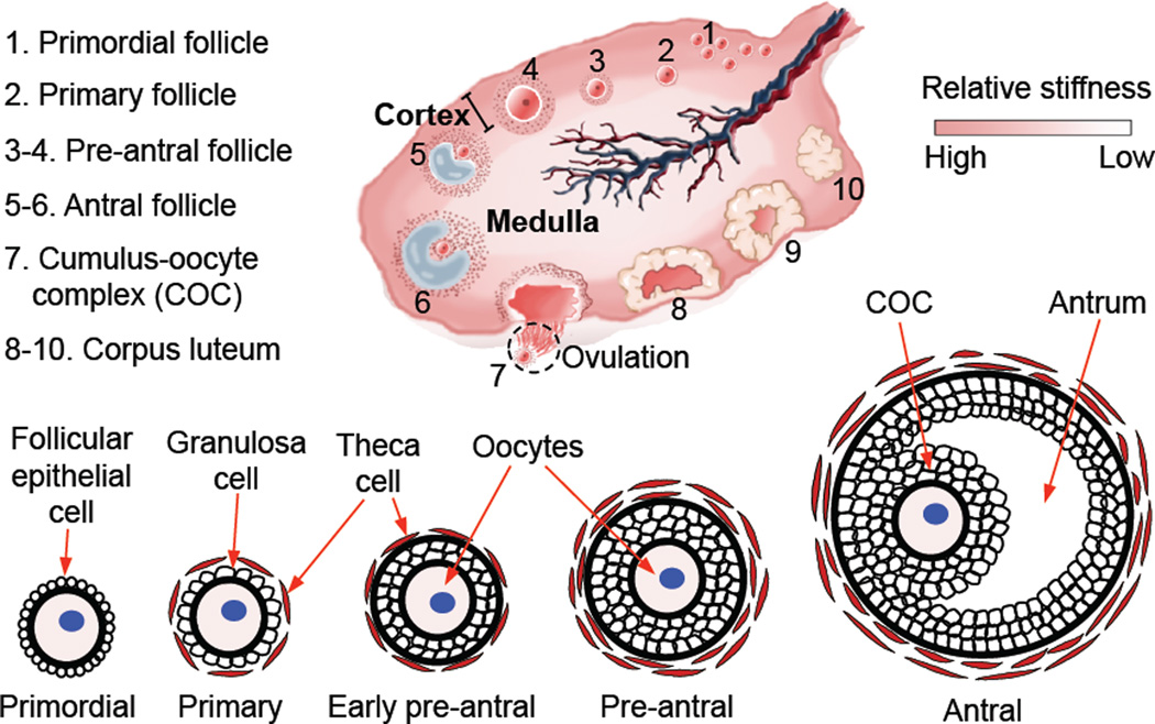
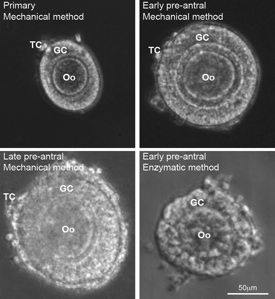
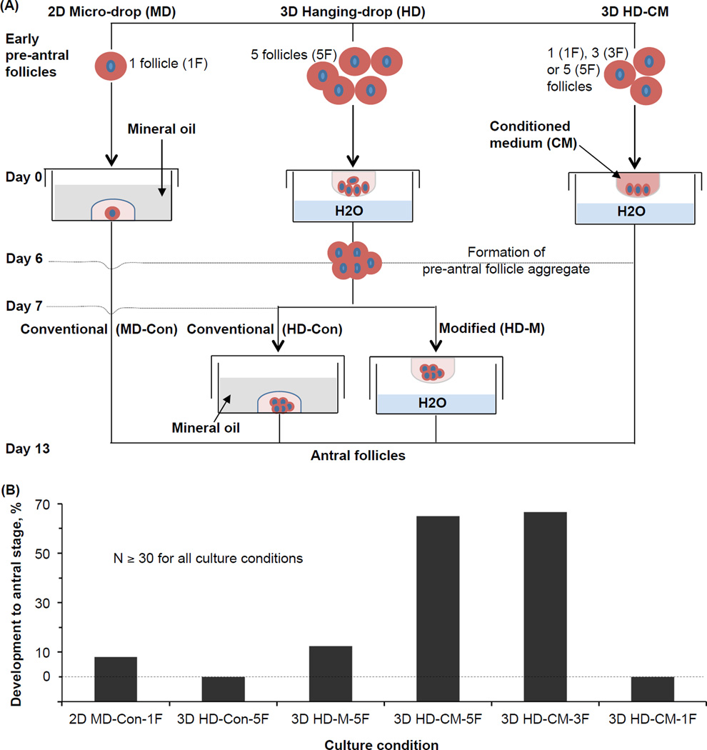
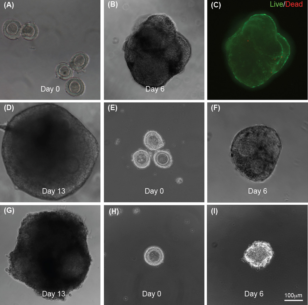
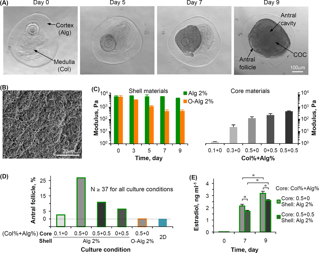
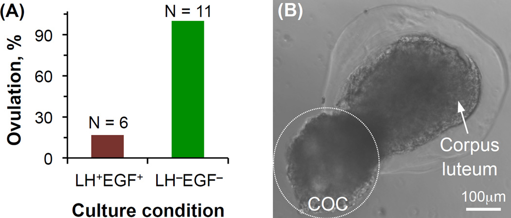
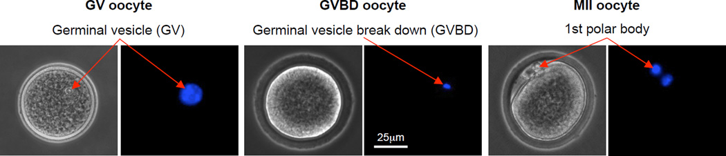
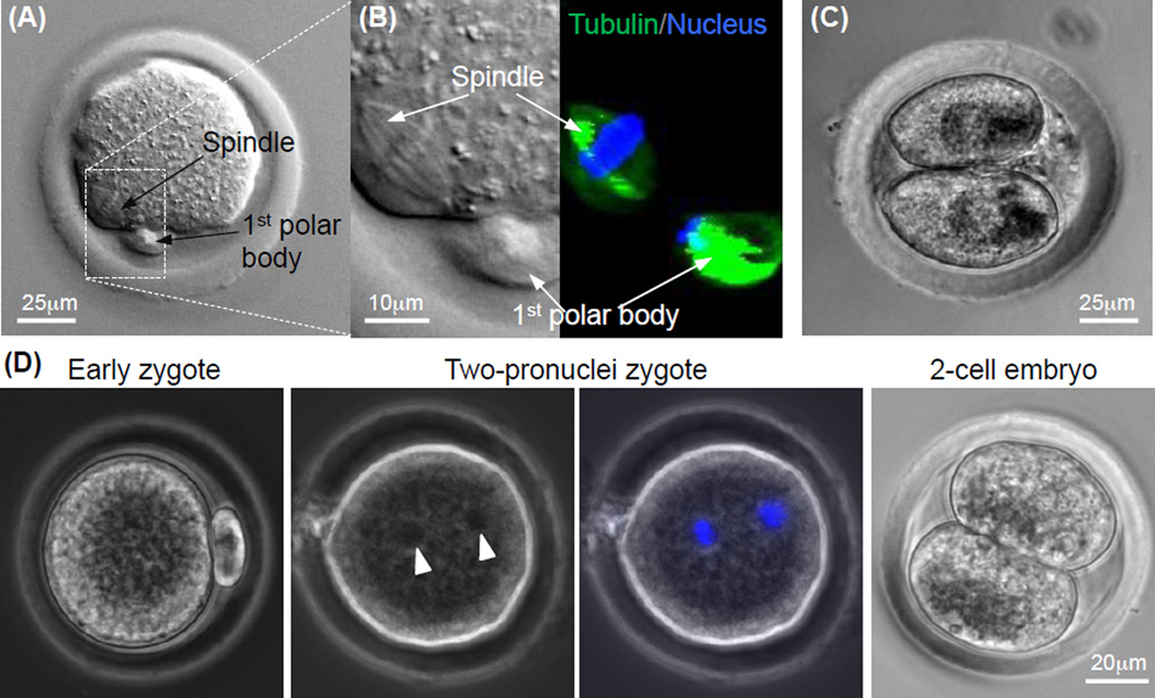
The ovarian follicle is the fundamental functional tissue unit of mammalian ovary. Each ovarian follicle contains one single oocyte. Isolation and in vitro culture of ovarian follicles to obtain fertilizable oocytes have been regarded as a promising strategy for women to combat infertility. The follicles from Peromyscus are considered as a better model than that from inbred mice for studying follicle culture. This is because Peromyscus mice are outbred (as with humans) with an increased life span. In this article, we reviewed studies on this subject conducted using Peromyscus follicles. These studies show that the conventional 2D micro-drop and 3D hanging-drop approaches established for in vitro culture of early preantral follicles from inbred mice are not directly applicable for cultivating the follicles from Peromyscus. However, the efficiency could be significantly improved by culturing multiple early preantral follicles in one hanging drop of Peromyscus ovarian cell-conditioned medium. It is further revealed that the mechanical heterogeneity in the extracellular matrix of ovary is crucial for developing early preantral follicles to the antral stage and for the subsequent ovulation to release cumulus-oocyte complex. These findings may provide valuable guidance for furthering the technology of in vitro follicle culture to restore fertility in the clinic.

Keywords: Alginate; Hanging drop; Mechanobiology; Microcapsule; Microfluidics; Ovulation.


Figures

Figure 1
Figure 1
A schematic illustration of the mammalian ovary showing the difference in mechanical properties between the ovarian cortex and medulla, the morphology of ovarian follicles at various developmental stages, and the ovulation of cumulus-oocyte complex (COC) from antral follicles leaving behind the corpus luteum. The schematic of mammalian ovary is reprinted and redrawn from reference [47] with permission from Elsevier.
Figure 2
Figure 2
Images showing the typical morphology of primary (75–99 µm), early preantral (100–125 µm), and late preantral (126–180 µm) follicles retrieved from ovaries of Peromyscus using the mechanical method together with that of early preantral follicle obtained using the enzymatic method: The follicles retrieved by the mechanical method have an intact outer membrane of theca cells (TCs) and an intact layer(s) of granulosa cells (GCs) in the middle together with a primary oocyte (Oo) in the center. In contrast, the middle and particularly, the outer layer of the follicles retrieved by the enzymatic method were severely compromised. The figure is reprinted and redrawn from reference [46] with permission from Mary Ann Liebert Inc.
Figure 3
Figure 3
In vitro culture of early preantral follicles of Peromyscus with 2D micro-drop and various versions of 3D hanging-drop methods: (A) A schematic illustration of the various methods used for in vitro culture of early preantral follicles, and (B) the quantitative data of development to the antral stage using the various methods. N: number of early preantral follicles. The figure is reprinted and redrawn from reference [46] with permission from Mary Ann Liebert Inc.
Figure 4
Figure 4
Typical micrographs showing development of early preantral follicles obtained mechanically from the Peromyscus ovaries under in vitro culture in hanging drop of ovarian cell-conditioned medium: Five single follicles per drop on day 0 (A), one single aggregate formed from the five follicles on day 6 (B), high viability (green) of the follicle aggregate on day 6 (C), and the aggregate of antral follicles developed from the five early preantral follicles after continuous culture in hanging drop for 13 days (D); three follicles per drop on days 0 (E) and 6 (F) and the aggregate of antral follicles developed from the three early preantral follicles after continuous culture in hanging drop for 13 days (G); and only one follicle per hanging drop on day 0 (H) and the degenerated follicle on day 6 (I). The figure is reprinted and redrawn from reference [46] with permission from Mary Ann Liebert Inc.
Figure 5
Figure 5
Development of early preantral follicles under miniaturized biomimetic 3D culture in core-shell microcapsules showing the crucial role of mechanical heterogeneity in regulating the follicle development: (A) Typical images showing the growth of an early preantral follicle in the biomimetic microtissue with a 2% alginate shell and 0.5% collagen core, (B) a typical scanning electron microscopy (SEM) image showing the collagen fiber in the 0.5% collagen core, (C) elastic modulus of various shell and core materials used for fabricate the ovarian microtissue, (D) quantitative data of the percentage of development to the antral stage of early preantral follicles cultured under various conditions collected from references [47,48], and (E) production of estradiol by the growing follicles under the two best culture conditions. Alg: alginate; Col: collagen; O-Alg: oxidized alginate; N: number of early preantral follicles; and *: p < 0.05. The figure is reprinted and redrawn from reference [47] with permission from Elsevier (for panel A) and from reference [48] with permission from John Wiley & Sons, Inc. (for panels B, C, and E).
Figure 6
Figure 6
Biomimetic ovulation in vitro via breaking apart the cortex (i.e., the alginate hydrogel shell) to release cumulus-oocyte complex (COC): (A) The effect of the combination of luteinizing hormone (LH) and epidermal growth factor (EGF) on the biomimetic ovulation, and (B) a typical image showing the ovulation of cumulus-oocyte complex (COC) from the biomimetic ovarian microtissue leaving behind a corpus luteum-like structure. N: number of antral follicles. The figure is reprinted and redrawn from reference [47] with permission from Elsevier
Figure 7
Figure 7
Typical phase contrast and fluorescence images of primary and secondary oocytes showing their morphology and nuclei distribution, respectively: The primary oocyte includes both GV (germinal vesicle) and GVBD (germinal vesicle breakdown) oocytes, while the MII (metaphase II) oocyte is secondary. A germinal vesicle containing all nuclear materials can be clearly seen in the GV oocyte, the germinal vesicle breaks down with the nuclear stain not as clearly identifiable in the GVBD oocyte, and the nuclear materials condense at two different locations with one being in the first polar body and the other in the cytoplasm of the MII oocyte. The figure is reprinted and redrawn from reference [46] with permission from Mary Ann Liebert Inc.
Figure 8
Figure 8
Quality of MII oocytes obtained by In vitro maturation of cumulus-oocyte complex from antral follicles developed from early preantral follicles by in vitro culture: (A) Typical image of a MII oocyte obtained from the antral follicles showing the characteristic 1st polar body and mitotic spindle, (B) nuclear and tubulin stains for visualizing the 1st polar body and mitotic spindle in the MII oocyte, (C) image of a two-cell embryo developed from the MII oocyte after parthenogenetic activation, and (D) typical micrographs showing development of MII oocyte after in vitro fertilization (early zygote) to the two-pronuclei and two-cell stages. The two pronuclei visible in the phase contrast image (arrow head) were further confirmed using fluorescence stain of the pronuclei (blue stains). The figure is reprinted and redrawn from reference [48] with permission from John Wiley & Sons, Inc. (for panels A–C) and from reference [46] with permission from Mary Ann Liebert Inc. (for panel D).

References

    1. De Vos M, Smitz J, Woodruff TK. Fertility preservation in women with cancer. Lancet. 2014;384:1302–1310. - PMC - PubMed
    1. Telfer EE, Zelinski MB. Ovarian follicle culture: advances and challenges for human and nonhuman primates. Fertil Steril. 2013;99:1523–1533. - PMC - PubMed
    1. Andersen CY, Kristensen SG, Greve T, Schmidt KT. Cryopreservation of ovarian tissue for fertility preservation in young female oncological patients. Future Oncol. 2012;8:595–608. - PubMed
    1. Jin M, Yu Y, Huang H. An update on primary ovarian insufficiency. Sci China Life Sci. 2012;55:677–686. - PubMed
    1. Donnez J, Dolmans MM. Cryopreservation and transplantation of ovarian tissue. Clin Obstet Gynecol. 2010;53:787–796. - PubMed

Publication types

LinkOut - more resources