

Effects of Antioxidant N-acetylcysteine Against Paraquat-Induced Oxidative Stress in Vital Tissues of Mice
- PMID: 27398384
- PMCID: PMC4936834
Effects of Antioxidant N-acetylcysteine Against Paraquat-Induced Oxidative Stress in Vital Tissues of Mice
Abstract
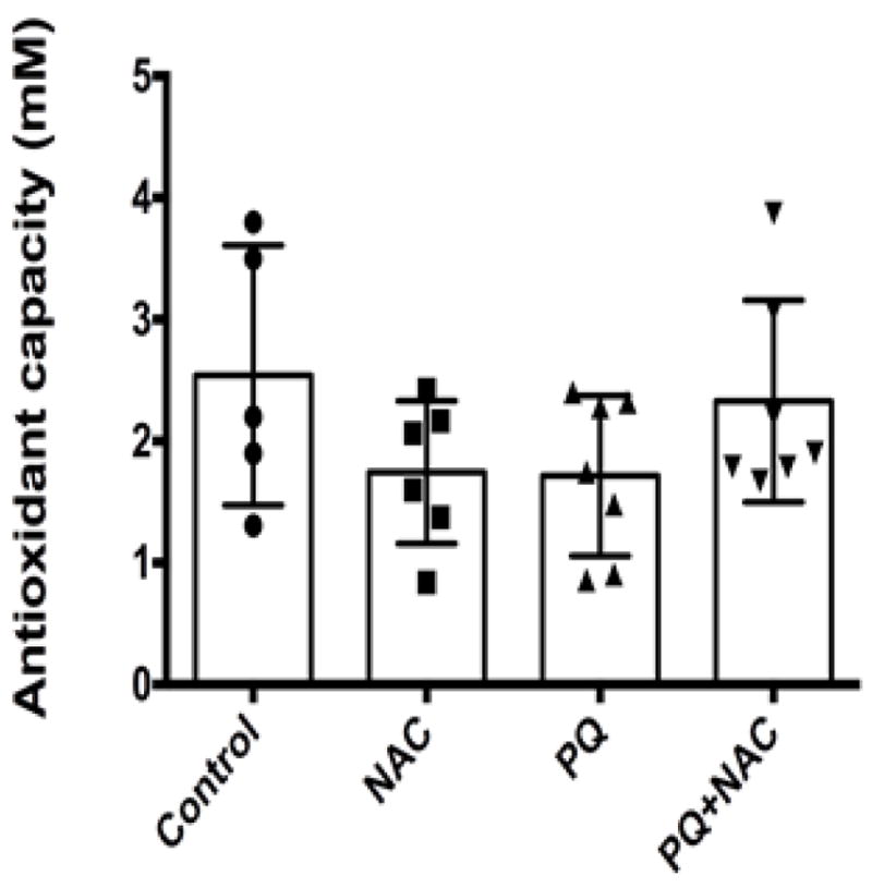
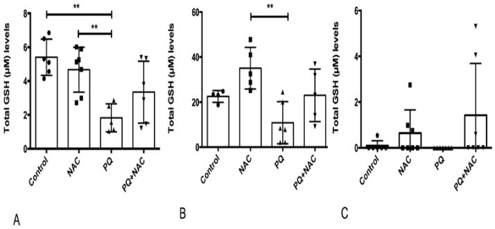
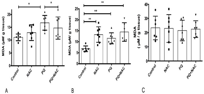
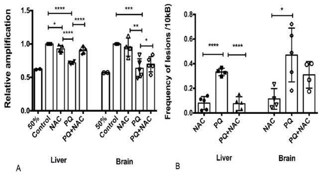
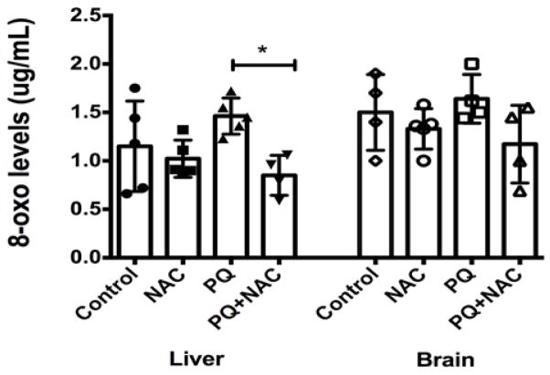
Paraquat (PQ) is a commonly used herbicide that induces oxidative stress via reactive oxygen species (ROS) generation. This study aimed to investigate the effects of the antioxidant N-acetylcysteine (NAC) against PQ-induced oxidative stress in mice. Male Balb/C mice (24) were randomly divided into 4 groups and treated for 3 weeks: 1) control (saline), 2) NAC (0.5% in diet), 3) PQ (20 mg/kg, IP) and 4) combination (PQ + NAC). Afterwards mice were sacrificed and oxidative stress markers were analyzed. Our data showed no significant change in serum antioxidant capacity. PQ enhanced lipid peroxidation (MDA) levels in liver tissue compared to control whereas NAC decreased MDA levels (p<0.05). NAC significantly increased MDA in brain tissue (p<0.05). PQ significantly depleted glutathione (GSH) levels in liver (p=0.001) and brain tissue (p<0.05) but non-significant GSH depletion in lung tissue. NAC counteracted PQ, showing a moderate increase GSH levels in liver and brain tissues. PQ significantly increased 8-oxodeoxyguanosine (8-OH-dG) levels (p<0.05) in liver tissue compared to control without a significant change in brain tissue. NAC treatment ameliorated PQ-induced oxidative DNA damage in the liver tissue. PQ significantly decreased the relative mtDNA amplification and increased the frequency of lesions in liver and brain tissue (p<0.0001), while NAC restored the DNA polymerase activity in liver tissue but not in brain tissue. In conclusion, PQ induced lipid peroxidation, oxidative nuclear DNA and mtDNA damage in liver tissues and depleted liver and brain GSH levels. NAC supplementation ameliorated the PQ-induced oxidative stress response in liver tissue of mice.
Keywords: Antioxidant Capacity; DNA Integrity; Glutathione levels; MDA levels; Mice; Oxidative Stress; Reactive Oxygen Species.
Figures
Similar articles
-
Comparison of oxidative stress status in the kidney tissue of male rats treated with paraquat and nanoparaquat.Sci Rep. 2025 Jan 2;15(1):389. doi: 10.1038/s41598-024-83156-2. Sci Rep. 2025. PMID: 39747891 Free PMC article.
-
Hepatoprotective effect of N-acetylcystein loaded niosomes on liver function in paraquat-induced acute poisoning.Pestic Biochem Physiol. 2019 Oct;160:146-153. doi: 10.1016/j.pestbp.2019.08.001. Epub 2019 Aug 14. Pestic Biochem Physiol. 2019. PMID: 31519249
-
Biochemical and molecular mechanisms of N-acetyl cysteine and silymarin-mediated protection against maneb- and paraquat-induced hepatotoxicity in rats.Chem Biol Interact. 2013 Jan 25;201(1-3):9-18. doi: 10.1016/j.cbi.2012.10.027. Epub 2012 Nov 16. Chem Biol Interact. 2013. PMID: 23159886
-
Protective Effects of N-acetylcysteine Niosome Nanoparticles on Paraquatinduced Nephrotoxicity in Male Rats.Pharm Nanotechnol. 2022;10(2):137-145. doi: 10.2174/2211738510666220214102034. Pharm Nanotechnol. 2022. PMID: 35156589
-
The manganese-salen compound EUK-134 and N-acetyl cysteine rescue from zinc- and paraquat-induced toxicity in rat polymorphonuclear leukocytes.Chem Biol Interact. 2015 Apr 25;231:18-26. doi: 10.1016/j.cbi.2015.02.012. Epub 2015 Feb 24. Chem Biol Interact. 2015. PMID: 25724285
Cited by
-
Bacopaside-I Alleviates the Detrimental Effects of Acute Paraquat Intoxication in the Adult Zebrafish Brain.Neurochem Res. 2021 Nov;46(11):3059-3074. doi: 10.1007/s11064-021-03416-9. Epub 2021 Aug 6. Neurochem Res. 2021. PMID: 34357519
-
Paraquat Preferentially Induces Apoptosis of Late Stage Effector Lymphocyte and Impairs Memory Immune Response in Mice.Int J Environ Res Public Health. 2019 Jun 11;16(11):2060. doi: 10.3390/ijerph16112060. Int J Environ Res Public Health. 2019. PMID: 31212664 Free PMC article.
-
Suppression of cirrhosis-related renal injury by N-acetyl cysteine.Curr Res Pharmacol Drug Discov. 2020 Oct 13;1:30-38. doi: 10.1016/j.crphar.2020.100006. eCollection 2020 Apr. Curr Res Pharmacol Drug Discov. 2020. PMID: 34909640 Free PMC article.
-
Mn Inhibits GSH Synthesis via Downregulation of Neuronal EAAC1 and Astrocytic xCT to Cause Oxidative Damage in the Striatum of Mice.Oxid Med Cell Longev. 2018 Aug 30;2018:4235695. doi: 10.1155/2018/4235695. eCollection 2018. Oxid Med Cell Longev. 2018. PMID: 30228854 Free PMC article.
-
N-Acetylcysteine as a treatment for sulphur mustard poisoning.Free Radic Biol Med. 2020 Dec;161:305-320. doi: 10.1016/j.freeradbiomed.2020.09.020. Epub 2020 Sep 25. Free Radic Biol Med. 2020. PMID: 32980537 Free PMC article. Review.
References
-
- Anderson ME, Luo JL. Glutathione therapy: from prodrugs to genes. Semin Liver Dis. 1998;18:415–424. - PubMed
-
- Balcerczyk A, Bartosz G. Thiols are main determinants of total antioxidant capacity of cellular homogenates. Free Radical Research. 2003;37:537–541. - PubMed
-
- Banaclocha MM, Martinez N. N-acetylcysteine elicited increase in cytochrome c oxidase activity in mice synaptic mitochondria. Brain Res. 1999;842:249–251. - PubMed
Grants and funding
LinkOut - more resources
Full Text Sources