
EPHB4 kinase-inactivating mutations cause autosomal dominant lymphatic-related hydrops fetalis

Silvia Martin-Almedina et al. J Clin Invest. .

Abstract

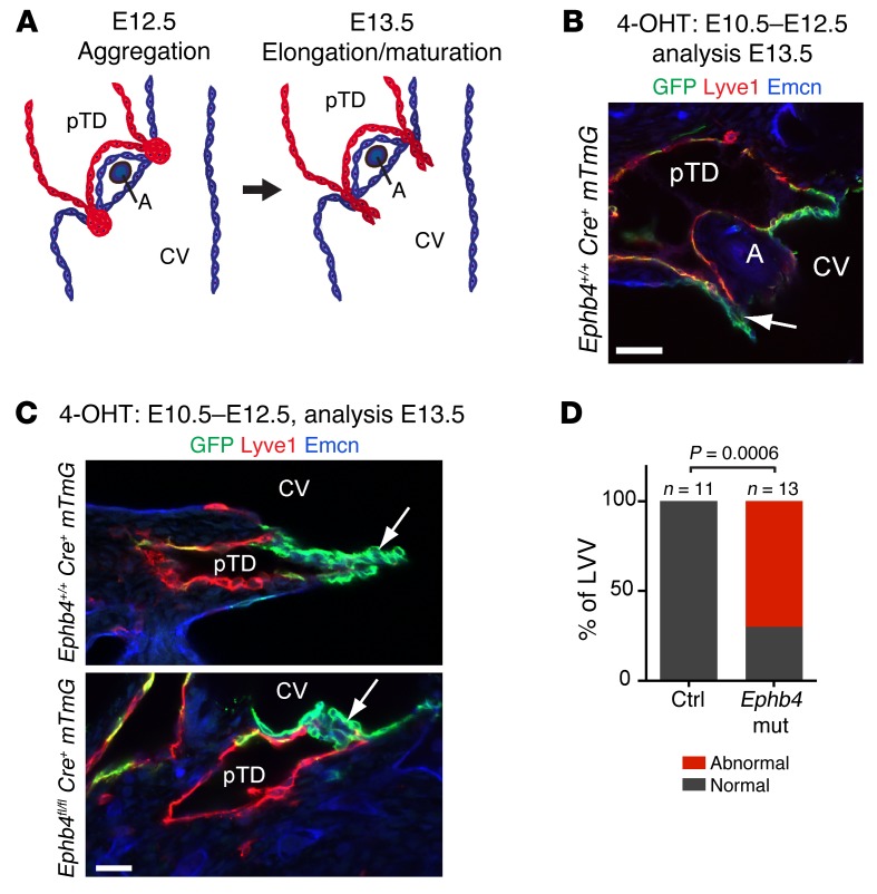
Hydrops fetalis describes fluid accumulation in at least 2 fetal compartments, including abdominal cavities, pleura, and pericardium, or in body tissue. The majority of hydrops fetalis cases are nonimmune conditions that present with generalized edema of the fetus, and approximately 15% of these nonimmune cases result from a lymphatic abnormality. Here, we have identified an autosomal dominant, inherited form of lymphatic-related (nonimmune) hydrops fetalis (LRHF). Independent exome sequencing projects on 2 families with a history of in utero and neonatal deaths associated with nonimmune hydrops fetalis uncovered 2 heterozygous missense variants in the gene encoding Eph receptor B4 (EPHB4). Biochemical analysis determined that the mutant EPHB4 proteins are devoid of tyrosine kinase activity, indicating that loss of EPHB4 signaling contributes to LRHF pathogenesis. Further, inactivation of Ephb4 in lymphatic endothelial cells of developing mouse embryos led to defective lymphovenous valve formation and consequent subcutaneous edema. Together, these findings identify EPHB4 as a critical regulator of early lymphatic vascular development and demonstrate that mutations in the gene can cause an autosomal dominant form of LRHF that is associated with a high mortality rate.


Figures

Figure 1
Figure 1. Mutations in EPHB4 cause LRHF.
(A) Pedigree of GLDUK family. (B) Pedigree of GLDNOR family. Stars indicate which samples have been exome sequenced. Genotypes indicated by minus signs (–) represent the WT allele, and plus signs represent (+) the mutant allele. Top row of genotypes in GLDUK genogram shows EPHB4, and bottom row of genotypes shows MIER2. Triangles, first trimester miscarriages; IUD, intrauterine death. GLDNOR:III.3 had trisomy 18.
Figure 2
Figure 2. Imaging of the lymphatic system in LRHF.
Anterior view of lower limb lymphoscintigraphy 2 hours after injection with radionuclide. (A) GLDUK:I.2, rerouting through skin and superficial tissues in the right leg and markedly reduced transport in the left leg. (B) GLDUK:II.4, normal uptake of tracer in the lymph nodes in the groin area, but with some rerouting in the calves (seen as the dark shading; arrows). (C) Unaffected subject with symmetrical transport of radionuclide within collecting lymph vessels in the leg.
Figure 3
Figure 3. Effect of p.Arg739Glu and p.Ile782Ser mutations on EPHB4 tyrosine phosphorylation in HEK293T cells.
(A) HEK293T cells were transfected with expression plasmids for EPHB4 WT and p.Arg739Glu and p.Ile782Ser mutants. Receptor phosphorylation was analyzed by immunoprecipitation and Western blotting using anti–p-tyrosine (p-Tyr, upper panel) and EPHB4 (lower panel) antibodies. The positions of molecular mass markers (in kDa) are indicated to the right of the gels. (B) EPHB4 WT and p.Arg739Glu and p.Ile782Ser mutants were cotransfected into HEK293T cells in different ratios of WT to mutant plasmid. Receptor phosphorylation was analyzed as above. One representative experiment (n = 3) is shown.
Figure 4
Figure 4. Effect of p.Arg739Glu and p.Ile782Ser mutations on EPHB4 tyrosine phosphorylation in LECs after Ephrin B2 stimulation.
LECs were transfected with expression plasmids for Myc-DDK–tagged EPHB4 WT and p.Arg739Glu and p.Ile782Ser mutants. The cells were stimulated with 1 μg/ml clustered Ephrin B2/Fc (EB2/Fc) or Fc alone. Receptor phosphorylation was analyzed by immunoprecipitation with anti-DDK antibody and Western blotting using anti–p-tyrosine (upper panel) and EPHB4 (lower panel) antibodies. The positions of molecular mass markers (in kDa) are indicated to the right of the gels. One representative experiment (n = 2) is shown.
Figure 5
Figure 5. Early embryonic deletion of Ephb4 leads to subcutaneous edema and abnormal dermal lymphatic vasculature.
(A) Schematic of the transgenes and 4-OHT administration (Cre induction; red arrowheads) schedule used for Ephb4 deletion in the lymphatic vasculature. Timing of primitive lymphatic vessel formation and time point for analysis are indicated. PLLV, peripheral longitudinal lymphatic vessel; LV, lymphatic vessel. (B) Edema in E15.5 control and Ephb4 mutant embryos after different 4-OHT treatments. P values determined by Fisher’s exact test. (C and D) Left panels: E15.5 Ephb4fl/+ and Ephb4fl/fl Prox1-CreERT2 R26-mTmG embryos. Most mutants treated with 4-OHT at E10.5–E14.5 showed subcutaneous edema (white arrowhead) and blood-filled lymphatic vessels (black arrowheads). Boxed area indicates the area of the skin imaged on the right. Right panels: whole-mount immunofluorescence of E15.5 thoracic skin for NRP2 (red) and GFP (green) to stain lymphatic vessels and gene-targeted cells, respectively. Scale bars: 200 μm (C and D).
Figure 6
Figure 6. Early embryonic deletion of Ephb4 leads to a failure of LVV formation.
(A) Schematic representation of LVV formation. CV, cardinal vein; A, artery. Adapted with permission from Developmental Biology (ref. ; Creative Commons user license available at http://www.sciencedirect.com/science/article/pii/S0012160615301032). (B) Whole-mount immunofluorescence of a transverse section of E13.5 Ephb4+/+ Prox1-CreERT2 R26-mTmG embryo for indicated proteins. GFP shows efficient Cre-mediated recombination in the LVV that extends to the lumen of the cardinal vein (arrow). (C) Whole-mount immunofluorescence of E13.5 LVVs showing extension of the valve leaflets to the lumen of the cardinal vein (arrow) in control (upper panel, n = 11 out of 11) but not in Ephb4 mutant (Ephb4 mut) embryo (lower panel, n = 9 out of 13). (D) Quantification of LVV morphology in control and Ephb4 mutant embryos. Normal, elongated leaflets; abnormal, no leaflets. P value determined by Fisher’s exact test. Scale bars: 50 μm (B); 25 μm (C).

References

    1. Bellini C, et al. Etiology of non-immune hydrops fetalis: an update. Am J Med Genet A. 2015;167A(5):1082–1088. - PubMed
    1. Connell FC, et al. The classification and diagnostic algorithm for primary lymphatic dysplasia: an update from 2010 to include molecular findings. Clin Genet. 2013;84(4):303–314. doi: 10.1111/cge.12173. - DOI - PubMed
    1. Alders M, et al. Mutations in CCBE1 cause generalized lymph vessel dysplasia in humans. Nat Genet. 2009;41(12):1272–1274. doi: 10.1038/ng.484. - DOI - PubMed
    1. Connell F, et al. Linkage and sequence analysis indicate that CCBE1 is mutated in recessively inherited generalised lymphatic dysplasia. Hum Genet. 2010;127(2):231–241. doi: 10.1007/s00439-009-0766-y. - DOI - PubMed
    1. Alders M, et al. Hennekam syndrome can be caused by FAT4 mutations and be allelic to Van Maldergem syndrome. Hum Genet. 2014;133(9):1161–1167. doi: 10.1007/s00439-014-1456-y. - DOI - PubMed

Publication types

MeSH terms

Substances