. 2016 Oct;5(10):1394-1405.
doi: 10.5966/sctm.2015-0371. Epub 2016 Jul 8.

High-Efficiency Serum-Free Feeder-Free Erythroid Differentiation of Human Pluripotent Stem Cells Using Small Molecules

Affiliations

High-Efficiency Serum-Free Feeder-Free Erythroid Differentiation of Human Pluripotent Stem Cells Using Small Molecules

Emmanuel N Olivier et al. Stem Cells Transl Med. 2016 Oct.

Abstract

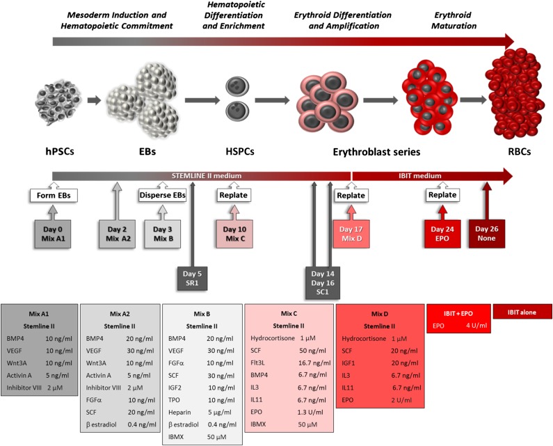
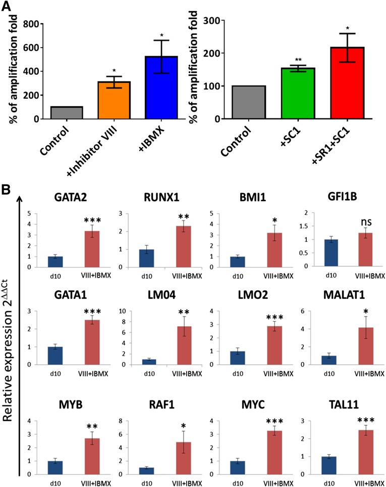
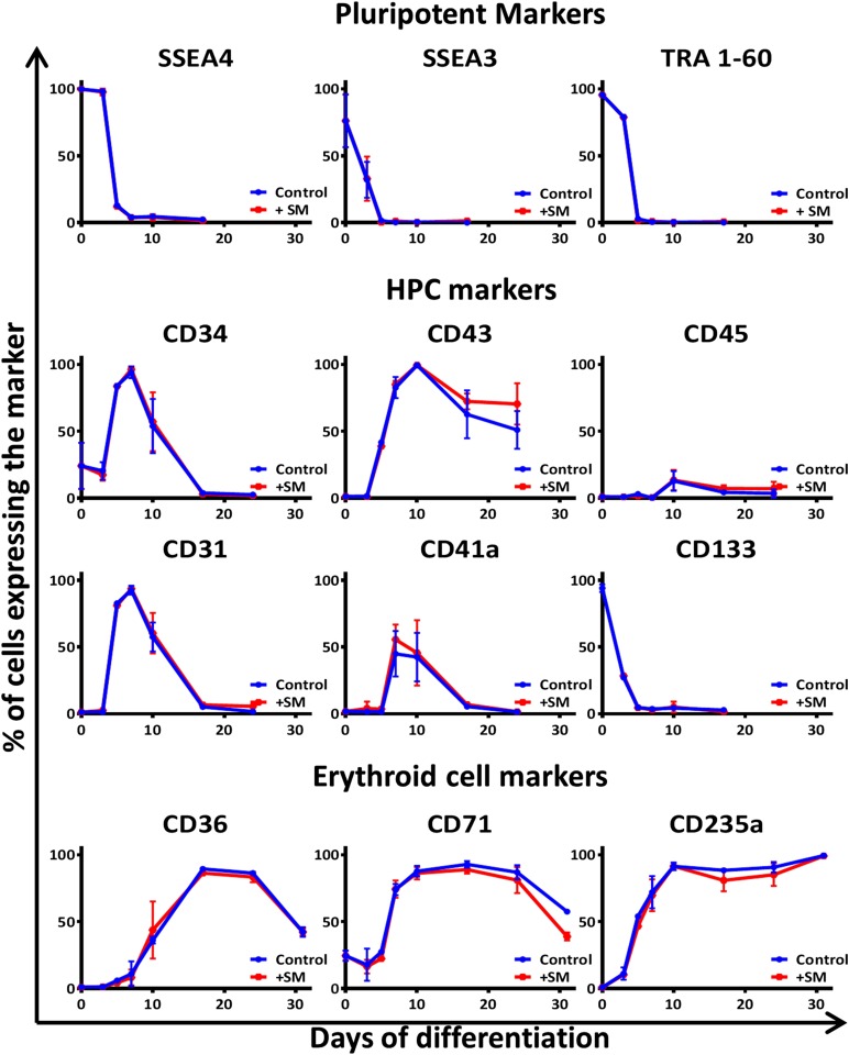
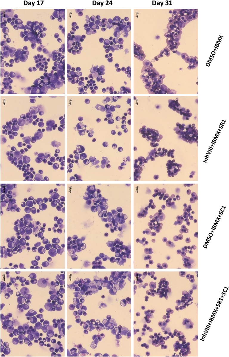
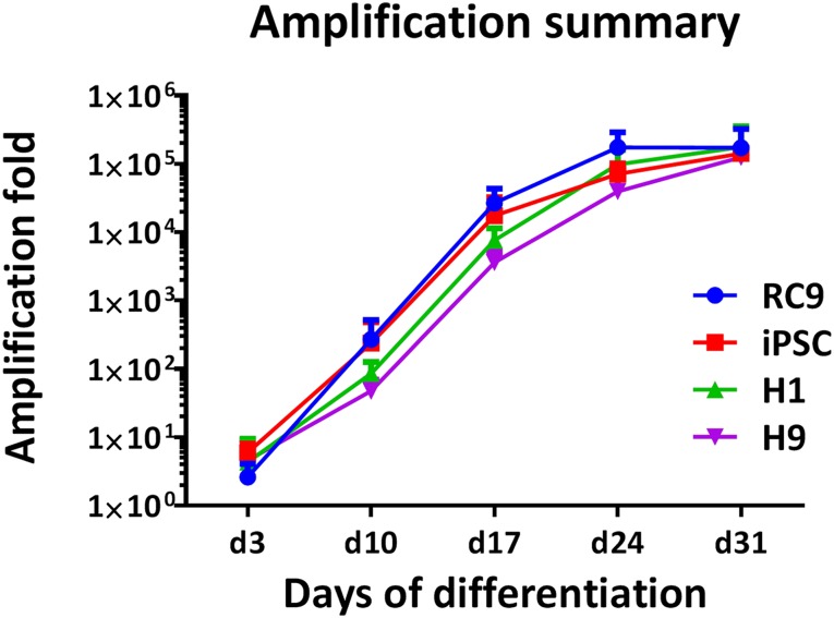
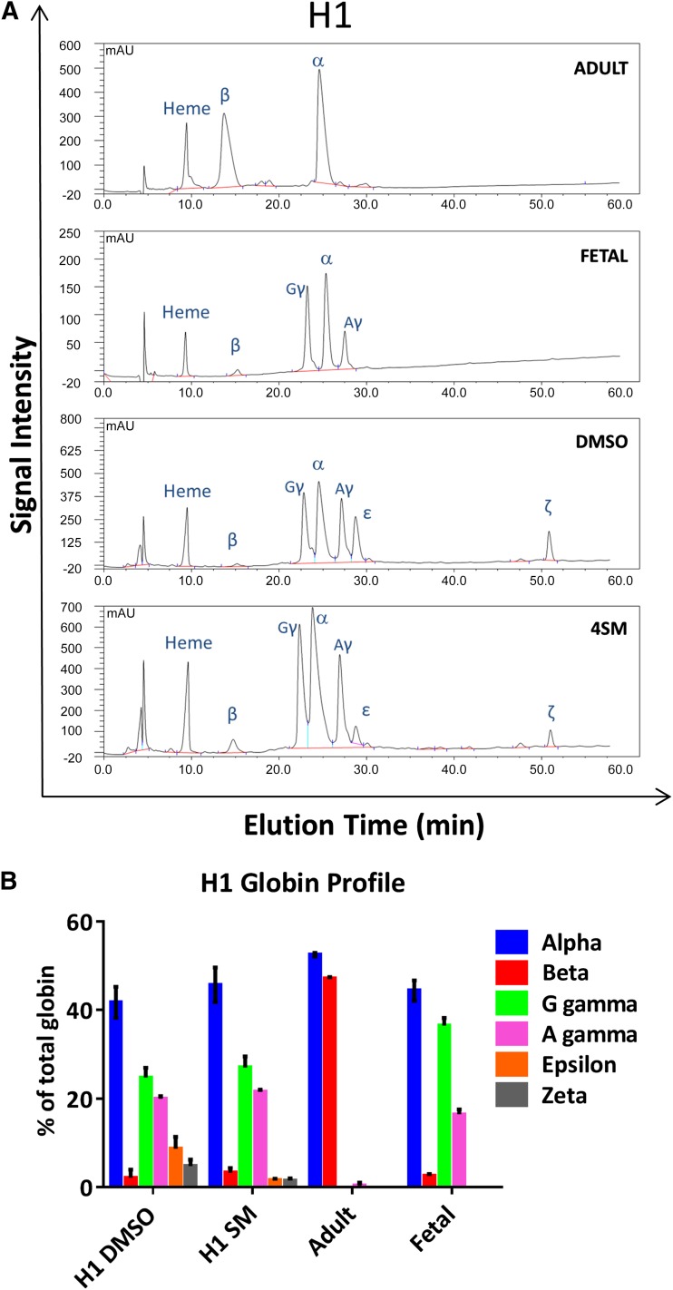
: This article describes a good manufacturing practice (GMP)-compatible, feeder-free and serum-free method to produce large numbers of erythroid cells from human pluripotent stem cells (hPSCs), either embryonic or induced. This multistep protocol combines cytokines and small molecules to mimic and surpass the early stages of development. It produces, without any selection or sorting step, a population of cells in which 91.8% ± 5.4% express CD34 at day 7, 98.6% ± 1.3% express CD43 at day 10, and 99.1% ± 0.95% of cells are CD235a positive by day 31 of the differentiation process. Moreover, this differentiation protocol supports extensive expansion, with a single hPSC producing up to 150 hematopoietic progenitor cells by day 10 and 50,000-200,000 erythroid cells by day 31. The erythroid cells produced exhibit a definitive fetal hematopoietic type, with 90%-95% fetal globin and variable proportion of embryonic and adult globin at the protein level. The presence of small molecules during the differentiation protocol has quantitative and qualitative effects; it increases the proportion of adult globin and decreases the proportion of embryonic globin. Given its level of definition, this system provides a powerful tool for investigation of the mechanisms governing early hematopoiesis and erythropoiesis, including globin switching and enucleation. The early stages of the differentiation protocol could also serve as a starting point for the production of endothelial cells and other hematopoietic cells, or to investigate the production of long-term reconstituting hematopoietic stem cells from hPSCs.

Significance: This differentiation protocol allows the production of a large amount of erythroid cells from pluripotent stem cells. Its efficiency is compatible with that of in vitro red blood cell production, and it can be a considerable asset for studying developmental erythropoiesis and red blood cell enucleation, thereby aiding both basic and translational research. In addition to red cells, the early stages of the protocol could also be used as a starting point for the large-scale production of other hematopoietic cell types, including the ultimate goal of generating long-term reconstituting hematopoietic stem cells.

Keywords: Cytokines; Erythropoiesis; Hematopoiesis; Pluripotent stem cells; Small molecule.


Figures

Figure 1.
Figure 1.
Diagram representing the feeder-free and serum-free erythroid differentiation of hPSCs augmented by the addition of small molecules. Abbreviations: BMP, bone morphogenic protein; EBs, embryoid bodies; EPO, erythropoietin; FGF, fibroblast growth factor; Flt3L, Flt3-ligand; hPSCs, human pluripotent stem cells; HSPCs, hematopoietic stem and progenitor cells; IBIT, IMDM + bovine serum albumin, insulin, transferrin; IBMX, isobutyl methyl xanthine; IGF, insulin-like growth factor; IL, interleukin; RBCs, red blood cells; SCF, stem cell factor; TPO, thrombopoietin; VEGF, vascular endothelial growth factor 165.
Figure 2.
Figure 2.
Analysis of the quantitative effects of the small molecules used on erythroid differentiation of H1 embryonic stem cells (ESCs). (A): Additional use of small molecules increased the yield of erythroid cells. Histograms representative of the small molecule effect on the yield of erythroid cells from the H1 hESC line, expressed as percentage of total amplification relative to the control. Left panel, effect of inhibitor VIII and IBMX assessed at day 24 of the differentiation. n = 3; mean ± SD. Unpaired t test; ∗, p < .05. Right panel, effect of SR1 and SR1 + SC1 assessed on day 31 of the differentiation, when used in addition of Inhibitor VIII and IBMX. n ≥ 4; mean ± SD. Unpaired t test; ∗, p < .05, ∗∗, p < .01. (B): Inhibitor VIII and IBMX addition increase the transcription of key genes involved in hematopoiesis. Quantitative polymerase chain reaction analysis of the effect of inhibitor VIII and IBMX at day 10 of the differentiation on genes known to be expressed during hematopoietic and erythroid development. n = 4 performed in duplicate; mean ± SD. Unpaired t test; ∗, p < .05; ∗∗, p < .01; ∗∗∗, p < .001. Abbreviations: IBMX, isobutyl methyl xanthine; ns, not significantly different.
Figure 3.
Figure 3.
Analysis of the qualitative effects of the small molecules used on erythroid differentiation of H1 embryonic stem cells (ESCs). Addition of small molecules does not modify the surface marker profile of differentiating cells. Antigenic profile of the culture during the erythroid differentiation of H1 human ESCs with (red lines) or without (Control; blue lines) small molecules (+SM; inhibitor VIII + IBMX + SR1 + SC1). The pluripotent markers show the loss of pluripotency during the first 5 days. The HPC markers show the emergence of the hematopoietic cells with a peak of hemangioblastic markers CD34, CD31, and CD41a at day 7. The erythroid cell markers CD36 (denoting the commitment toward erythroid lineage) and CD235a (also known as glycophorin A) confirm the evolution of the culture toward an almost pure erythroid population between days 17 and 31. Abbreviations: HPC, hematopoietic progenitor cell; SM, small molecules. An example of individual flow cytometry plots is shown in supplemental online Fig. 7.
Figure 4.
Figure 4.
Erythroid cell morphology progresses over time in the differentiating cultures and in the late stages is substantially improved by the addition of different combinations of SMs as labeled. Photographs of rapid Romanowsky staining of cytospin preparations at different stages of the differentiation that show the effects of different combination of small molecules on the morphology of the differentiating and differentiated cells at days 17, 24, and 31 of the protocol. Original magnification ×400; scale bars = 10 μm. For reference, images of the morphologically distinct stages of erythroid differentiation are within Figures 4 and 5 of Hu et al. [44]. Abbreviations: DMSO, dimethyl sulfoxide; IBMX, isobutyl methyl xanthine; SM, small molecules.
Figure 5.
Figure 5.
Illustration of the yield achieved with different cell lines. Fold amplification of the initial input of human pluripotent stem cells (hPSCs) to the erythroid stage obtained with four different hPSC lines: RC9, n = 5; H1, n = 4; H9, n = 3; iPSC 33D6, n = 5. Abbreviation: iPSC, induced pluripotent stem cell.
Figure 6.
Figure 6.
High-performance liquid chromatography (HPLC) analysis of the globin chains expressed at day 31 of H1 human embryonic stem cell (hESC) differentiation. (A): Example of elution profiles obtained by HPLC of H1 hESC-derived erythroid cells with (SM) and without (DMSO) small molecules and of the adult and fetal control used to determine the elution time of the different globin chains. (B): Globin chains were assessed at the protein level after erythroid differentiation with (SM) or without (DMSO) the 4 small molecules added (inhibitor VIII + isobutyl methyl xanthine + SR1 + SC1). Lysates from adult peripheral blood and fetal liver red blood cells were used as reference controls to establish the elution time of each peak. Abbreviations: DMSO, dimethyl sulfoxide; SM, small molecules.

References

    1. Thomson JA, Itskovitz-Eldor J, Shapiro SS, et al. Embryonic stem cell lines derived from human blastocysts. Science. 1998;282:1145–1147. - PubMed
    1. Kaufman DS, Hanson ET, Lewis RL, et al. Hematopoietic colony-forming cells derived from human embryonic stem cells. Proc Natl Acad Sci USA. 2001;98:10716–10721. - PMC - PubMed
    1. Vodyanik MA, Bork JA, Thomson JA, et al. Human embryonic stem cell-derived CD34+ cells: Efficient production in the coculture with OP9 stromal cells and analysis of lymphohematopoietic potential. Blood. 2005;105:617–626. - PubMed
    1. Olivier EN, Qiu C, Velho M, et al. Large-scale production of embryonic red blood cells from human embryonic stem cells. Exp Hematol. 2006;34:1635–1642. - PubMed
    1. Zambidis ET, Peault B, Park TS, et al. Hematopoietic differentiation of human embryonic stem cells progresses through sequential hematoendothelial, primitive, and definitive stages resembling human yolk sac development. Blood. 2005;106:860–870. - PMC - PubMed

Publication types