. 2017 Nov;22(11):1576-1584.
doi: 10.1038/mp.2016.103. Epub 2016 Jul 12.

Deficient autophagy in microglia impairs synaptic pruning and causes social behavioral defects

Affiliations

Deficient autophagy in microglia impairs synaptic pruning and causes social behavioral defects

H-J Kim et al. Mol Psychiatry. 2017 Nov.

Abstract

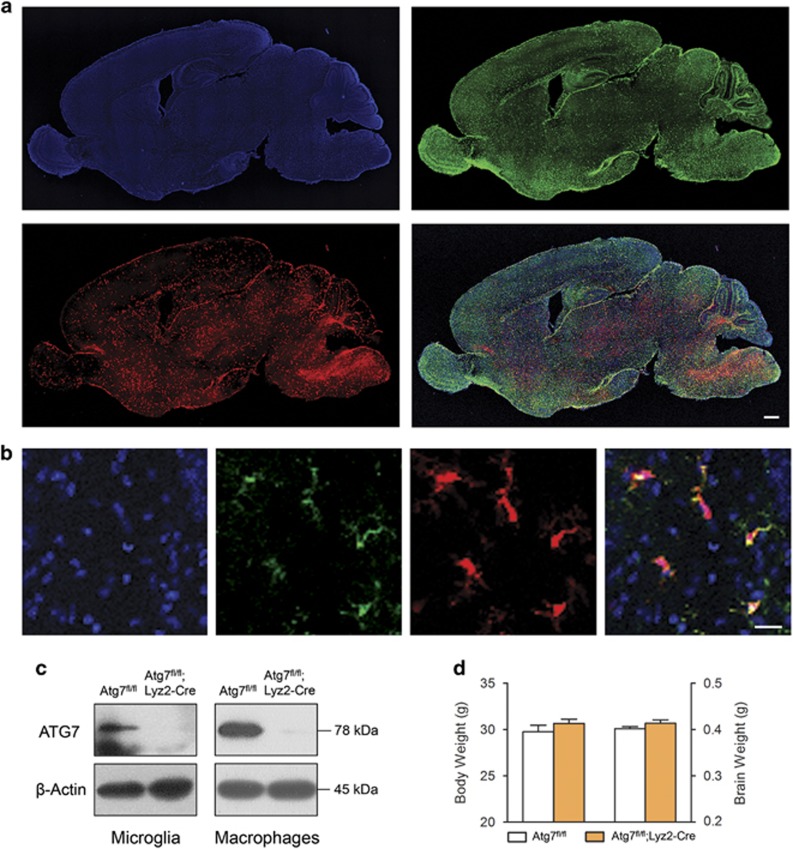
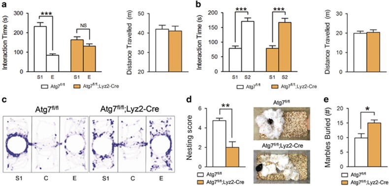
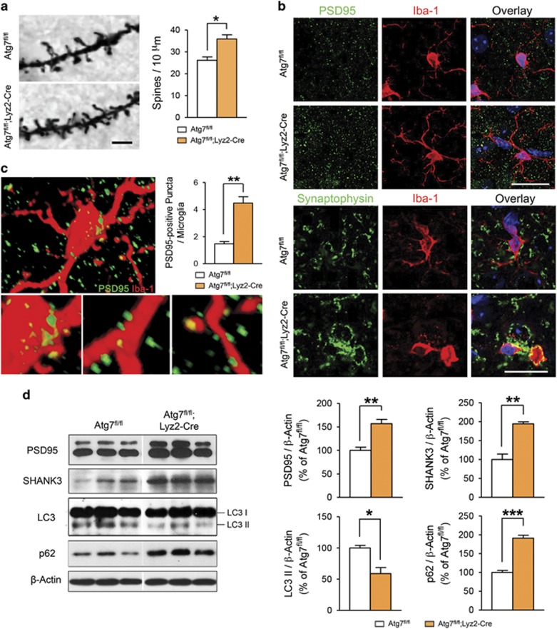
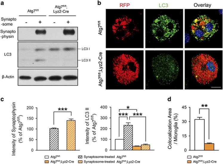
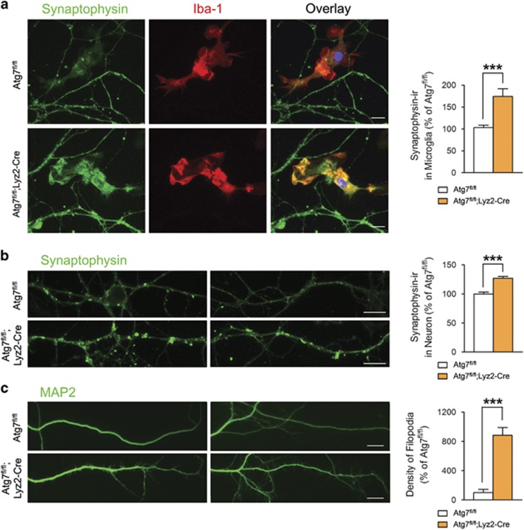
Autism spectrum disorders (ASDs) are neurodevelopmental disorders caused by various genetic and environmental factors that result in synaptic abnormalities. ASD development is suggested to involve microglia, which have a role in synaptic refinement during development. Autophagy and related pathways are also suggested to be involved in ASDs. However, the precise roles of microglial autophagy in synapses and ASDs are unknown. Here, we show that microglial autophagy is involved in synaptic refinement and neurobehavior regulation. We found that deletion of atg7, which is vital for autophagy, from myeloid cell-specific lysozyme M-Cre mice resulted in social behavioral defects and repetitive behaviors, characteristic features of ASDs. These mice also had increases in dendritic spines and synaptic markers and altered connectivity between brain regions, indicating defects in synaptic refinement. Synaptosome degradation was impaired in atg7-deficient microglia and immature dendritic filopodia were increased in neurons co-cultured with atg7-deficient microglia. To our knowledge, our results are the first to show the role of microglial autophagy in the regulation of the synapse and neurobehaviors. We anticipate our results to be a starting point for more comprehensive studies of microglial autophagy in ASDs and the development of putative therapeutics.


Conflict of interest statement

The authors declare no conflict of interest.

Figures

Figure 1
Figure 1
Phenotypes of mice with Atg7-deficient microglia. (a) Sagittal images of Atg7fl/+;Lyz2Cre/+;ROSA26-tdTomato mouse brain at postnatal day 7 stained with an antibody against Iba-1 (green) and Hoechst 33342 (blue). Tomato expression (red) was detected in all brain regions. Scale bar=500 μm. (b) Most of the Tomato expression (red) was co-localized with Iba-1-positive microglia (green). Scale bar=20 μm. (c) Atg7 protein was not detected in the lysate of primary microglia cultures or in peritoneal macrophages collected from Atg7fl/fl;Lyz2-Cre mouse. (d) Body and brain weights of adult Atg7fl/fl;Lyz2-Cre mice were similar to those of controls (n=12 for each group).
Figure 2
Figure 2
Autistic behaviors in mice with Atg7-deficient microglia. (a) In the three-chamber sociability test, Atg7fl/fl;Lyz2-Cre mice did not show any preference for stranger 1 (S1), whereas Atg7fl/fl mice spent more time in contact with S1 than an empty cup (E). No difference was observed in the total distance travelled between groups for 10 min. n=12 for each group. (b) Both Atg7fl/fl and Atg7fl/fl;Lyz2-Cre mice interacted with stranger 2 (S2) more than with S1 during a social novelty test. n=12 for each group. (c) Representative tracks during the sociability test are displayed. (d) In the nest-building test, Atg7fl/fl;Lyz2-Cre mice had lower scores than controls. n=7 for Atg7fl/fl, n=8 for Atg7fl/fl;Lyz2-Cre. (e) Atg7fl/fl;Lyz2-Cre mice buried more marbles than control mice in a 30-min period in the marble-burying test. n=7 for each group. *P<0.05, **P<0.01, ***P<0.001. NS, not significant (two-tailed Student’s t-test).
Figure 3
Figure 3
Increased dendritic spine density in mice with Atg7-deficient microglia. (a) Representative images of dendrites in the somatosensory 2 (S2) brain region from Atg7fl/fl and Atg7fl/fl;Lyz2-Cre mouse after Golgi-Cox staining. The number of dendritic spines in Atg7fl/fl;Lyz2-Cre mice was significantly increased compared with control mice at postnatal day 15. n=150 segments for Atg7fl/fl, n=60 segments for Atg7fl/fl;Lyz2-Cre. Scale bar=2 μm. (b) PSD95 and synaptophysin immunoreactivity was more frequently co-localized with Iba-1 immunoreactivity in the brain of Atg7fl/fl;Lyz2-Cre mice than that of Atg7fl/fl mice. Representative images from each group are shown. Scale bar=20 μm. (c) Three-dimensional confocal image, constructed from serial confocal z-stack images, of Iba-1-positive microglia (red) from an Atg7fl/fl;Lyz2-Cre mouse. High-magnitude images of PSD95-positive puncta (green) engulfed by Iba-1-positive microglia are shown in the lower panel. The number of PSD95-positive puncta per microglia (yellow) was significantly increased in Atg7fl/fl;Lyz2-Cre mice compared with controls at postnatal day 12. n=40 cells for each group. (d) Western blot images of synaptic markers (PSD95 and SHANK3) and autophagy-related proteins (LC3-II and p62). Intensities of both PSD95 and SHANK3 were significantly higher in Atg7fl/fl;Lyz2-Cre mice than in the controls. In Atg7fl/fl;Lyz2-Cre mice, the LC3-II intensity was decreased and that of p62 is increased compared with Atg7fl/fl mice. n=5 for each group. *P<0.05, **P<0.01, ***P<0.001 (two-tailed Student’s t-test).
Figure 4
Figure 4
Impaired degradation of the synaptosome by atg7-deficient microglia. (a, c) Primary microglia from Atg7fl/fl or Atg7fl/fl;Lyz2-Cre mice were cultured and isolated synaptosomes from red fluorescent protein (RFP)-expressing mouse brain were added to the microglial cultures. Western blots showed that synaptophysin is increased and LC3-II is decreased in atg7-deficient microglia compared with wild-type microglia. One-way analysis of variance with Tukey’s post hoc test. F(3,11)=355.40, P<0.001 (synaptophysin). F(3,11)=57.19, P<0.001 (LC3-II). n=3 for each group. (b, d) Immunocytochemistry of microglia showing that punctated forms of LC3 (green) were increased in wild-type microglia, with co-localization (yellow) of RFP-positive synaptosomes (red). LC3 signals were diffuse and the RFP signal was increased in atg7-deficient microglia compared with wild-type microglia. Scale bar=20 μm. *P<0.05, **P<0.01, ***P<0.001 (two-tailed Student’s t-test).
Figure 5
Figure 5
Neuronal changes after their co-culture with atg7-deficient microglia. (a) Primary mouse cortical neurons (18 days in vitro) were co-cultured with primary microglia from Atg7fl/fl or Atg7fl/fl;Lyz2-Cre mice for 2 weeks and then immunostained with antibodies against synaptophysin (green) and Iba-1 (red). Greater accumulation of synaptophysin was seen in atg7-deficient Iba-1-positive microglia than in wild-type microglia. Scale bar=20 μm. (b) Synaptophysin puncta were increased in neurons co-cultured with atg7-deficient microglia. Scale bar=20 μm. (c) MAP2-positive dendrites showed increased filopodia in neurons co-cultured with atg7-deficient microglia. Scale bar=20 μm. ***P<0.001 (two-tailed Student’s t-test).

References

    1. King BH, Navot N, Bernier R, Webb SJ. Update on diagnostic classification in autism. Curr Opin Psychiatry 2014; 27: 105–109. - PMC - PubMed
    1. Bourgeron T. A synaptic trek to autism. Curr Opin Neurobiol 2009; 19: 231–234. - PubMed
    1. Peca J, Feng G. Cellular and synaptic network defects in autism. Curr Opin Neurobiol 2012; 22: 866–872. - PMC - PubMed
    1. Hutsler JJ, Zhang H. Increased dendritic spine densities on cortical projection neurons in autism spectrum disorders. Brain Res 2010; 1309: 83–94. - PubMed
    1. Zoghbi HY, Bear MF. Synaptic dysfunction in neurodevelopmental disorders associated with autism and intellectual disabilities. Cold Spring Harb Perspect Biol 2012; 4: a009886. - PMC - PubMed

Publication types