. 2016 Nov;24(11):1913-1925.
doi: 10.1038/mt.2016.114. Epub 2016 Jun 6.

Elimination of Latently HIV-infected Cells from Antiretroviral Therapy-suppressed Subjects by Engineered Immune-mobilizing T-cell Receptors

Affiliations

Elimination of Latently HIV-infected Cells from Antiretroviral Therapy-suppressed Subjects by Engineered Immune-mobilizing T-cell Receptors

Hongbing Yang et al. Mol Ther. 2016 Nov.

Abstract

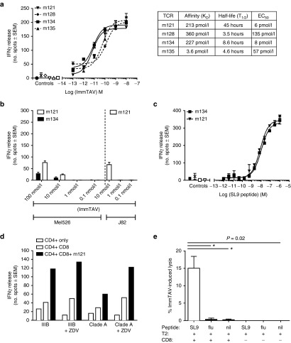
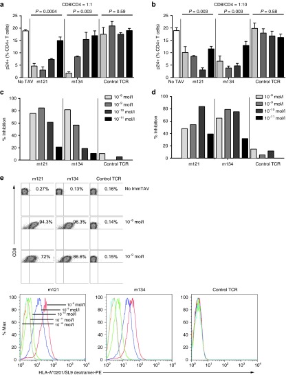
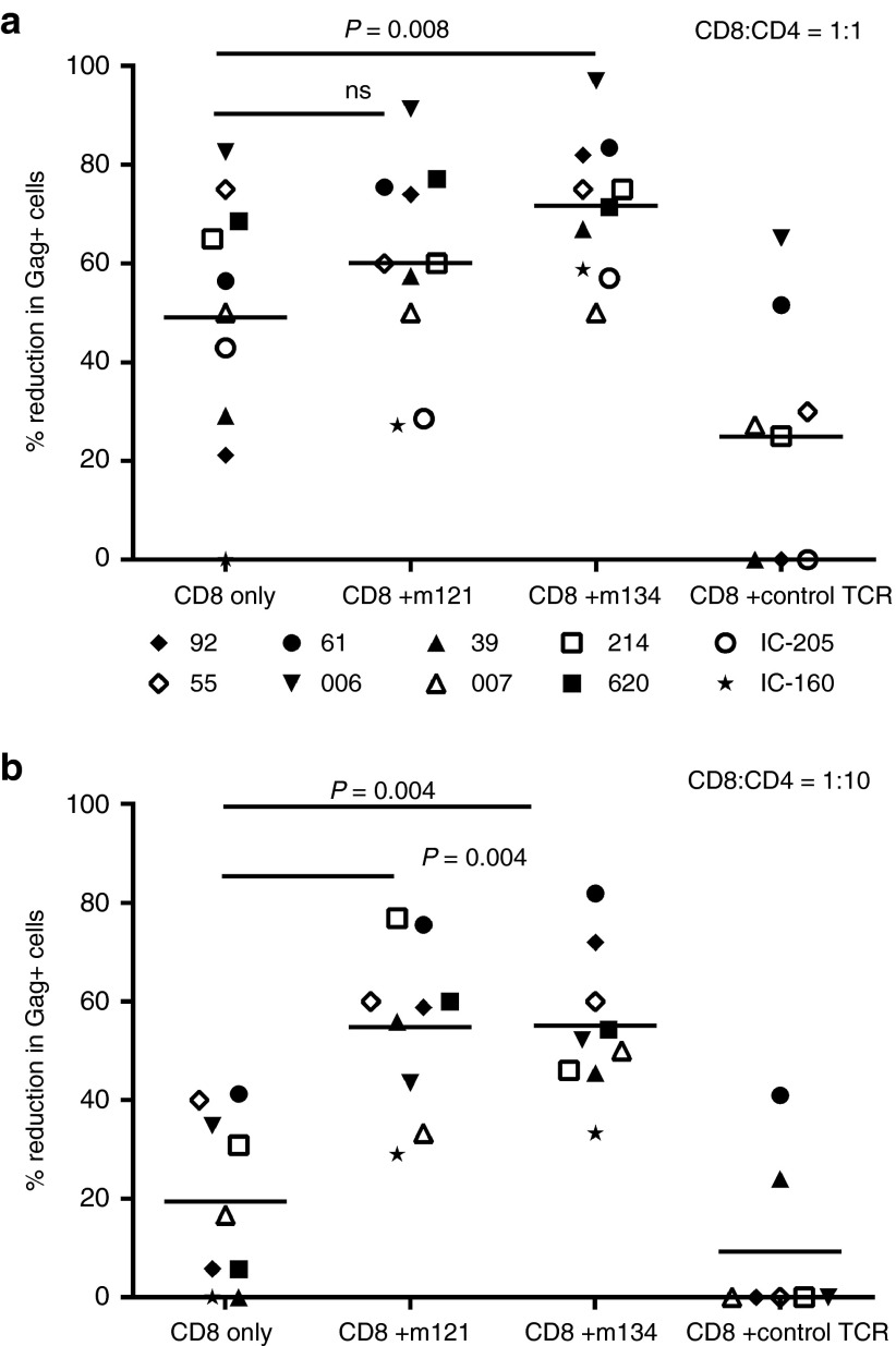
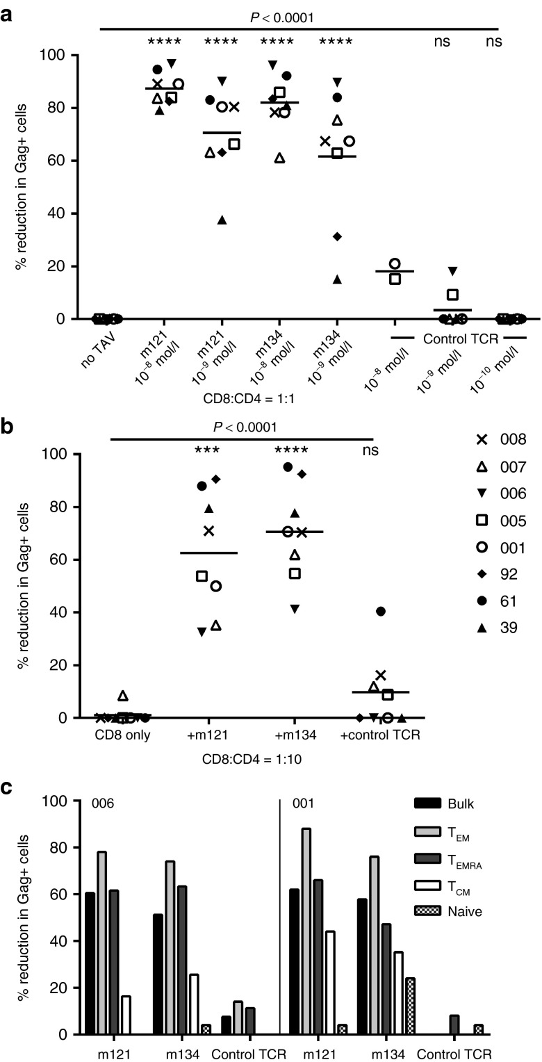
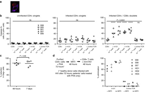
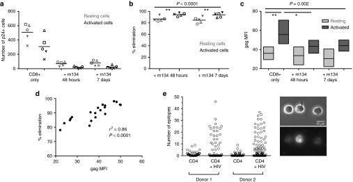
Persistence of human immunodeficiency virus (HIV) in a latent state in long-lived CD4+ T-cells is a major barrier to eradication. Latency-reversing agents that induce direct or immune-mediated cell death upon reactivation of HIV are a possible solution. However, clearance of reactivated cells may require immunotherapeutic agents that are fine-tuned to detect viral antigens when expressed at low levels. We tested the antiviral efficacy of immune-mobilizing monoclonal T-cell receptors against viruses (ImmTAVs), bispecific molecules that redirect CD8+ T-cells to kill HIV-infected CD4+ T-cells. T-cell receptors specific for an immunodominant Gag epitope, SL9, and its escape variants were engineered to achieve supraphysiological affinity and fused to a humanised CD3-specific single chain antibody fragment. Ex vivo polyclonal CD8+ T-cells were efficiently redirected by immune-mobilising monoclonal T-cell receptors against viruses to eliminate CD4+ T-cells from human histocompatibility leukocyte antigen (HLA)-A*0201-positive antiretroviral therapy-treated patients after reactivation of inducible HIV in vitro. The efficiency of infected cell elimination correlated with HIV Gag expression. Immune-mobilising monoclonal T-cell receptors against viruses have potential as a therapy to facilitate clearance of reactivated HIV reservoir cells.


Figures

Figure 1
Figure 1
Biophysical and functional analysis of affinity-matured immune-mobilising monoclonal T-cell receptors against viruses (ImmTAVs) specific for SL9 epitope. (a) Left: Interferon (IFN)-γ responses of CD8+ T-cells from a healthy donor (8 × 104 cells/well) mediated by titrated concentrations of four SL9-directed ImmTAVs (m121, m128, m134, and m135) when cultured with T2 cells pulsed with 10–9 mol/l SL9 peptide (5 × 104 cells/well), quantified by ELISpot. Negative controls for each ImmTAV at 10–9 mol/l were T2 cells pulsed with an irrelevant peptide (open symbols), CD8+ T-cells in the absence of ImmTAV (cross symbol), and T2 cells only (closed circle). Right: Binding parameters (affinity (KD) and half-life (T1/2)) of each of the four ImmTAVs to SL9 peptide, obtained by surface plasmon resonance analysis. (b) Reactivity of ImmTAVs m121 and m134, against Gag SL9-negative, human histocompatibility leukocyte antigen (HLA) -A*0201-expressing human melanoma cells (Mel526) (5 × 104 cells/well). m121 was also tested against malignant human urothelial cells (J82). ImmTAVs were used at concentrations indicated, with CD8+ T-cells from a healthy donor (8 × 104 cells/well). (c). IFN-γ ELISpot assays showing sensitivity of ImmTAVs, m121, and m134 (10–9 mol/l ImmTAV), to T2 cells (5 × 104/well) pulsed with titrated concentrations of SL9 peptide. Healthy donor CD8+ T-cells and controls were as described in a. (d) Response to human immunodeficiency virus (HIV) -infected (IIIB or A isolates) primary CD4+ T-cells (5 × 104 cells/well) by healthy donor CD8+ T-cells (8 × 104 cells/well) when redirected by m121 ImmTAV (10–8 mol/l). Zidovudine (ZDV) (5 μg/ml) was added as indicated, as we observed that this reduced nonspecific IFN-γ release by CD4+ T-cells when infected with HIV in vitro. (e) CFSE-labelled T2 cells were pulsed with 10 μmol/l SL9 peptide, irrelevant (influenza) peptide or unpulsed, then cultured with healthy donor CD8+ T-cells with or without ImmTAV (1 nmol/l) for 24 hours and analyzed by flow cytometry.
Figure 2
Figure 2
Inhibition of exogenous HIV by ImmTAV-redirected CD8+ T-cells. Primary CD4+ T-cells from healthy HLA-A*0201-positive donors were infected with HIV-IIIB and cultured alone (5 × 104 cells/well), with autologous CD8+ T-cells only or with CD8+ T-cells plus HIV ImmTAVs, m121 and m134, or an irrelevant TCR-anti-CD3 scFV fusion (control TCR). (a, b). Frequencies of HIV-infected (p24 Ag+) CD4+ T-cells and (c, d) percent inhibition after 7 days' culture at CD8+/CD4+ cell ratios of 1:1 a, c and 1:10 b, d; ImmTAVs were tested at concentrations ranging from 10–8 – 10–11 mol/l. Data shown are mean ± SD and are representative of three experiments. Percent inhibition was calculated as described in Materials and Methods. (e) Saturation of CD8+ T-cells with ImmTAVs: CD8+ T-cells were incubated with ImmTAVs at concentrations indicated, before staining with an HLA-A*0201/SL9 dextramer and analysis by flow cytometry. Dextramer binding is shown in dot plots (top) at ImmTAV concentrations of 0, 10–8, and 10–9 mol/l and in histograms (bottom) at ImmTAV concentrations of 10–8 – 10–12 mol/l for each TCR. HLA, human histocompatibility leukocyte antigen; HIV, human immunodeficiency virus; TCR, T-cell receptor.
Figure 3
Figure 3
ImmTAV-mediated inhibition of autologous HIV spread in CD4+ T-cells from HIV-infected subjects. Purified CD4+ T-cells (1 × 105) from 10 HIV-positive HLA-A*0201+ subjects (8 ART-treated and 2 ART-naive, represented by symbols shown above) were stimulated with PHA for 72 hours to promote replication of endogenous HIV, then cultured with autologous CD8+ T-cells at CD8+/CD4+ cell ratios of 1:1 (a) and 1:10 (b), either alone or with ImmTAVs as indicated (ImmTAVs at 10–8 mol/l for the 1:1 ratio, 10–11 mol/l for the 1:10 ratio). The irrelevant TCR-anti-CD3 scFV fusion (control TCR) was tested at 10–9 mol/l and 10–12 mol/l for the 1:1 and 1:10 ratios respectively because of nonspecific cell death at higher concentrations. Reduction in Gag+ cells was determined on day 7 of coculture. Horizontal lines indicate mean values. HLA, human histocompatibility leukocyte antigen; HIV, human immunodeficiency virus; ImmTAV, immune-mobilising monoclonal T-cell receptors against virus; TCR, T-cell receptor.
Figure 4
Figure 4
Antiviral potency of ImmTAV-redirected healthy donor CD8+ T-cells against HIV-infected CD4+ T-cells from patients. Purified primary CD4+ T-cells (1 × 105) from 8 HIV-positive HLA-A*0201-positive ART-treated subjects were stimulated with PHA to reactivate latent HIV, then cultured with CD8+ T-cells from an HIV-negative donor at (a) a CD8+/CD4+ cell ratio of 1:1, alone or with ImmTAVs at the concentrations indicated; (b) a CD8+/CD4+ ratio of 1:10, alone or with m121 and m134 − 10–11 mol/l, irrelevant TCR-anti-CD3 scFV fusion (control TCR) − 10–12 mol/l. The same subjects are represented in a and b by the symbols in the legend. Reduction in Gag+ cells was determined on day 7 of coculture. Horizontal lines indicate mean values. Note that inhibition in the presence of the control TCR was negligible even when used at the same concentration as the HIV ImmTAVs. (c) Healthy donor CD8+ T-cells were sorted into subsets according to expression of CCR7 and CD45RA and cultured with purified PHA-activated CD4+ T-cells from two ART-treated patients at a ratio of 1:2, in the presence or absence of ImmTAVs (all at 10–9 mol/l) for 7 days. TEM – effector memory, CCR7−/CD45RA−; TEMRA – terminally differentiated effector, CCR7-/CD45RA+; TCM – central memory, CCR7+/CD45RA-; naive, CCR7+/CD45RA+. Reduction in Gag+ cells was determined on day 7 of co-culture. ART, antiretroviral therapy; PHA, phytohemagglutinin; HLA, human histocompatibility leukocyte antigen; HIV, human immunodeficiency virus; ImmTAV, immune-mobilising monoclonal T-cell receptors against virus; TCR, T-cell receptor.
Figure 5
Figure 5
ImmTAVs mediate killing of infected CD4+ T-cells. (a) Confocal image of a conjugate between an HIV-infected CD4+ T-cell and healthy donor CD8+ T-cells in the presence of an ImmTAV (m121, 10–9 mol/l). Red – p24 Ag; magenta – CD8. (b) Purified activated CD4+ T-cells from 5 HLA-A*0201-positive ART-treated patients were cultured with healthy donor CD8+ T-cells alone or with ImmTAVs at the concentrations indicated. Caspase-3 expression in CD4+ T-cells that were uninfected (left), HIV-infected singlets (middle) or HIV-infected and forming conjugates with CD8+ T-cells (right) was determined by flow cytometric analysis (gating strategy in Supplementary Figure S2). (c) Purified activated CD4+ T-cells from the same five ART-treated patients were cultured with healthy donor CD8+ T-cells (CD8+/CD4+ cell ratio = 1:1) alone or with an ImmTAV (m134, 10–8 mol/l), which was either present throughout the culture period or washed out by replacing the culture medium after 48 hours. Reduction in Gag+ cells, normalized to CD8-only values, on day 7 of coculture is shown. In b and c, horizontal lines indicate mean values. (d) Schema (left) to illustrate the same coculture assay with NRTI-treated CD4+ T-cells. Tenofovir (10 µmol/l) was added to PHA-activated CD4+ T-cells from three ART-treated patients (006, 007, 008, black symbols, immediately after harvesting) and three healthy donors (HD1, HD2, HD3, gray symbols, immediately after spinoculation with HIV IIIB). After 48 hours of drug / mock drug exposure, healthy donor CD8+ T-cells and ImmTAVs were added and cultures were maintained for a further 48 hours. Percent elimination of HIV-infected cells normalized to no TCRs is shown (right). ART, ; PHA, ; HD, ; HIV, human immunodeficiency virus; ImmTAV, immune-mobilising monoclonal T-cell receptors against virus; TCR, T-cell receptor.
Figure 6
Figure 6
ImmTAV-mediated elimination of activated and resting HIV-infected CD4+ T-cells. Purified PHA-stimulated CD4+ T-cells from the same five HLA-A*0201-positive ART-treated patients as in Figure 5 were cultured with healthy donor CD8+ T-cells alone or with ImmTAV m134 (10–8 mol/l). The ImmTAV was either present for the duration of coculture or washed out after 48 hours. (a) The number of activated (CD25+/CD69+/HLA-DR+) and resting (CD25-/CD69-/HLA-DR-) Gag+ CD4+ T-cells remaining on day 7 of coculture and (b) percent elimination of infected cells, normalized to CD8-only values, within each subset is shown. (c) Mean fluorescence intensity (MFI, arbitrary units) of Gag expression in residual infected activated and resting CD4+ T-cells after culture under the conditions indicated (as for a and b; bars and horizontal lines represent range and mean respectively). Consistency in MFI values was ensured by acquiring all samples in a single run. (d). Correlation between Gag MFI (arbitrary units) and normalized percent elimination of infected cells. (e) Biotinylated m134 ImmTAV was detected by microscopy on the surface of individual HIV-1 IIIB-infected CD4+ T-cells from two healthy donors after staining with streptavidin-PE. Data shown in left panel represent counts of fluorescent spots; the total for each cell was obtained using Z-stack images. Uninfected cells were used to determine background staining. Right panel: representative phase-contrast (top) and corresponding fluorescence images (bottom) of infected CD4+ T-cells from donor 1 stained with m134 ImmTAV. Fluorescence images are 3D reconstructions of individual planes. The brightness/contrast of images was adjusted to optimize epitope visualization. The scale bar represents 10 µm. HLA, human histocompatibility leukocyte antigen; PHA, phytohemagglutinin; HIV, human immunodeficiency virus; ImmTAV, immune-mobilising monoclonal T-cell receptors against virus.
Figure 7
Figure 7
ImmTAV-mediated clearance of autologous HIV reservoir cells. (a). Schema illustrating assay to quantify ImmTAV-mediated elimination of resting HIV-infected Cd4+ T-cells from ART-treated patients after viral reactivation. CD25-/69-/HLA-DR- CD4+ cells were thoroughly washed after PHA treatment and cultured in duplicate at a density of 3 × 105 cells/well alone, with autologous CD8+ T-cells only or with healthy donor CD8+ T-cells plus ImmTAVs (10–9 mol/l). Irradiated allogeneic PBMC feeders (1.5–3 × 106) were added to all wells. CD8+/CD4+ cell ratios were 1:1 throughout. After 14 days, Gag+ cells were quantified by flow cytometry. (b) Number of Gag+ cells remaining at day 14 of culture (mean of duplicate wells). (c) Percent elimination of Gag+ cells, determined by normalizing to no TCRs. In all subjects, culture supernatants from CD4+ cell-only wells were positive for free p24 Ag and ImmTAV-treated wells were negative (determined by enzyme-linked immunosorbent assay, cut-off 1500 pg/ml). (d) CD25-/69-/HLA-DR- CD4+ cells from five ART-treated HLA-A*0201-positive subjects were thoroughly washed after LRA (bryostatin / romidepsin) treatment and cultured in duplicate with healthy donor CD8+ T-cells (1:1 ratio) ± ImmTAV m121 or control TCR (10–9 mol/l) for a further 42 hours. Supernatants were harvested and viral outgrowth was determined by quantification of HIV RNA, as described in Materials and Methods. LRA, latency-reversing agent; HLA, human histocompatibility leukocyte antigen; ART, antiretroviral therapy; PHA, phytohemagglutinin; HIV, human immunodeficiency virus; ImmTAV, immune-mobilising monoclonal T-cell receptors against virus; PBMC, peripheral blood mononuclear cells; TCR, T-cell receptor.

Comment in

References

    1. Allers, K, Hütter, G, Hofmann, J, Loddenkemper, C, Rieger, K, Thiel, E et al. (2011). Evidence for the cure of HIV infection by CCR5Δ32/Δ32 stem cell transplantation. Blood 117: 2791–2799. - PubMed
    1. Shan, L, Deng, K, Shroff, NS, Durand, CM, Rabi, SA, Yang, HC et al. (2012). Stimulation of HIV-1-specific cytolytic T lymphocytes facilitates elimination of latent viral reservoir after virus reactivation. Immunity 36: 491–501. - PMC - PubMed
    1. Autran, B, Descours, B and Bacchus, C (2013). Immune control of HIV-1 reservoirs. Curr Opin HIV AIDS 8: 204–210. - PubMed
    1. Archin, NM, Liberty, AL, Kashuba, AD, Choudhary, SK, Kuruc, JD, Crooks, AM et al. (2012). Administration of vorinostat disrupts HIV-1 latency in patients on antiretroviral therapy. Nature 487: 482–485. - PMC - PubMed
    1. Vanham, G and Van Gulck, E (2012). Can immunotherapy be useful as a “functional cure” for infection with Human Immunodeficiency Virus-1? Retrovirology 9: 72. - PMC - PubMed

MeSH terms