

Paclitaxel causes degeneration of both central and peripheral axon branches of dorsal root ganglia in mice
- PMID: 27401104
- PMCID: PMC4940970
- DOI: 10.1186/s12868-016-0285-4
Paclitaxel causes degeneration of both central and peripheral axon branches of dorsal root ganglia in mice
Abstract
Background: Peripheral neuropathy is a common and dose-limiting side effect of many cancer chemotherapies. The taxane agents, including paclitaxel (Taxol(®)), are effective chemotherapeutic drugs but cause degeneration of predominantly large myelinated afferent sensory fibers of the peripheral nervous system in humans and animal models. Dorsal root ganglia (DRG) neurons are sensory neurons that have unipolar axons each with two branches: peripheral and central. While taxane agents induce degeneration of peripheral axons, whether they also cause degeneration of central nervous system axons is not clear. Using a mouse model of paclitaxel-induced neuropathy, we investigated the effects of paclitaxel on the central branches of sensory axons.
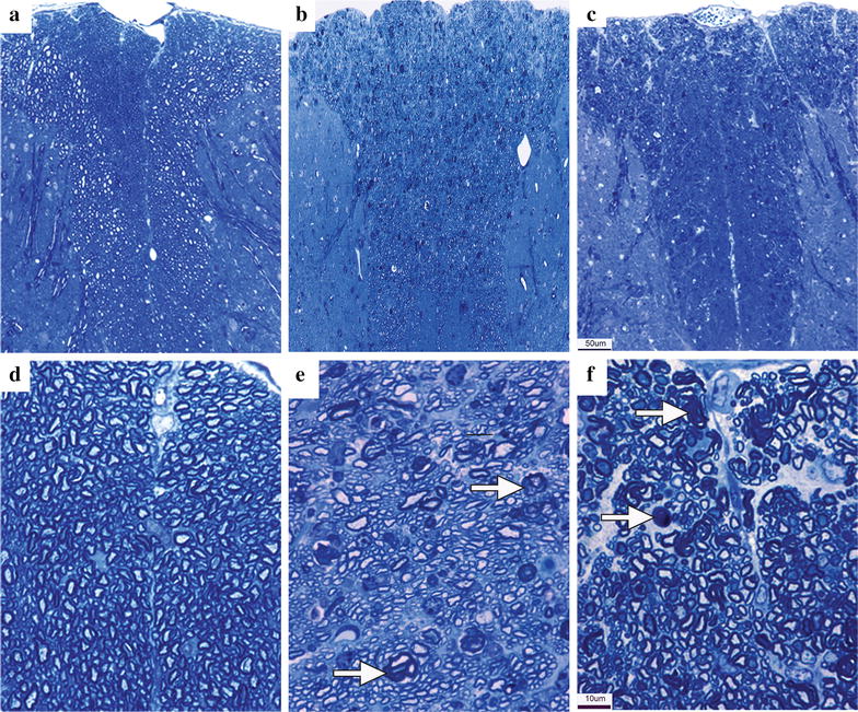
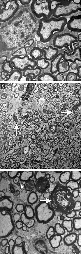
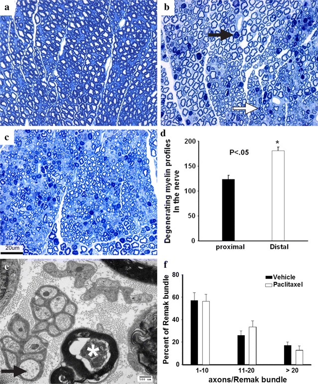
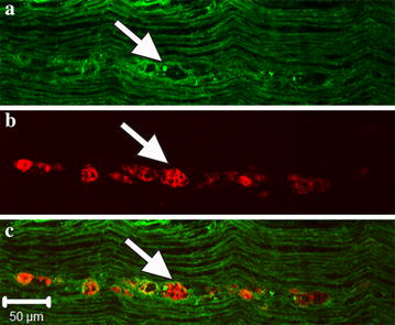
Results: We observed that in the spinal cords of paclitaxel-intoxicated mice, degenerated axons were present in the dorsal columns, where the central branches of DRG axons ascend rostrally. In the peripheral nerves, degenerated myelinated fibers were present in significantly greater numbers in distal segments than in proximal segments indicating that this model exhibits the distal-to-proximal degeneration pattern generally observed in human peripheral nerve disorders.
Conclusions: We conclude that paclitaxel causes degeneration of both the peripheral and central branches of DRG axons, a finding that has implications for the site and mode of action of chemotherapy agents on the nervous system.
Keywords: Activated macrophages; Axonal degeneration; Neuropathy.
Figures

Similar articles
-
Targeting Axon Integrity to Prevent Chemotherapy-Induced Peripheral Neuropathy.Mol Neurobiol. 2019 May;56(5):3244-3259. doi: 10.1007/s12035-018-1301-8. Epub 2018 Aug 16. Mol Neurobiol. 2019. PMID: 30117103
-
Suramin-induced neuropathy in an animal model.J Neurol Sci. 2001 Nov 15;192(1-2):71-80. doi: 10.1016/s0022-510x(01)00633-5. J Neurol Sci. 2001. PMID: 11701155
-
Compartmentalized microfluidic culture platform to study mechanism of paclitaxel-induced axonal degeneration.Exp Neurol. 2009 Jul;218(1):124-8. doi: 10.1016/j.expneurol.2009.04.017. Epub 2009 May 3. Exp Neurol. 2009. PMID: 19409381 Free PMC article.
-
Rodent models of chemotherapy-induced peripheral neuropathy.ILAR J. 2014;54(3):273-81. doi: 10.1093/ilar/ilt053. ILAR J. 2014. PMID: 24615440 Review.
-
A conditioning lesion induces changes in gene expression and axonal transport that enhance regeneration by increasing the intrinsic growth state of axons.Exp Neurol. 2010 May;223(1):11-8. doi: 10.1016/j.expneurol.2009.09.006. Epub 2009 Sep 17. Exp Neurol. 2010. PMID: 19766119 Review.
Cited by
-
Neuroprotective Effect of Ramipril Is Mediated by AT2 in a Mouse MODEL of Paclitaxel-Induced Peripheral Neuropathy.Pharmaceutics. 2022 Apr 12;14(4):848. doi: 10.3390/pharmaceutics14040848. Pharmaceutics. 2022. PMID: 35456682 Free PMC article.
-
Human Intravenous Immunoglobulin Alleviates Neuropathic Symptoms in a Rat Model of Paclitaxel-Induced Peripheral Neurotoxicity.Int J Mol Sci. 2021 Jan 21;22(3):1058. doi: 10.3390/ijms22031058. Int J Mol Sci. 2021. PMID: 33494384 Free PMC article.
-
TMI-1, TNF-α-Converting Enzyme Inhibitor, Protects Against Paclitaxel-Induced Neurotoxicity in the DRG Neuronal Cells In Vitro.Front Pharmacol. 2022 Feb 16;13:842779. doi: 10.3389/fphar.2022.842779. eCollection 2022. Front Pharmacol. 2022. PMID: 35250589 Free PMC article.
-
7-Chloro-4-(Phenylselanyl) Quinoline Is a Novel Multitarget Therapy to Combat Peripheral Neuropathy and Comorbidities Induced by Paclitaxel in Mice.Mol Neurobiol. 2022 Oct;59(10):6567-6589. doi: 10.1007/s12035-022-02991-4. Epub 2022 Aug 15. Mol Neurobiol. 2022. PMID: 35965270
-
Comparative Analysis of Chemotherapy-Induced Peripheral Neuropathy in Bioengineered Sensory Nerve Tissue Distinguishes Mechanistic Differences in Early-Stage Vincristine-, Cisplatin-, and Paclitaxel-Induced Nerve Damage.Toxicol Sci. 2021 Feb 26;180(1):76-88. doi: 10.1093/toxsci/kfaa186. Toxicol Sci. 2021. PMID: 33410881 Free PMC article.
References
Publication types
MeSH terms
Substances
Grants and funding
LinkOut - more resources
Full Text Sources
Other Literature Sources
