

Adenosine Selectively Depletes Alloreactive T Cells to Prevent GVHD While Conserving Immunity to Viruses and Leukemia
- PMID: 27401140
- PMCID: PMC5113112
- DOI: 10.1038/mt.2016.147
Adenosine Selectively Depletes Alloreactive T Cells to Prevent GVHD While Conserving Immunity to Viruses and Leukemia
Abstract
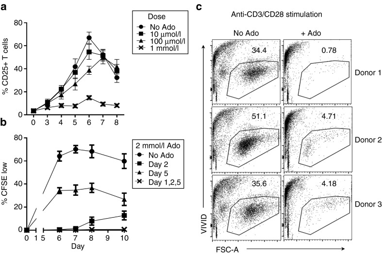
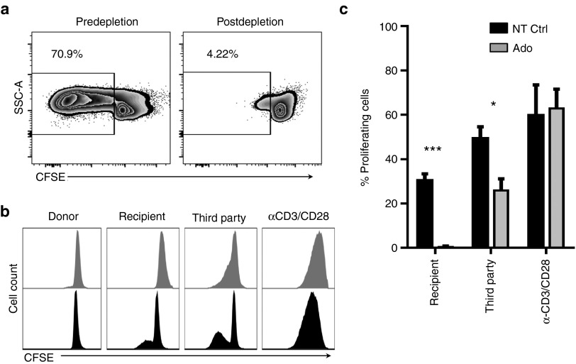
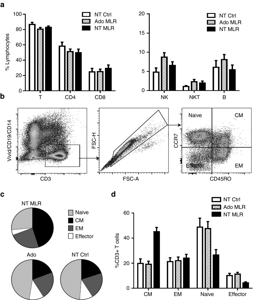
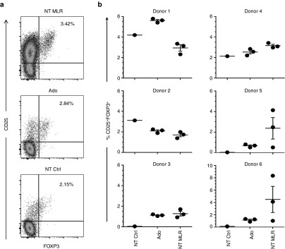
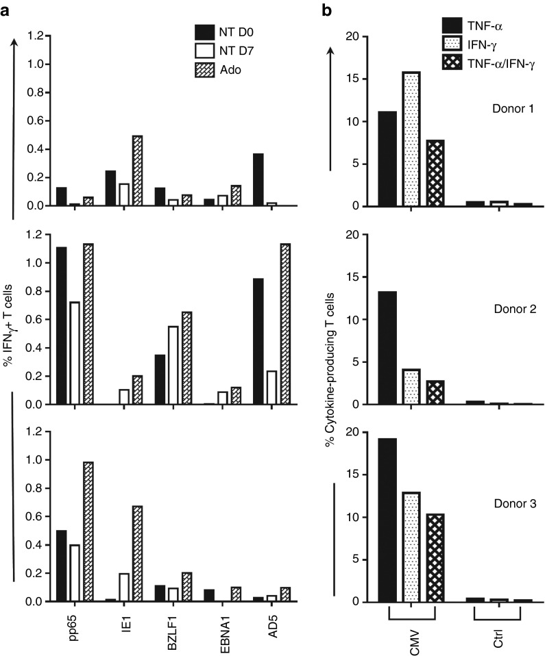
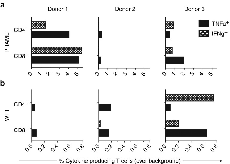
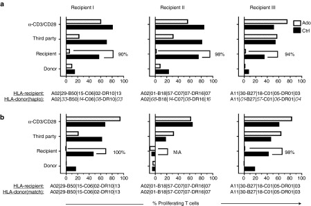
Selective depletion (SD) of alloreactive T cells from allogeneic hematopoeitic stem cell transplants to prevent graft-versus-host disease (GVHD) without compromising immune reconstitution and antitumor responses remains a challenge. Here, we demonstrate a novel SD strategy whereby alloreacting T cells are efficiently deleted ex vivo with adenosine. SD was achieved in human leukocyte antigen (HLA) mismatched cocultures by multiple exposures to 2 mmol/l adenosine over 7 days. Adenosine depleted greater than to 90% of alloproliferating T cells in mismatched, haploidentical, and matched sibling pairs while conserving response to third-party antigens. Alloreactive CD4 and CD8 T cells were targeted for depletion while NK and B cells were preserved. Our novel approach also preserved nonalloreactive naive, central, and effector memory T-cell subsets, Tregs, and notably preserved T-cell responses against DNA viruses that contribute to transplant related mortality after allogeneic hematopoeitic stem cell transplants. Additionally, T cells recognizing leukemia-associated antigens were efficiently generated in vitro from the cell product post-SD. This study is the first to demonstrate that adenosine depletion of alloactivated T cells maintains a complete immune cell profile and recall viral responses. Expansion of tumor antigen-specific subsets postdepletion opens the possibility of generating T-cell products capable of graft-versus-tumor responses without causing GVHD.
Figures
Similar articles
-
TcRαβ-depleted haploidentical transplantation results in adult acute leukemia patients.Hematology. 2017 Apr;22(3):136-144. doi: 10.1080/10245332.2016.1238182. Epub 2016 Oct 10. Hematology. 2017. PMID: 27724812
-
CD6+ T cell depleted allogeneic bone marrow transplantation from genotypically HLA nonidentical related donors.Biol Blood Marrow Transplant. 1997 Apr;3(1):11-7. Biol Blood Marrow Transplant. 1997. PMID: 9209736
-
Clinical-scale selective depletion of alloreactive T cells using an anti-CD25 immunotoxin.Neoplasma. 2003;50(4):296-9. Neoplasma. 2003. PMID: 12937844
-
Selective T-cell depletion for haplotype-mismatched allogeneic stem cell transplantation.Semin Oncol. 2012 Dec;39(6):674-82. doi: 10.1053/j.seminoncol.2012.09.004. Semin Oncol. 2012. PMID: 23206844 Review.
-
Haploidentical hematopoietic stem cell transplantation with a megadose T-cell-depleted graft: harnessing natural and adaptive immunity.Semin Oncol. 2012 Dec;39(6):643-52. doi: 10.1053/j.seminoncol.2012.09.002. Semin Oncol. 2012. PMID: 23206841 Review.
Cited by
-
Regulation of Treg cells by cytokine signaling and co-stimulatory molecules.Front Immunol. 2024 May 13;15:1387975. doi: 10.3389/fimmu.2024.1387975. eCollection 2024. Front Immunol. 2024. PMID: 38807592 Free PMC article. Review.
-
Cell therapies for hematological malignancies: don't forget non-gene-modified t cells!Blood Rev. 2018 May;32(3):203-224. doi: 10.1016/j.blre.2017.11.004. Epub 2017 Nov 27. Blood Rev. 2018. PMID: 29198753 Free PMC article. Review.
-
Haploidentical- versus identical-sibling transplant for high-risk pediatric AML: A multi-center study.Cancer Commun (Lond). 2020 Mar;40(2-3):93-104. doi: 10.1002/cac2.12014. Epub 2020 Mar 16. Cancer Commun (Lond). 2020. PMID: 32175698 Free PMC article.
-
Mesenchymal Stromal Cells: What Is the Mechanism in Acute Graft-Versus-Host Disease?Biomedicines. 2017 Jul 1;5(3):39. doi: 10.3390/biomedicines5030039. Biomedicines. 2017. PMID: 28671556 Free PMC article. Review.
-
Developing T-cell therapies for lymphoma without receptor engineering.Hematology Am Soc Hematol Educ Program. 2017 Dec 8;2017(1):622-631. doi: 10.1182/asheducation-2017.1.622. Hematology Am Soc Hematol Educ Program. 2017. PMID: 29222313 Free PMC article. Review.
References
-
- Martelli, MF, Di Ianni, M, Ruggeri, L, Falzetti, F, Carotti, A, Terenzi, A et al. (2014). HLA-haploidentical transplantation with regulatory and conventional T-cell adoptive immunotherapy prevents acute leukemia relapse. Blood 124: 638–644. - PubMed
-
- Huang, XJ, Liu, DH, Liu, KY, Xu, LP, Chen, H, Han, W et al. (2009). Treatment of acute leukemia with unmanipulated HLA-mismatched/haploidentical blood and bone marrow transplantation. Biol Blood Marrow Transplant 15: 257–265. - PubMed
-
- Luznik, L, O'Donnell, PV, Symons, HJ, Chen, AR, Leffell, MS, Zahurak, M et al. (2008). HLA-haploidentical bone marrow transplantation for hematologic malignancies using nonmyeloablative conditioning and high-dose, posttransplantation cyclophosphamide. Biol Blood Marrow Transplant 14: 641–650. - PMC - PubMed
Publication types
MeSH terms
Substances
LinkOut - more resources
Full Text Sources
Other Literature Sources
Medical
Research Materials
