Review
. 2016 Aug;11(2):87-97.
doi: 10.1007/s11751-016-0258-2. Epub 2016 Jul 11.

The management of paediatric diaphyseal femoral fractures: a modern approach

Affiliations
Review

The management of paediatric diaphyseal femoral fractures: a modern approach

Al-Achraf Khoriati et al. Strategies Trauma Limb Reconstr. 2016 Aug.

Abstract

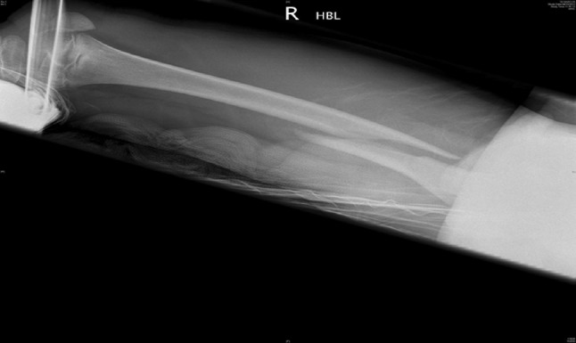
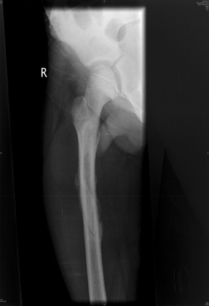
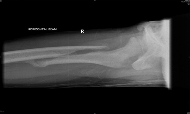
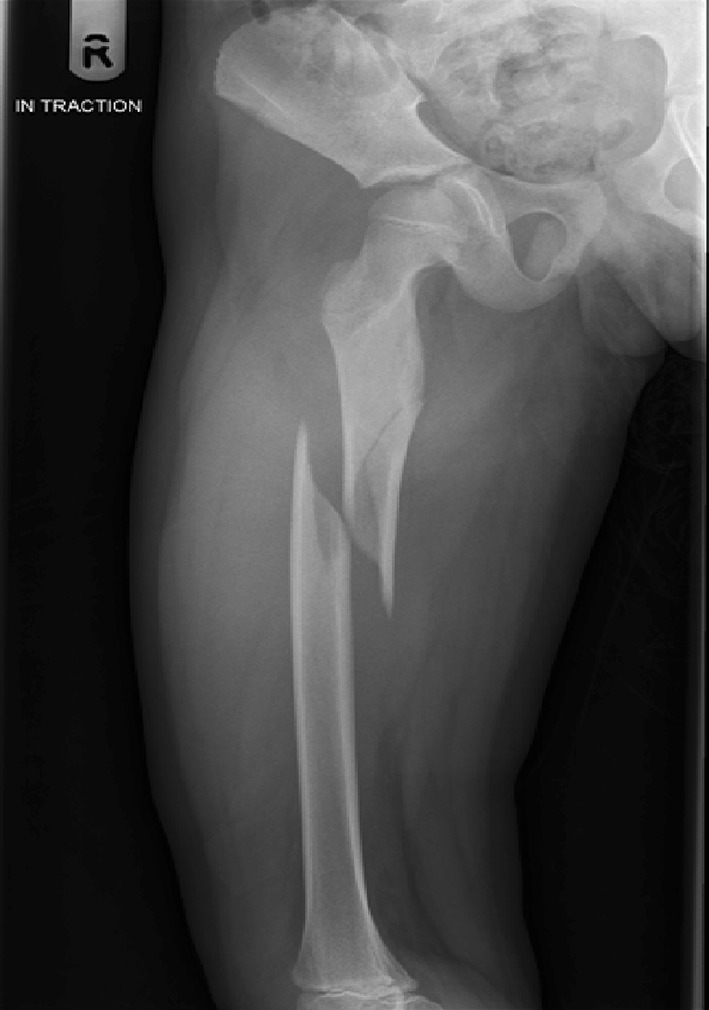
The definitive treatment of paediatric femoral diaphyseal fractures remains controversial. Modalities of treatment vary mostly according to age, with fracture pattern and site having a lesser impact. Current evidence is reflective of this variation with most evidence cited by the American Academy of Orthopedic Surgeons being level 4 or 5. The authors present a review of the most up-to-date evidence relating to the treatment of these fractures in each age group. In an attempt to clarify the current trends, we have produced an algorithm for decision-making based on the experience from our own tertiary referral level 1 major trauma centre.

Keywords: Evidence; Femur; Fracture; Management; Paediatric; Review; Trauma.
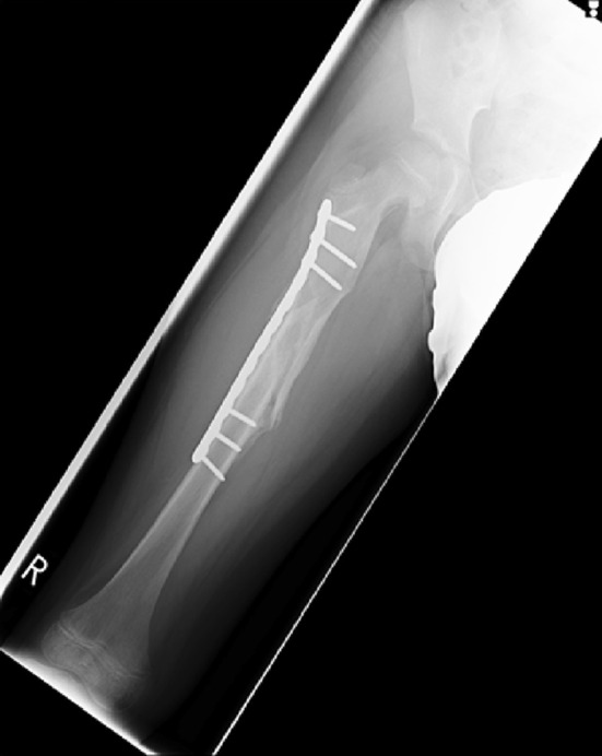
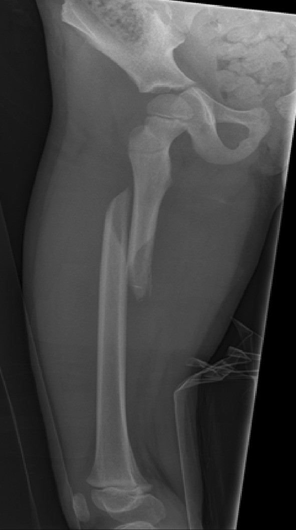
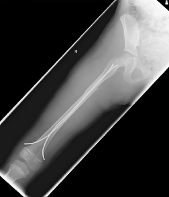
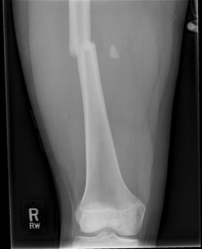

Figures

Fig. 1
Fig. 1
Paediatric diaphyseal classification system
Fig. 2
Fig. 2
AP radiograph demonstrating a distal 1/3 spiral femoral diaphyseal fracture in a 6-year-old child
Fig. 3
Fig. 3
AP radiograph of the same child as shown in Fig. 2, taken at 8 weeks, showing solid union and acceptable alignment after an initial treatment of 3- to 4-week in-line traction
Fig. 4
Fig. 4
AP radiographs demonstrating a proximal 1/3 spiral femoral diaphyseal fracture in a 6-year-old child
Fig. 5
Fig. 5
Lateral radiographs demonstrating a proximal 1/3 spiral femoral diaphyseal fracture in a 6-year-old child
Fig. 6
Fig. 6
After 4 weeks in traction, a healthy callus is seen to form in children aged 5–12 years
Fig. 7
Fig. 7
After 4 weeks in traction, a healthy callus is seen to form in children aged 5–12 years
Fig. 8
Fig. 8
AP radiograph demonstrating a proximal third diaphyseal fracture of the femur in an 8-year-old. Note the spiral fracture with a butterfly fragment—elastic nails may be unstable here
Fig. 9
Fig. 9
AP radiograph showing the patient from Fig. 8, treated with a submuscular 3.5-mm bridge plate. At just 8 weeks, there is abundant callus and the patient is fully weight bearing
Fig. 10
Fig. 10
AP radiograph demonstrating a spiral femoral fracture ideal for treatment with flexible nails
Fig. 11
Fig. 11
AP radiograph showing satisfactory restoration of length, rotation and alignment with the use of titanium elastic nails, of the fracture in Fig. 10
Fig. 12
Fig. 12
Treatment of a femoral diaphyseal fracture in a 16-year-old girl. Note is made of the subtle nuances such as the narrow canal and non-fused physis which must be considered in the management of these fractures
Fig. 13
Fig. 13
Treatment of a femoral diaphyseal fracture in a 16-year-old girl. Note is made of the subtle nuances such as the narrow canal and non-fused physis which must be considered in the management of these fractures
Fig. 14
Fig. 14
Algorithm summarising the management pathways for paediatric femoral diaphyseal fractures

References

    1. Bridgman S, Wilson R. Epidemiology of femoral fractures in children in the West Midlands region of England 1991–2001. J Bone Joint Surg Br. 2004;86(8):1152–1157. doi: 10.1302/0301-620X.86B8.14810. - DOI - PubMed
    1. Harvey AR, Bowyer GW, Clarke NMP. The management of paediatric femoral shaft fractures. Curr Orthop. 2002;16(4):293–299. doi: 10.1054/cuor.2002.0254. - DOI
    1. Schwend RM, Werth C, Johnston A. Femur shaft fractures in toddlers and young children: rarely from child abuse. J Pediatr Orthop. 2000;20(4):475–481. - PubMed
    1. Pierce MC, Bertocci GE, Janosky JE, Aguel F, Deemer E, Moreland M, et al. Femur fractures resulting from stair falls among children: an injury plausibility model. Pediatrics. 2005;115(6):1712–1722. doi: 10.1542/peds.2004-0614. - DOI - PubMed
    1. Wallace ME, Hoffman EB. Remodelling of angular deformity after femoral shaft fractures in children. J Bone Joint Surg Br. 1992;74(5):765–769. - PubMed

LinkOut - more resources