

Pittsburgh compound B imaging and cerebrospinal fluid amyloid-β in a multicentre European memory clinic study
- PMID: 27401520
- PMCID: PMC4995359
- DOI: 10.1093/brain/aww160
Pittsburgh compound B imaging and cerebrospinal fluid amyloid-β in a multicentre European memory clinic study
Abstract
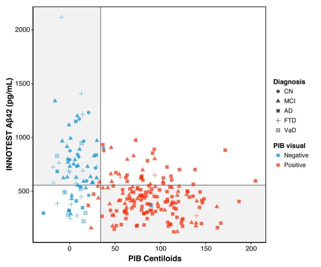
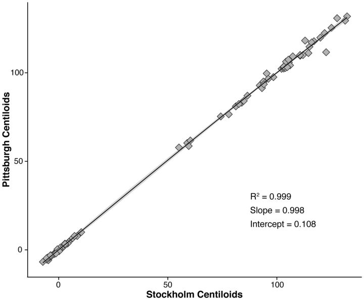
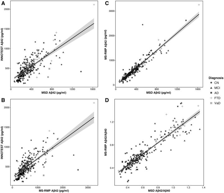
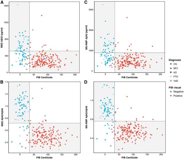
The aim of this study was to assess the agreement between data on cerebral amyloidosis, derived using Pittsburgh compound B positron emission tomography and (i) multi-laboratory INNOTEST enzyme linked immunosorbent assay derived cerebrospinal fluid concentrations of amyloid-β42; (ii) centrally measured cerebrospinal fluid amyloid-β42 using a Meso Scale Discovery enzyme linked immunosorbent assay; and (iii) cerebrospinal fluid amyloid-β42 centrally measured using an antibody-independent mass spectrometry-based reference method. Moreover, we examined the hypothesis that discordance between amyloid biomarker measurements may be due to interindividual differences in total amyloid-β production, by using the ratio of amyloid-β42 to amyloid-β40 Our study population consisted of 243 subjects from seven centres belonging to the Biomarkers for Alzheimer's and Parkinson's Disease Initiative, and included subjects with normal cognition and patients with mild cognitive impairment, Alzheimer's disease dementia, frontotemporal dementia, and vascular dementia. All had Pittsburgh compound B positron emission tomography data, cerebrospinal fluid INNOTEST amyloid-β42 values, and cerebrospinal fluid samples available for reanalysis. Cerebrospinal fluid samples were reanalysed (amyloid-β42 and amyloid-β40) using Meso Scale Discovery electrochemiluminescence enzyme linked immunosorbent assay technology, and a novel, antibody-independent, mass spectrometry reference method. Pittsburgh compound B standardized uptake value ratio results were scaled using the Centiloid method. Concordance between Meso Scale Discovery/mass spectrometry reference measurement procedure findings and Pittsburgh compound B was high in subjects with mild cognitive impairment and Alzheimer's disease, while more variable results were observed for cognitively normal and non-Alzheimer's disease groups. Agreement between Pittsburgh compound B classification and Meso Scale Discovery/mass spectrometry reference measurement procedure findings was further improved when using amyloid-β42/40 Agreement between Pittsburgh compound B visual ratings and Centiloids was near complete. Despite improved agreement between Pittsburgh compound B and centrally analysed cerebrospinal fluid, a minority of subjects showed discordant findings. While future studies are needed, our results suggest that amyloid biomarker results may not be interchangeable in some individuals.
Keywords: Alzheimer’s disease; CSF Aβ42; CSF Aβ42/40; Centiloids; PiB PET.
© The Author (2016). Published by Oxford University Press on behalf of the Guarantors of Brain.
Figures


Similar articles
-
Concordance Between Different Amyloid Immunoassays and Visual Amyloid Positron Emission Tomographic Assessment.JAMA Neurol. 2017 Dec 1;74(12):1492-1501. doi: 10.1001/jamaneurol.2017.2814. JAMA Neurol. 2017. PMID: 29114726 Free PMC article.
-
Regional dynamics of amyloid-β deposition in healthy elderly, mild cognitive impairment and Alzheimer's disease: a voxelwise PiB-PET longitudinal study.Brain. 2012 Jul;135(Pt 7):2126-39. doi: 10.1093/brain/aws125. Epub 2012 May 23. Brain. 2012. PMID: 22628162
-
Accuracy of brain amyloid detection in clinical practice using cerebrospinal fluid β-amyloid 42: a cross-validation study against amyloid positron emission tomography.JAMA Neurol. 2014 Oct;71(10):1282-9. doi: 10.1001/jamaneurol.2014.1358. JAMA Neurol. 2014. PMID: 25155658
-
Subjective cognitive decline in conjunction with cerebrospinal fluid anti-ATP1A3 autoantibodies and a low amyloid β 1-42/1-40 ratio: Report and literature review.Behav Brain Res. 2025 May 8;485:115541. doi: 10.1016/j.bbr.2025.115541. Epub 2025 Mar 16. Behav Brain Res. 2025. PMID: 40101839 Review.
-
Update on amyloid imaging: from healthy aging to Alzheimer's disease.Curr Neurol Neurosci Rep. 2009 Sep;9(5):345-52. doi: 10.1007/s11910-009-0051-4. Curr Neurol Neurosci Rep. 2009. PMID: 19664363 Free PMC article. Review.
Cited by
-
The Cerebrospinal Fluid Aβ1-42/Aβ1-40 Ratio Improves Concordance with Amyloid-PET for Diagnosing Alzheimer's Disease in a Clinical Setting.J Alzheimers Dis. 2017;60(2):561-576. doi: 10.3233/JAD-170327. J Alzheimers Dis. 2017. PMID: 28869470 Free PMC article.
-
Serum Beta-Secretase 1 Activity Is a Potential Marker for the Differential Diagnosis between Alzheimer's Disease and Frontotemporal Dementia: A Pilot Study.Int J Mol Sci. 2024 Jul 30;25(15):8354. doi: 10.3390/ijms25158354. Int J Mol Sci. 2024. PMID: 39125924 Free PMC article.
-
Discordant amyloid-β PET and CSF biomarkers and its clinical consequences.Alzheimers Res Ther. 2019 Sep 12;11(1):78. doi: 10.1186/s13195-019-0532-x. Alzheimers Res Ther. 2019. PMID: 31511058 Free PMC article.
-
Retinal texture biomarkers may help to discriminate between Alzheimer's, Parkinson's, and healthy controls.PLoS One. 2019 Jun 21;14(6):e0218826. doi: 10.1371/journal.pone.0218826. eCollection 2019. PLoS One. 2019. PMID: 31226150 Free PMC article.
-
Multicenter Analytical Validation of Aβ40 Immunoassays.Front Neurol. 2017 Jul 3;8:310. doi: 10.3389/fneur.2017.00310. eCollection 2017. Front Neurol. 2017. PMID: 28725210 Free PMC article.
References
-
- Andreasson U, Vanmechelen E, Shaw LM, Zetterberg H, Vanderstichele H. Analytical aspects of molecular Alzheimer's disease biomarkers. Biomark Med 2012; 6: 377–89. - PubMed
-
- Ayakta N, Lockhart S, O'Neill J, Ossenkoppele R, Reed B, Olichney J, et al. Centiloid thresholds for amyloid positivity derived from autopsy-proven cases. In: Human Amyloid Imaging Conference Book of Abstracts, ID PP89, 29–30, Miami, FL, Jan 13–15, 2016, World Events Forum, Inc.
-
- Bacskai BJ, Frosch MP, Freeman SH, Raymond SB, Augustinack JC, Johnson KA, et al. Molecular imaging with Pittsburgh compound B confirmed at autopsy: a case report. Arch Neurol 2007; 64: 431–4. - PubMed
-
- Benaglia T, Chauvreau D, Hunter DR, Young DS. mixtools: an R package for analyzing finite mixture models. J Stat Softw 2009; 32: 1–29.
Publication types
MeSH terms
Substances
LinkOut - more resources
Full Text Sources
Other Literature Sources
Medical
